

Do Individuals with History of Patellofemoral Pain Walk and Squat Similarly to Healthy Controls? A 3D Kinematic Analysis During Pain Remission Phase
- PMID: 35136687
- PMCID: PMC8805120
- DOI: 10.26603/001c.31044
Do Individuals with History of Patellofemoral Pain Walk and Squat Similarly to Healthy Controls? A 3D Kinematic Analysis During Pain Remission Phase
Abstract
Background: Patellofemoral pain (PFP) is typically accompanied by changes in movement pattern. However, it is unclear if these changes persist in the remission phase of symptoms. Investigating movement patterns in individuals in remission phase of PFP may help to further guide the rehabilitation process and to understand whether changes are due to high levels of pain or related to other factors.
Purpose: To compare 3D kinematics during walking and the single leg squat (SLS) between individuals with history of PFP in remission phase and a control group without history of lower limb injuries and PFP.
Study design: Cross-sectional case-control study.
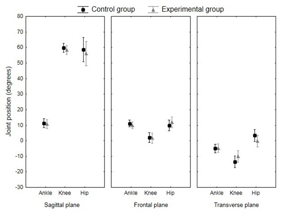
Methods: Individuals with onset of PFP for at least one year and in phase of remission of symptoms (experimental group [EG]; n=13, 30±8 years) were compared to a control group (CG, n=13, 28±7 years). A 10-camera motion analysis system (Vicon-Nexus®) was used to record 3D ankle, knee, hip and trunk angles during walking and SLS.
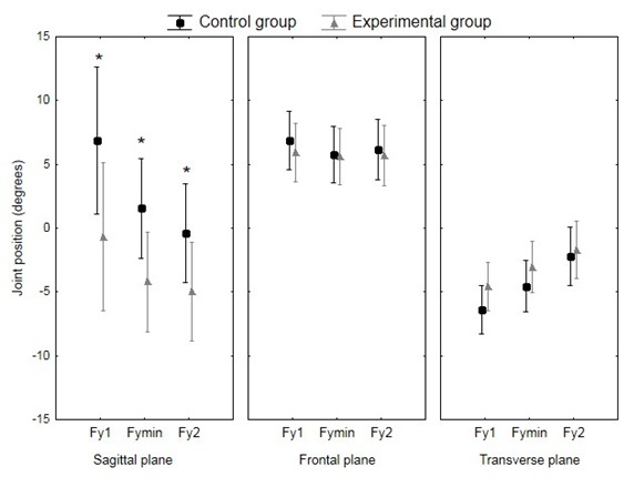
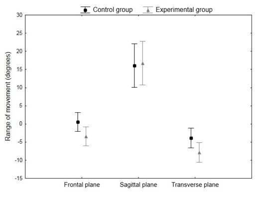
Results: The EG presented less ankle dorsiflexion, knee and hip flexion during the stance phase of walking compared to the CG (p=0.005, large effect size ηp2 = 0.141). During the SLS, no between-group differences were observed for the ankle, knee and hip angles at the peak of knee flexion (p>0.05). A trend for increased trunk range of movement in the EG compared to the CG was observed (p=0.075, medium effect size ηp2 = 0.127).
Conclusion: The results of this study indicate less movement in the sagittal plane during walking, and a trend towards more movement of the trunk during SLS in the EG compared to the CG. The participants of the EG had minimal symptoms, to the point of not classifying them as pathological. However, the between-group differences suggest that even in the remission phase, kinematic differences persist for some reason and may contribute to the recurring pain in PFP individuals.
Level of evidence: Level 3.
Keywords: biomechanical phenomena; knee; lower extremity; patellofemoral pain syndrome.
Conflict of interest statement
The authors report no conflicts of interest or bias in this work.
Figures
References
-
- Crossley Kay M, Stefanik Joshua J, Selfe James, Collins Natalie J, Davis Irene S, Powers Christopher M, McConnell Jenny, Vicenzino Bill, Bazett-Jones David M, Esculier Jean-Francois, Morrissey Dylan, Callaghan Michael J. British Journal of Sports Medicine. 14. Vol. 50. BMJ; 2016 Patellofemoral pain consensus statement from the 4th International Patellofemoral Pain Research Retreat, Manchester. Part 1: Terminology, definitions, clinical examination, natural history, patellofemoral osteoarthritis and patient-reported outcome measures; pp. 839–843. - DOI - DOI - PMC - PubMed
-
- Care-seeking behaviour of adolescents with knee pain: a population-based study among 504 adolescents. Rathleff Michael S, Skuldbøl Sune K, Rasch Mads N B, Roos Ewa M, Rasmussen Sten, Olesen Jens L. Jul 30;2013 BMC Musculoskeletal Disorders. 14(1):225. doi: 10.1186/1471-2474-14-225. doi: 10.1186/1471-2474-14-225. - DOI - DOI - PMC - PubMed
-
- Incidence and prevalence of patellofemoral pain: a systematic review and meta-analysis. Smith Benjamin E., Selfe James, Thacker Damian, Hendrick Paul, Bateman Marcus, Moffatt Fiona, Rathleff Michael Skovdal, Smith Toby O., Logan Pip. Screen Hazel RC., editor. Jan 11;2018 PLOS ONE. 13(1):e0190892. doi: 10.1371/journal.pone.0190892. doi: 10.1371/journal.pone.0190892. - DOI - DOI - PMC - PubMed
-
- Effectiveness of manual therapy combined with physical therapy in treatment of patellofemoral pain syndrome: systematic review. Espí-López Gemma Victoria, Arnal-Gómez Anna, Balasch-Bernat Mercè, Inglés Marta. Jun;2017 Journal of Chiropractic Medicine. 16(2):139–146. doi: 10.1016/j.jcm.2016.10.003. doi: 10.1016/j.jcm.2016.10.003. - DOI - DOI - PMC - PubMed
LinkOut - more resources
Full Text Sources
Research Materials