. 2022 Apr 4;220(4):iyac010.
doi: 10.1093/genetics/iyac010.

Hox proteins interact to pattern neuronal subtypes in Caenorhabditis elegans males

Affiliations

Hox proteins interact to pattern neuronal subtypes in Caenorhabditis elegans males

Andrea K Kalis et al. Genetics. .

Abstract

Hox transcription factors are conserved regulators of neuronal subtype specification on the anteroposterior axis in animals, with disruption of Hox gene expression leading to homeotic transformations of neuronal identities. We have taken advantage of an unusual mutation in the Caenorhabditis elegans Hox gene lin-39, lin-39(ccc16), which transforms neuronal fates in the C. elegans male ventral nerve cord in a manner that depends on a second Hox gene, mab-5. We have performed a genetic analysis centered around this homeotic allele of lin-39 in conjunction with reporters for neuronal target genes and protein interaction assays to explore how LIN-39 and MAB-5 exert both flexibility and specificity in target regulation. We identify cis-regulatory modules in neuronal reporters that are both region-specific and Hox-responsive. Using these reporters of neuronal subtype, we also find that the lin-39(ccc16) mutation disrupts neuronal fates specifically in the region where lin-39 and mab-5 are coexpressed, and that the protein encoded by lin-39(ccc16) is active only in the absence of mab-5. Moreover, the fates of neurons typical to the region of lin-39-mab-5 coexpression depend on both Hox genes. Our genetic analysis, along with evidence from Bimolecular Fluorescence Complementation protein interaction assays, supports a model in which LIN-39 and MAB-5 act at an array of cis-regulatory modules to cooperatively activate and to individually activate or repress neuronal gene expression, resulting in regionally specific neuronal fates.

Keywords: Caenorhabditis elegans; BiFC; Hox genes; TALE homeodomain proteins; male-specific neurons; neurogenesis; ventral cord neurons.


Figures

Fig. 1.
Fig. 1.
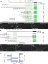
Hox and TALE transcription factors define 3 anteroposterior zones of CP neuron fate in C. elegans males. a) The serotonergic reporter tph-1::mCherry is expressed in CPs 1–6 in a wild-type male; flp-21::gfp is expressed in CPs 7-9. Expression of tph-1::mCherry is relatively less intense in zone 1 CPs 1–4 than in zone 2 CPs 5–6 (see Fig. 3a). b) Schematic showing zone-specific and hox-dependent expression of CP neuron reporters in wild-type, lin-39 null mutant, and mab-5 null mutant males. Bar colors indicate zone-typical reporter expression, such that zone 1 = blue, zone 2 = green, zone 3 = yellow. c–f) TALE factors ceh-20 and unc-62 are required for normal tph-1::mCherry expression in Zones 1 and 2, but not for flp-21::gfp expression in zone 3. c) Expression of tph-1::mCherry (red) in CPs 1–6 and flp-21::gfp (green) in CPs 7–9 in a male treated with control (empty vector) RNAi. d and e) Reduced expression of tph-1::mCherry and expanded expression of flp-21::gfp in males treated with ceh-20 RNAi (d) or unc-62 RNAi (e). c–e) Filled circle colors summarize zone identities of CP neurons. Scale bar = 50 µM. f)Summary of tph-1::mCherry and flp-21::gfp expression in CPs in males treated with control, ceh-20, and unc-62 RNAi; CPs+ denotes mean number of fluor-positive CPs/worm in each zone. Bar colors indicate zone identities of CP neurons; error bars indicate standard error of the mean (SEM); number of ventral cords scored is indicated at right; *P < 0.05, unpaired t-test, compared to control RNAi.
Fig. 2.
Fig. 2.
Cis-regulatory elements in tph-1 and flp-22 drive zone-specific and Hox-responsive reporter expression in male CP neurons. a) Identification of tph-1 CRMs driving CP5-6 (zone 2)-specific expression of GFP in the ventral cord. All lines scored were extrachromosomal arrays, except for the integrated array cccIs2 [tph-1(CP5-6)::gfp]. Potential Hox- and TALE-binding sites, as identified by cisBP analysis, are indicated for the region encompassed by the tph-1(CP5-6)::gfp reporter. b–e) The tph-1(CP5-6)::gfp reporter, which contains upstream sequences from –585 to –91, requires both lin-39 and mab-5 for expression in CPs 5–6. b) Expression of tph-1(CP5-6)::gfp in CPs 5–6 in a wild-type male. c and d) tph-1(CP5-6)::gfp is not expressed in ventral cord neurons in lin-39(e1760) or mab-5(e1239) mutants. e) Identification of flp-22 CRMs driving CP 1–4 (zone 1)-specific expression of GFP in the ventral cord. All lines scored were extrachromosomal arrays, except for the integrated array ynIs80. Potential Hox- and TALE-binding sites, as identified by cisBP analysis, are indicated for the region encompassed by the flp-22(–2,060/–1,533)::gfp reporter which contains upstream sequences from –2,060 to –1,533. f–h) The flp-22(–2,060/–1,533)::gfp reporter requires lin-39 and mab-5 for CP1-4-specific expression. (f) Expression of flp-22(–2,060/–1,533)::gfp in CPs 1–4 in a wild-type male. g) flp-22(–2,060/–1,533)::gfp is not expressed in ventral cord neurons in lin-39(e1760) mutants. h) flp-22(–2,060/–1,533)::gfp is expanded posteriorly, and encompasses CPs 1–6 in mab-5(e1239) mutants. i) Summary of tph-1(CP5–6)::gfp and flp-22(–2,060–1,533)::gfp in CPs in wild-type and Hox mutant males. CPs+ denotes mean number of fluor-positive CPs/worm in each zone; error bars indicate standard error of the mean (SEM); number of ventral cords scored is indicated at right; *P < 0.05, unpaired t-test, compared to WT. In a and e) reporter boundaries are indicated in base pairs, relative to the first coding exon of tph-1 and flp-22. + indicates GFP expression in the indicated CP neurons in all transgenic lines examined; ± indicates expression in some lines examined; – indicates no GFP expression in any lines examined. Reporters labeled pes-10::gfp included the pes-10 minimal promoter. Scale bars = 100 µM.
Fig. 3.
Fig. 3.
Anterior expansion of MAB-5 specifies zone 2-like fates in zone 1 CPs. a–d) CP reporter expression in wild-type (left) and mab-5(e1751gf) (right) males. Filled circle colors summarize zone identities of CP neurons; scale bar = 100 µM. a) tph-1::mCherry expression in CPs 1–6 in wild-type and mab-5(e1751) males. Insets show more intense relative expression in CPs 5–6 than in CPs 1–4 in wild-type (P < 0.0001, n = 15, unpaired t-test), but equally intense expression throughout CPs 1–6 in mab-5(e1751gf) (P > 0.7, n = 17, unpaired t-test). b) flp-22::gfp is expressed in CPs 1–4 in a wild-type male, but is not expressed in CPs in a mab-5(e1751gf) male. c) flp-21::gfp is expressed in CPs 7–9 in both wild-type and mab-5(e1751gf) males. mab-5(e1751) males also display expression of flp-21::gfp in ectopic anterior CPs. d) tph-1(CP5-6)::gfp is expressed in CPs 5–6 in a wild-type male, but expands to CPs 1–6 in a mab-5(e1751gf) male. e) Summary of tph-1::m Cherry, flp-22::gfp, flp-21::gfp, and tph-1(CP5-6)::gfp expression in CPs (CPs+ denotes mean number of fluor-positive CPs/worm in each zone) in wild-type and mab-5(e1751gf) males. Bar colors indicate zone identities of CP neurons; error bars indicate standard error of the mean (SEM); number of ventral cords scored is indicated at right; *P < 0.05, unpaired t-test, compared to WT. f) Diagram representing CP neuron homeotic transformations in mab-5(e1751gf), in which zone 1 neurons (CPs 1–4) assume fates normally reserved for zone 2 neurons (CP 5–6).
Fig. 4.
Fig. 4.
The lin-39(ccc16) mutation affects zone 2 CP fates specifically and in a mab-5-dependent manner. a–f) CP reporter expression in lin-39(ccc16) and lin-39(ccc16) mab-5(e1239) males; refer to Fig. 3 for wild-type expression of CP reporters. tph-1::mCherry is expressed in CPs 1–4, but not CPs 5–6 in a lin-39(ccc16) male a), but is expressed in CPs 1–6 in a lin-39(ccc16) mab-5(e1239) male d). flp-22::gfp is expressed in CPs 1–4 in a lin-39(ccc16) male b), and in CPs 1–6 in a lin-39(ccc16) mab-5(e1239) male e). flp-21::gfp is expressed in CPs 5–9 in lin-39(ccc16) c), but is not expressed in CPs in a lin-39(ccc16) mab-5(e1239) f) male. Filled circle colors summarize zone identities of CP neurons; scale bar = 100 µM. g) Summary of tph-1::mCherry, Serotonin (SER) antibody staining, tph-1(CP5-6)::gfp, flp-22::gfp, and flp-21::gfp expression in CPs (CPs+ denotes mean number of fluor-positive CPs/worm in each zone) in wild-type, lin-39(ccc16), and lin-39(ccc16) mab-5(e1239) males. Bar colors indicate zone identities of CP neurons; error bars indicate standard error of the mean (SEM); number of ventral cords scored is indicated at right; *P < 0.05, unpaired t-test, compared to WT. h) Diagram representing CP neuron homeotic transformations in lin-39(ccc16) and lin-39(ccc16) mab-5(e1239) mutants. In lin-39(ccc16), zone 2 neurons (CPs 5–6) assume fates normally reserved for zone 3 CP neurons (7–9). In lin-39(ccc16) mab-5(e1239), the LIN-39(ccc16) protein is able to specify zone 1 fates in zone 2 CPs 5–6 in the absence of mab-5. lin-39* denotes the ccc16 isoform of lin-39.
Fig. 5.
Fig. 5.
lin-39(ccc16) disrupts sequences C-terminal to the homeodomain. a) Gene structure of lin-39 indicating protein domains and alleles. Previously identified alleles of lin-39 are located either before or in the homeodomain: n709 and n1872 are splice acceptor and donor mutations, respectively, n1880 and n1760 are substitutions that result in a stop codon, and n2110 a substitution. DNA sequencing indicates that ccc16 is a g->a exon 5 splice acceptor mutation. b) 3′ RACE reveals 2 ccc16 splice isoforms. Isoform lin-39(ccc16in) uses an intronic splice acceptor resulting in 7 amino acids before a stop codon (*). Isoform lin-39(ccc16ex) exon 5 splice acceptor results in a frameshift and early stop codon (*). c) CRISPR-engineered changes to lin-39 exon 5 or splice acceptor. Sequence changes are indicated in red. ex, truncated exon variant; in, intronic splice variant; TxV, valine substitution for threonine in consensus MAPK site. d) Reduced expression of tph-1::mCherry in CPs 5–6 in lin-39(ccc21) and lin-39(ccc22). Filled circle colors summarize zone identities of CP neurons; scale bar = 100 µM. e) Summary of tph-1::mCherry expression in CPs 1–6 (CPs+ denotes mean number of fluor-positive CPs/worm in each zone) in lin-39 mutant alleles. lin-39 temperature-sensitive alleles were scored at both the permissive (P) temperature of 16C and restrictive (R) temperature of 25°C. Significant differences of tph-1::mCherry expression in CPs 5–6 are indicated for comparisons between lin-39(ccc16) and other C-terminal alleles (ordinary one-way ANOVA with multiple comparisons, *P < 0.05, ****P < 0.0001, ns = not significant). Bar colors indicate zone identities of CP neurons; error bars indicate standard error of the mean (SEM); number of ventral cords scored is indicated at right. In e), for CPs 1–4, all means are significantly different from WT at P < 0.05 (unpaired t-test) except for ccc19, ccc22, and ccc23; for CPs 5–6, all means are significantly different from WT at P < 0.05 except for ccc23.
Fig. 6.
Fig. 6.
Bimolecular fluorescence complementation (BiFC) ectopic protein interaction assay reveals interaction between Hox and TALE proteins. LIN-39, LIN-39(ccc16ex), LIN-39(ccc16in), MAB-5, or CEH-20 were fused in frame with the Venus fragments VN173 or VC155, and expressed in transgenic C. elegans hermaphrodites via the hsp16.41 heat-shock promoter. a–h) GFP images of worms expressing CEH-20 or MAB-5 with LIN-39, empty vector, LIN-39(ccc16ex), or LIN-39(ccc16in). Ectopic in vivo interaction between LIN-39 and both CEH-20 a) and MAB-5 e) reconstitutes Venus fluorescence prominently in intestinal nuclei. b–d and f–h) No interaction or weaker interaction is observed between CEH-20 or MAB-5 and empty vector or LIN-39 C-terminal mutants. Scale bar = 75um. i–j and m) Quantification of Venus fluorescence for each pair of proteins. One dot represents the average of 6 intestinal nuclei per worm, bars represent the mean of all worms for a given pair. i and j) For each pairwise combination, 30 worms in each of 3 extrachromosomal lines were examined (see also Supplementary Fig. 1). i) CEH-20 reconstitutes fluorescence with LIN-39 and MAB-5; additionally, LIN-39 interacts with MAB-5 (nested unpaired t-test). j) LIN-39 C-terminal mutations disrupt LIN-39 and MAB-5 interactions but only partially disrupt LIN-39 and CEH-20 interactions (nested one way ANOVA). k–m) RNAi of ceh-20 significantly disrupts reconstitution of fluorescence between LIN-39 and MAB-5 in 2 extrachromosomal lines, scuEx27 and scuEx28 (n = 60 for each, unpaired t-test). *P < 0.05, **P < 0.01, ****P < 0.0001, A.U., arbitrary unit, ns, not significant.
Fig. 7.
Fig. 7.
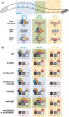
Model of Hox and TALE regulation of CP neuronal subtype specification along the A–P axis. a) In wild-type males, CRM 1 is active in CPs 1–4, CRM 2 is active in CPs 5–6, and CRM 3 is active in CPs 7–9. b) Summary of regional (CPs 1–4, CPs 5–6, and CPs 7–9) expression at each CRM (1, 2, and 3) in Wild Type, lin-39(0), lin-39(ccc16), lin-39(ccc16) mab-5(0), mab-5(0), mab-5(gf), and ceh-20(RNAi) or unc-62(RNAi). a and b) Bar colors indicate zone-typical reporter expression, such that zone 1 = blue, zone 2 = green, zone 3 = yellow, no Zone specified = gray. Green arrows = active expression, red inhibitor = repression, CRM = cis-regulatory module, gf = gain-of-function, 0 = null, dark blue L39 = LIN-39, light blue L39 = LIN-39(ccc16), gold C20 = CEH-20, orange U62 = UNC-62, red M5 = MAB-5. Created with BioRender.com.

Similar articles

Cited by

References

    1. Alper S, Kenyon C.. The zinc finger protein REF-2 functions with the Hox genes to inhibit cell fusion in the ventral epidermis of C. elegans. Development. 2002;129(14):3335–3348. - PubMed
    1. Araya CL, Kawli T, Kundaje A, Jiang L, Wu B, Vafeados D, Terrell R, Weissdepp P, Gevirtzman L, Mace D, et al.Regulatory analysis of the C. elegans genome with spatiotemporal resolution. Nature. 2014;512(7515):400–405. - PMC - PubMed
    1. Arribere JA, Bell RT, Fu BXH, Artiles KL, Hartman PS, Fire AZ.. Efficient marker-free recovery of custom genetic modifications with CRISPR/Cas9 in Caenorhabditis elegans. Genetics. 2014;198(3):837–846. - PMC - PubMed
    1. Baek M, Enriquez J, Mann RS.. Dual role for Hox genes and Hox co-factors in conferring leg motoneuron survival and identity in Drosophila. Development. 2013;140(9):2027–2038. - PMC - PubMed
    1. Bridoux L, Zarrineh P, Mallen J, Phuycharoen M, Latorre V, Ladam F, Losa M, Baker SM, Sagerstrom C, Mace KA, et al.HOX paralogs selectively convert binding of ubiquitous transcription factors into tissue-specific patterns of enhancer activation. PLoS Genet. 2020;16(12):e1009162. - PMC - PubMed

Publication types

MeSH terms