. 2022 Jul 5;77(7):1279-1286.
doi: 10.1093/gerona/glac034.

DNA Damage in Circulating Hematopoietic Progenitor Stem Cells as Promising Biological Sensor of Frailty

Affiliations

DNA Damage in Circulating Hematopoietic Progenitor Stem Cells as Promising Biological Sensor of Frailty

Chiara Grasselli et al. J Gerontol A Biol Sci Med Sci. .

Abstract

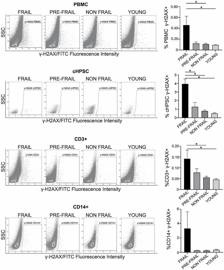
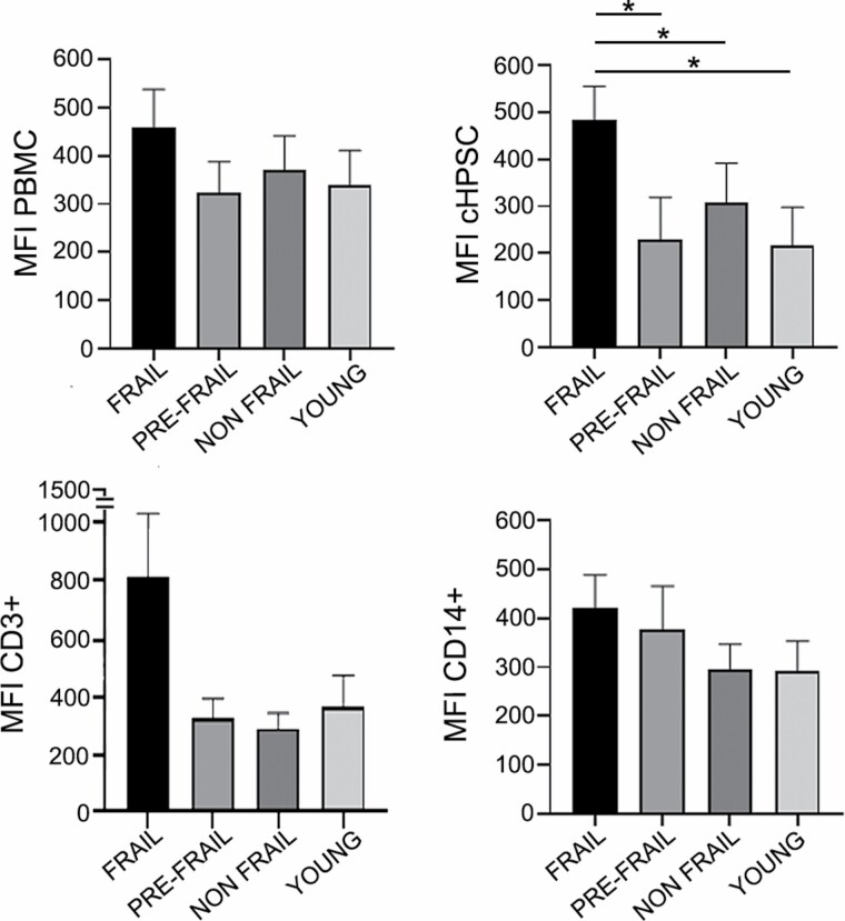
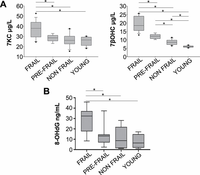
Frailty is an age-related syndrome that exposes individuals to increased vulnerability. Although it is potentially reversible, in most cases it leads to negative outcomes, including mortality. The different methods proposed identify frailty after the onset of clinical manifestations. An early diagnosis might make it possible to manage the frailty progression better. The frailty pathophysiology is still unclear although mechanisms, in particular, those linked to inflammation and immunosenescence, have been investigated. A common feature of several clinical aspects involved in senescent organisms is the increase of oxidative stress, described as one of the major causes of deoxyribonucleic acid (DNA) damage accumulation in aged cells including the adult stem cell compartment. Likely, this accumulation is implicated in frailty status. The oxidative status of our frail, pre-frail, and non-frail population was characterized. In addition, the DNA damage in hematopoietic cells was evidenced by analyzing the peripheral blood mononuclear cell and their T lymphocyte, monocyte, circulating hematopoietic progenitor stem cell (cHPSC) subpopulations. The phosphorylation of C-terminal of histone H2AX at amino acid Ser 139 (γ-H2AX), which occurs at the DNA double-strand break focus, was evaluated. In our frail population, increased oxidative stress and a high level of DNA damage in cHPSC were found. This study may have potential implications because the increment of DNA damage in cHPSC could be suggestive of an organism impairment preceding the evident frailty. In addition, it may open the possibility for attenuation of frailty progression throughout specific drugs acting on preventing DNA damage or removing damaged cells.

Keywords: Biology of aging; Cellular senescence; Oxidative stress; γ-H2AX.


Figures

Figure 1.
Figure 1.
DNA damage in PBMC isolated by Ficoll-Paque and in the respective cell subpopulations: cHPSC CD34+/CD3-/CD14-, T lymphocytes CD3+, monocytes CD14+. Representative FACS analysis of γ-H2AX dot plots of the different studied groups (40 frail, 13 pre-frail, 32 non-frail, 46 young samples). Histograms represent the percentage of cells positive for DNA damage (γ-H2AX) in the different groups. SSC: side scatter. Data are expressed as means ± SEM. *p < .05.
Figure 2.
Figure 2.
Mean fluorescence intensity (MFI) of DNA damage in peripheral blood mononuclear cell (PBMC) isolated by Ficoll-Paque and in the respective cell subpopulations: cHPSC CD34+/CD3-/CD14-, T lymphocytes CD3+, monocytes CD14+. Histograms represent the averaged median values of MFI in the different study groups (40 frail, 13 pre-frail, 32 non-frail, 46 young samples). Data are expressed as means ± SEM. *p < .05.
Figure 3.
Figure 3.
Representation of oxidative stress evaluated in plasma. (A) Level of 7-keto-cholesterol (7KC, left) and 7β-hydroxycholesterol (7βOHC, right) in 17 frail, 9 pre-frail, 18 non-frail, 10 young samples. (B) Level of 8-hydroxy-2-deoxy Guanosine (8-OH-dG) in 10 frail, 9 pre-frail, 10 non-frail, 10 young samples. *p < .01. Data are expressed as means ± SD.

References

    1. Dent E, Martin FC, Bergman H, Woo J, Romero-Ortuno R, Walston JD. Management of frailty: opportunities, challenges, and future directions. Lancet. 2019;394:1376–1386. doi:10.1016/S0140-6736(19)31785-4 - DOI - PubMed
    1. Vermeiren S, Vella-Azzopardi R, Beckwee D, et al. . Frailty and the prediction of negative health outcomes: a meta-analysis. J Am Med Dir Assoc. 2016;17:1163.e1–1163.e17. doi:10.1016/j.jamda.2016.09.010 - DOI - PubMed
    1. Lang PO, Michel JP, Zekry D. Frailty syndrome: a transitional state in a dynamic process. Gerontology. 2009;55(5):539–549. doi:10.1159/000211949 - DOI - PubMed
    1. Fried LP, Tangen CM, Walston J, et al. . Frailty in older adults: evidence for a phenotype. J Gerontol A Biol Sci Med Sci 2001;56(3):M146–M157. doi:10.1093/gerona/56.3.m146 - DOI - PubMed
    1. Collerton J, Martin-Ruiz C, Davies K, et al. . Frailty and the role of inflammation, immunosenescence and cellular ageing in the very old: cross-sectional findings from the Newcastle 85+ Study. Mech Ageing Dev. 2012;133(6):456–466. doi:10.1016/j.mad.2012.05.005 - DOI - PubMed

Publication types