Case Reports
. 2022 May 17;107(6):1714-1726.
doi: 10.1210/clinem/dgac079.

Approach to the Patient With Lipodystrophy

Affiliations
Case Reports

Approach to the Patient With Lipodystrophy

Lindsay T Fourman et al. J Clin Endocrinol Metab. .

Abstract

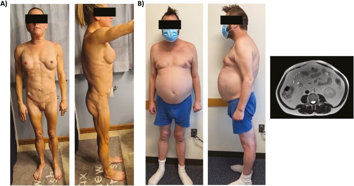
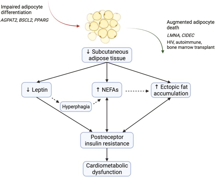
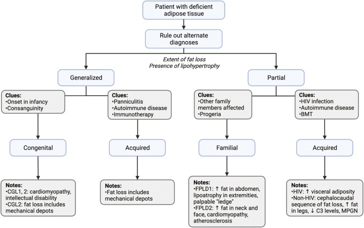
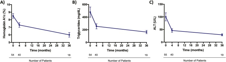
Lipodystrophy constitutes a spectrum of diseases characterized by a generalized or partial absence of adipose tissue. Underscoring the role of healthy fat in maintenance of metabolic homeostasis, fat deficiency in lipodystrophy typically leads to profound metabolic disturbances including insulin resistance, hypertriglyceridemia, and ectopic fat accumulation. While rare, recent genetic studies indicate that lipodystrophy is more prevalent than has been previously thought, suggesting considerable underdiagnosis in clinical practice. In this article, we provide an overview of the etiology and management of generalized and partial lipodystrophy disorders. We bring together the latest scientific evidence and clinical guidelines and expose key gaps in knowledge. Through improved recognition of the lipodystrophy disorders, patients (and their affected family members) can be appropriately screened for cardiometabolic, noncardiometabolic, and syndromic abnormalities and undergo treatment with targeted interventions. Notably, insights gained through the study of this rare and extreme phenotype can inform our knowledge of more common disorders of adipose tissue overload, including generalized obesity.

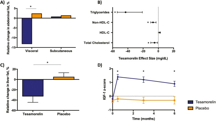
Keywords: Dunnigan variety lipodystrophy; HIV-associated lipodystrophy; lipodystrophy; metreleptin; tesamorelin.


Figures

Figure 1.
Figure 1.
Clinical cases. A, Case 1: The patient has lipoatrophy of the limbs and torso with sparing of the face and mons pubis. She also has increased muscularity of her upper and lower extremities, which is most evident on the lateral view. B, Case 2: The patient has mild lipoatrophy of the lower extremities and buttocks with profound abdominal lipohypertrophy. Cross-sectional magnetic resonance imaging at L4 revealed significant visceral adiposity and a high visceral to subcutaneous fat ratio. Both patients provided written consent for their photographs to be published.
Figure 2.
Figure 2.
Pathophysiology of cardiometabolic dysfunction in lipodystrophy. Lipodystrophy is broadly defined by a generalized or partial absence of adipose tissue, except in some patients with HIV-associated lipodystrophy, who may present with lipohypertrophy alone. In general, lack of fat may stem from impaired adipocyte differentiation or augmented adipocyte death secondary to genetic or acquired factors. Deficient adipose tissue may lead to a complex cascade of altered adipokines, metabolites, and energy flux, which collectively promotes insulin resistance and resultant cardiometabolic dysfunction. AGPAT2, 1-acylglycerol-3-phosphate O-acyltransferase 2; BSCL2, Berardinelli-Seip congenital lipodystrophy 2 protein; CIDEC, cell death–inducing DFFA-like effector C; LMNA, lamin A/C; NEFAs, nonesterified fatty acids; PPARG, peroxisome proliferator-activated receptor γ. Figure was created with BioRender.com.
Figure 3.
Figure 3.
Conceptual approach to lipodystrophy diagnosis. Lipodystrophy is categorized into 4 broad groups according to extent of fat loss (generalized vs partial) and etiology (genetic vs acquired). This flowchart provides a conceptual approach to the patient with suspected lipodystrophy, paying particular attention to classic presentations of the most common lipodystrophy subtypes. Not all lipodystrophy subtypes and clinical presentations are shown. Progeroid features in the presence of partial lipodystrophy should lead to consideration of familial etiologies as indicated by the flow diagram, but it should be noted that progeria may also be seen in association with generalized forms of disease. BMT, bone marrow transplantation; C3, complement 3; CGL, congenital generalized lipodystrophy; FPLD, familial partial lipodystrophy; MPGN, membranoproliferative glomerulonephritis. Figure was created with BioRender.com.
Figure 4.
Figure 4.
Metabolic effects of metreleptin in non–HIV-associated lipodystrophy. Metreleptin leads to sustained improvements in glycemic control, hypertriglyceridemia, and hepatic steatosis among individuals with non–HIV-associated lipodystrophy. In the representative data shown, patients with generalized or partial lipodystrophy who had metabolic dysfunction and low baseline leptin levels (< 8.0 ng/mL in males; < 12.0 ng/mL in females) were treated with open-label metreleptin for up to 3 years. Metreleptin statistically significantly reduced A, HbA1c (–2.1% ± 0.5%); B, triglycerides (–35.4% ± 13.7%); and C, ALT (–45 ± 19 U/L). The most rapid decrements occurred in the first 4 months of therapy followed by slower but continued improvements throughout the follow-up period (57). Mean and SEM are shown. ALT, alanine transaminase; HbA1c, glycated hemoglobin A1c.
Figure 5.
Figure 5.
Metabolic effects of tesamorelin in HIV-associated lipodystrophy. A, In 2 large, phase 3, placebo-controlled trials of patients with HIV and increased abdominal adiposity, tesamorelin led to a statistically significant reduction in visceral fat (–15.4%) while preserving subcutaneous fat over 26 weeks (62). B, Tesamorelin also led to declines in triglycerides (–43 mg/dL; 95% CI, –54 to –21 mg/dL), total cholesterol (–5 mg/dL; 95% CI, –10 to –1 mg/dL) and non-HDL cholesterol (–7 mg/dL; 95% CI, –10 to –3 mg/dL) vs placebo (62). C, In patients with HIV and nonalcoholic fatty liver disease, tesamorelin-treated individuals experienced a substantial decline in liver fat compared to placebo-treated controls over 1 year (–37%; 95% CI, –67% to –7%) (66). D, Insulin-like growth factor 1 (IGF-1) levels among individuals with HIV-associated abdominal fat accumulation have been shown to increase relative to placebo, but typically remain within the normal physiologic range (z score < 2.0) (63). Data are mean difference and 95% CI in B and mean and SEM in C and D. Asterisks indicate statistical significance as defined by P less than .05. HDL-C, high-density lipoprotein cholesterol; non–HDL-C, non–high-density lipoprotein cholesterol.

References

    1. Brown RJ, Araujo-Vilar D, Cheung PT, et al. . The diagnosis and management of lipodystrophy syndromes: a multi-society practice guideline. J Clin Endocrinol Metab. 2016;101(12):4500-4511. - PMC - PubMed
    1. Agarwal AK, Garg A. Genetic disorders of adipose tissue development, differentiation, and death. Annu Rev Genomics Hum Genet. 2006;7:175-199. - PubMed
    1. Gonzaga-Jauregui C, Ge W, Staples J, et al. . Clinical and molecular prevalence of lipodystrophy in an unascertained large clinical care cohort. Diabetes. 2020;69(2):249-258. - PubMed
    1. Frayn KN. Adipose tissue as a buffer for daily lipid flux. Diabetologia. 2002;45(9):1201-1210. - PubMed
    1. Stern JH, Rutkowski JM, Scherer PE. Adiponectin, leptin, and fatty acids in the maintenance of metabolic homeostasis through adipose tissue crosstalk. Cell Metab. 2016;23(5):770-784. - PMC - PubMed

Publication types