Dickkopf‑3 and β‑catenin play opposite roles in the Wnt/β‑catenin pathway during the abnormal subchondral bone formation of human knee osteoarthritis
- PMID: 35137918
- PMCID: PMC8904073
- DOI: 10.3892/ijmm.2022.5103
Dickkopf‑3 and β‑catenin play opposite roles in the Wnt/β‑catenin pathway during the abnormal subchondral bone formation of human knee osteoarthritis
Abstract
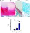
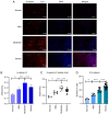
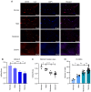
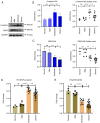
Osteoarthritis (OA) is condition which poses a main concern to the aging population and its severity is expected to increase with the increasing life expectancy. In the future, several possible targets for OA treatment need to be defined. Dickkopf‑related protein 3 (DKK3) is an atypical member of the Wnt‑antagonistic dickkopf‑related protein (DKK) family. The availability of research into the role of DKK3 in the abnormal remodeling of subchondral bone in human knee joints is currently limited. Thus, the aim of the present study was the evaluation of DKK3 expression in the abnormal bone remodeling of subchondral bone in human knee OA in order to clarify the role of DKK3 in subchondral bone remodeling and to acknowledge its potential relevance to β‑catenin. In total, 38 specimens were collected from osteotomies of the medial tibial plateau of the human knee. The patient samples were then divided into the normal, mild, moderate and severe symptom groups, according to the Osteoarthritis Research Society International (OARSI) score. Following hematoxylin and eosin (H&E) and Safranin O‑fast green staining for alkaline phosphatase (AZO method), changes in the distribution and number of osteocytes in the subchondral bone and the degree of sclerosis of the subchondral bone were observed. Immunohistochemical staining, immunofluorescence, western blot analysis and reverse‑transcription quantitative PCR (RT‑qPCR) were used for the detection of DKK3 and β‑catenin expression level changes in osteoblasts in the subchondral bone of the medial tibial plateau. H&E and alkaline phosphatase staining revealed that the total number of osteocytes in the subchondral bone increased with the severity of the disease. The samples were also evaluated using Safranin O‑Fast Green staining and were attributed a score according to the OARSI scoring system: The scoring number and cartilage damage increased along with OA severity. Immunohistochemistry and immunofluorescence assays demonstrated that β‑catenin expression in osteocytes increased from mild to moderate, whereas DKK3 expression decreased with the development of arthritis from normal, mild to moderate. According to the results of western blot analysis, β‑catenin expression was higher in moderate OA and then decreased in severe OA. On the other hand, the DKK3 levels decreased along with the progression from normal, mild to moderate OA. The results of RT‑qPCR demonstrated that β‑catenin and DKK3 gene expression differed with the degree of OA. On the whole, the present study demonstrates that DKK3 and β‑catenin may play opposite roles in OA subchondral bone remodeling.
Keywords: Dickkopf‑3; Wnt/β‑catenin signaling; osteoarthritis; osteoblasts; sclerosis; subchondral bone; β‑catenin.
Conflict of interest statement
The authors declare that they have no competing interests.
Figures


Similar articles
-
Sclerostin expression in the subchondral bone of patients with knee osteoarthritis.Int J Mol Med. 2016 Nov;38(5):1395-1402. doi: 10.3892/ijmm.2016.2741. Epub 2016 Sep 19. Int J Mol Med. 2016. PMID: 27665782 Free PMC article.
-
Control of Dkk-1 ameliorates chondrocyte apoptosis, cartilage destruction, and subchondral bone deterioration in osteoarthritic knees.Arthritis Rheum. 2010 May;62(5):1393-402. doi: 10.1002/art.27357. Arthritis Rheum. 2010. PMID: 20131282
-
The protective mechanism of SIRT1 on cartilage through regulation of LEF-1.BMC Musculoskelet Disord. 2021 Jul 27;22(1):642. doi: 10.1186/s12891-021-04516-x. BMC Musculoskelet Disord. 2021. PMID: 34315467 Free PMC article.
-
Modeling knee osteoarthritis pathophysiology using an integrated joint system (IJS): a systematic review of relationships among cartilage thickness, gait mechanics, and subchondral bone mineral density.Osteoarthritis Cartilage. 2018 Nov;26(11):1425-1437. doi: 10.1016/j.joca.2018.06.017. Epub 2018 Jul 26. Osteoarthritis Cartilage. 2018. PMID: 30056214
-
Microenvironment in subchondral bone: predominant regulator for the treatment of osteoarthritis.Ann Rheum Dis. 2021 Apr;80(4):413-422. doi: 10.1136/annrheumdis-2020-218089. Epub 2020 Nov 6. Ann Rheum Dis. 2021. PMID: 33158879 Free PMC article. Review.
Cited by
-
Osteoimmunology in Osteoarthritis: Unraveling the Interplay of Immunity, Inflammation, and Joint Degeneration.J Inflamm Res. 2025 Mar 19;18:4121-4142. doi: 10.2147/JIR.S514002. eCollection 2025. J Inflamm Res. 2025. PMID: 40125089 Free PMC article. Review.
-
Cuprorivaite microspheres inhibit cuproptosis and oxidative stress in osteoarthritis via Wnt/β-catenin pathway.Mater Today Bio. 2024 Oct 16;29:101300. doi: 10.1016/j.mtbio.2024.101300. eCollection 2024 Dec. Mater Today Bio. 2024. PMID: 39469313 Free PMC article.
References
-
- Omoumi P, Babel H, Jolles BM, Favre J. Relationships between cartilage thickness and subchondral bone mineral density in non-osteoarthritic and severely osteoarthritic knees: In vivo concomitant 3D analysis using CT arthrography. Osteoarthritis Cartilage. 2019;27:621–629. doi: 10.1016/j.joca.2018.12.014. - DOI - PubMed

