. 2022 Jun;94(6):2438-2452.
doi: 10.1002/jmv.27650. Epub 2022 Feb 19.

Construction and characterization of two SARS-CoV-2 minigenome replicon systems

Affiliations

Construction and characterization of two SARS-CoV-2 minigenome replicon systems

Hu Zhang et al. J Med Virol. 2022 Jun.

Abstract

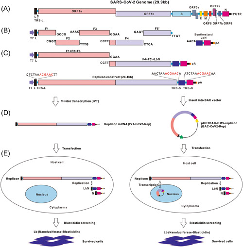
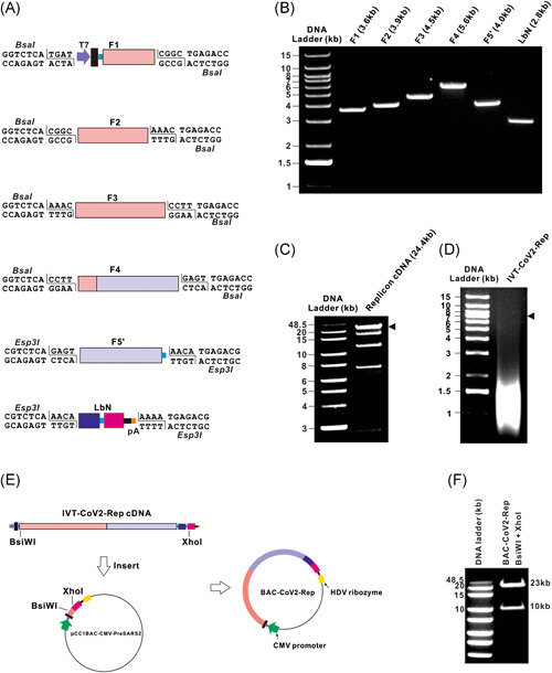
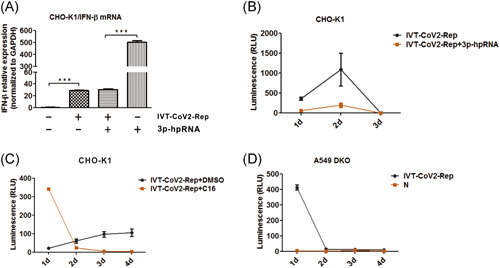
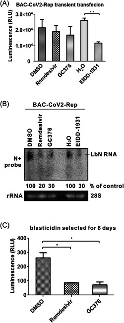
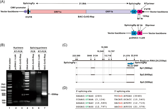
The ongoing COVID-19 pandemic severely impacts global public health and economies. To facilitate research on severe acute respiratory syndrome coronavirus 2 (SARS-CoV-2) virology and antiviral discovery, a noninfectious viral replicon system operating under biosafety level 2 containment is warranted. We report herein the construction and characterization of two SARS-CoV-2 minigenome replicon systems. First, we constructed the IVT-CoV2-Rep complementary DNA template to generate a replicon messenger RNA (mRNA) with nanoluciferase (NLuc) reporter via in vitro transcription (IVT). The replicon mRNA transfection assay demonstrated a rapid and transient replication of IVT-CoV2-Rep in a variety of cell lines, which could be completely abolished by known SARS-CoV-2 replication inhibitors. Our data also suggest that the transient phenotype of IVT-CoV2-Rep is not due to host innate antiviral responses. In addition, we have developed a DNA-launched replicon BAC-CoV2-Rep, which supports the in-cell transcription of a replicon mRNA as initial replication template. The BAC-CoV2-Rep transient transfection system exhibited a much stronger and longer replicon signal compared to the IVT-CoV2-Rep version. We also found that a portion of the NLuc reporter signal was derived from the spliced BAC-CoV2-Rep mRNA and was resistant to antiviral treatment, especially during the early phase after transfection. In summary, the established SARS-CoV-2 transient replicon systems are suitable for basic and antiviral research, and hold promise for stable replicon cell line development with further optimization.

Keywords: SARS coronavirus; antiviral agents; cellular effect; disease control; immune responses; innate immunity; mRNA/splicing; virus classification.


Conflict of interest statement

The authors declare no conflict of interests.

Figures

Figure 1
Figure 1
Schematic design of SARS‐CoV‐2 minigenome replicons. (A) The genome organization of SARS‐CoV‐2 is schematically presented with the major ORFs indicated as color blocks. (B) The synthesized DNA fragments of ORF1ab (F1ʹ–F5ʹ) and LbN cassette are shown with their ligatable cohesive ends. The T7 promoter sequence and a poly(A) tail are located at the 5ʹ terminus of F1 and 3ʹ terminus of LbN, respectively. (C) The DNA fragments are directionally ligated in vitro to assemble the IVT‐CoV2‐Rep cDNA. The nucleotides shown above the blue boxes are site‐specific viral transcription regulatory sequences (TRS). (D) IVT‐CoV2‐Rep cDNA serves as an IVT template to synthesize replicon mRNA or it is cloned into pCC1BAC‐CMV‐PreSARS2 vector to construct BAC‐CoV2‐Rep. (E) IVT‐CoV2‐Rep mRNA or BAC‐CoV‐Rep is transfected into mammalian cells for assessment of replicon replication and stable replicon cell line selection. See text for more details. BAC, bacterial artificial chromosome; cDNA, complementary DNA; CMV, cytomegalovirus; IVT, in vitro transcription; E, envelope gene; L, leader sequence; Lb, nanoluciferase (NLuc)‐blasticidin S‐resistance (BSR) fusion gene; M, membrane protein; mRNA, messenger RNA; N, nucleocapsid gene; ORF, open reading frame; S, spike gene; SARS‑CoV‑2, severe acute respiratory syndrome coronavirus 2
Figure 2
Figure 2
Assembly of SARS‐CoV‐2 replicons. (A) A diagram of each cDNA fragment with the terminal BsaI or Esp3I restriction site is shown. (B) Gel analysis of the purified cDNA fragments is presented. The individual cDNA fragments (F1 to LbN) were released from corresponding plasmids by BsaI or Esp3I as indicated in (A) and gel purified, followed by purity examination on a 0.8% agarose gel. A 1 kb plus DNA ladder served as size marker. (C) Gel analysis of the cDNA ligation products is shown. 1 µg of purified ligation product was analyzed on a 0.8% agarose gel. The triangle indicates the full‐length IVT‐CoV2‐Rep cDNA product. (D) Gel analysis of IVT RNA transcripts is shown. 1 µg of IVT‐CoV2‐Rep RNA was analyzed on a 0.8% native agarose gel. Note that the DNA size markers do not directly correlate with the RNA size. The triangle indicates the approximate position of the full‐length replicon transcript. (E) The strategy of construction of BAC‐CoV2‐Rep is shown. The assembled IVT‐CoV2‐Rep cDNA was digested by BsiWI/XhoI and inserted into the corresponding restriction site of pCC1BAC‐CMV‐PreSARS2 to generate BAC‐CoV2‐Rep. The CMV promoter and HDV ribozyme site carried by the vector backbone are indicated. (F) The positive clone of BAC‐CoV2‐Rep was verified by BsiWI/XhoI double digestion and analysis by agarose gel electrophoresis. BAC, bacterial artificial chromosome; cDNA, complementary DNA; CMV, cytomegalovirus; HDV, hepatitis delta virus; IVT, in vitro transcription; SARS‑CoV‑2, severe acute respiratory syndrome coronavirus 2
Figure 3
Figure 3
Detection of NLuc reporter signal in cells after IVT‐CoV2‐Rep RNA transfection. (A) Vero, (B,F) CHO‐K1, (C) Huh7.5, and (D,E) BHK‐21 cells were transfected with 200 ng IVT‐CoV2‐Rep RNA or N IVT RNA in a 96‐well plate. NLuc activities were measured at indicated time points and plotted as relative luminescence units (RLU) (mean ± SD, n = 2). IVT, in vitro transcription
Figure 4
Figure 4
Inhibition of IVT‐CoV2‐Rep by antiviral compounds. CHOK1 cells were transfected with 200 ng IVT‐CoV2‐Rep RNA in a 96‐well plate and subjected to the following treatments: (A) Solvent control (0.1% DMSO or H2O); (B) 5 μM remdesivir or GC376 with DMSO as a control; and (C) 5 μM EIDD‐1931 with H2O as a control. NLuc activities were measured at indicated time points (mean ± SD, n = 2). DMSO, dimethyl sulfoxide; IVT, in vitro transcription; RLU, relative luminescence unit
Figure 5
Figure 5
IVT‐CoV2‐Rep replication and cellular innate responses. (A) CHO‐K1 cells were left untransfected or transfected with IVT‐CoV2‐Rep only, IVT‐CoV2‐Rep + 3p‐hpRNA, or 3p‐hpRNA only. The cells were harvested 2 days later for total RNA extraction, followed by RT‐qPCR quantification of Chinese hamster IFN‐β and GAPDH mRNAs. The relative expression levels of IFN‐β mRNA compared to the untransfected control (set as 1) were normalized to GAPDH mRNA levels (mean ± SD, n = 3; ***p < 0.001). (B) CHO‐K1 cells were transfected with IVT‐CoV2‐Rep with or without 3p‐hpRNA. NLuc activity was monitored daily for 3 days (mean ± SD, n = 2). (C) CHO‐K1 cells were transfected with IVT‐CoV2‐Rep and simultaneously treated with PKR inhibitor C16 (1 μM). The NLuc activity was measured daily for 4 days (mean ± SD, n = 2). (D) A549 DKO cells were transfected with IVT‐CoV2‐Rep or N mRNA. NLuc activity was measured daily for 4 days (mean ± SD, n = 2). GAPDH, glyceraldehyde 3‐phosphate dehydrogenase; hpRNA, hairpin RNA; IFN‐β, interferon β; IVT, in vitro transcription; mRNA, messenger RNA; RLU, relative luminescence unit; RT‐qPCR, reverse transcription‐quantitative polymerase chain reaction
Figure 6
Figure 6
Replication of BAC‐CoV2‐Rep in cell culture. (A) CHO‐K1 cells were transfected with the indicated plasmids. NLuc activity was measured at indicated time points and plotted as RLU (mean ± SD, n = 2). (B) CHO‐K1 cells were transfected with control vector or BAC‐CoV2‐Rep. At 2 and 4 days posttransfection, the N protein was detected in the cells by immunofluorescence using a SARS‐CoV‐2 N protein‐specific antibody. (C) CHO‐K1 cells were transfected with the indicated plasmids for 2 days. The total intracellular RNA was analyzed by Northern blot using an N gene sense strand‐specific riboprobe. Ribosomal RNA (rRNA) served as a loading control. BAC, bacterial artificial chromosome; RLU, relative luminescence unit; SARS‑CoV‑2, severe acute respiratory syndrome coronavirus 2
Figure 7
Figure 7
Antiviral treatment of BAC‐CoV2‐Rep replication. CHO‐K1 cells were transfected with BAC‐CoV2‐Rep, followed by treatment with 10 μM remdesivir, GC376, or EIDD‐1931. 0.1% DMSO or H2O served as solvent treatment control. After treatment for 2 days, cells were subjected to (A) an NLuc assay (mean ± SD, n = 2; **p < 0.01) and (B) a viral RNA Northern blot assay. (C) CHO‐K1 cells were transfected with BAC‐CoV2‐Rep for 2 days, followed by blasticidin treatment (10 μg/ml) for 8 days. The surviving cells were pooled and treated with remdesivir (10 μM), GC376 (10 μM), or DMSO control for 2 days. The treated cells were lysed and subjected to NLuc assay (mean ± SD, n = 2; *p < 0.05). BAC, bacterial artificial chromosome; CMV, cytomegalovirus; DMSO, dimethyl sulfoxide; RLU, relative luminescence unit; rRNA, ribosomal RNA
Figure 8
Figure 8
Identification of alternative splicing products of BAC‐CoV2‐Rep RNA. (A) A schematic design of RT‐PCR detection of spliced replicon RNA is shown. The forward primer (Fp) targets an immediate downstream region of 5ʹUTR, the reverse primer (Rp) targets an internal sequence of Lb ORF. Nf and Nr are the primers for qPCR of N gene. The reverse transcription primer (Rtprimer) targets the 3ʹ end of N ORF. The primer sequences are listed in Table S1. (B) CHO‐K1 cells were transfected with pcDNA3.1‐LbN or BAC‐CoV2‐Rep for 2 days. Total RNA was extracted from the transfected cells and subjected to RT‐PCR using the indicated primers, followed by agarose gel analysis. RT‐PCR by N primers and direct PCR of BAC‐CoV2‐Rep by splicing primers were used to validate the RT reaction and primer design. A predominant BAC‐CoV2‐Rep‐derived RT‐PCR product of ∼550 bp is indicated by a triangle symbol. (C) The ∼550 bp RT‐PCR product was gel purified, cloned into vector, and sequenced. Three detected splicing variants of BAC‐CoV2‐Rep RNA, specifically Sp1, Sp2, and Sp3, are schematically illustrated underneath the full‐length replicon. The numbers indicate the nucleotide positions of splicing sites. (D) The sequences of identified 5ʹ and 3ʹ splicing sites are listed. The consensus sequences of canonical 5ʹ splice donor and 3ʹ acceptor motif GT_AG are in green and red color, respectively. BAC, bacterial artificial chromosome; CMV, cytomegalovirus; ORF, open reading frame; RT‐qPCR, reverse transcription‐quantitative polymerase chain reaction; UTR, untranslational region

Similar articles

Cited by

References

    1. Zhou P, Yang XL, Wang XG, et al. A pneumonia outbreak associated with a new coronavirus of probable bat origin. Nature. 2020;579(7798):270‐273. - PMC - PubMed
    1. Wu F, Zhao S, Yu B, et al. A new coronavirus associated with human respiratory disease in China. Nature. 2020;579(7798):265‐269. - PMC - PubMed
    1. Coronaviridae Study Group of the International Committee on Taxonomy of Viruses . The species Severe acute respiratory syndrome‐related coronavirus: classifying 2019‐nCoV and naming it SARS‐CoV‐2. Nat Microbiol. 2020;5:536‐544. - PMC - PubMed
    1. Harvey WT, Carabelli AM, Jackson B, et al. SARS‐CoV‐2 variants, spike mutations and immune escape. Nat Rev Microbiol. 2021;19(7):409‐424. - PMC - PubMed
    1. Gao SJ, Guo H, Luo G. Omicron variant (B.1.1.529) of SARS‐CoV‐2, a global urgent public health alert! J Med Virol. 2021. - PMC - PubMed

Publication types