Review
. 2022 Apr;9(10):e2105152.
doi: 10.1002/advs.202105152. Epub 2022 Feb 9.

Bioactive Materials Promote Wound Healing through Modulation of Cell Behaviors

Affiliations
Review

Bioactive Materials Promote Wound Healing through Modulation of Cell Behaviors

Ruotao Li et al. Adv Sci (Weinh). 2022 Apr.

Abstract

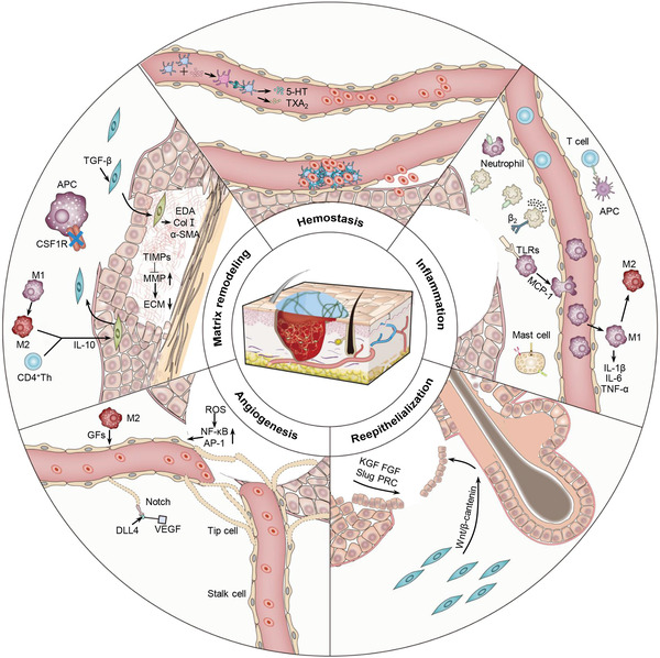
Skin wound repair is a multistage process involving multiple cellular and molecular interactions, which modulate the cell behaviors and dynamic remodeling of extracellular matrices to maximize regeneration and repair. Consequently, abnormalities in cell functions or pathways inevitably give rise to side effects, such as dysregulated inflammation, hyperplasia of nonmigratory epithelial cells, and lack of response to growth factors, which impedes angiogenesis and fibrosis. These issues may cause delayed wound healing or even non-healing states. Current clinical therapeutic approaches are predominantly dedicated to preventing infections and alleviating topical symptoms rather than addressing the modulation of wound microenvironments to achieve targeted outcomes. Bioactive materials, relying on their chemical, physical, and biological properties or as carriers of bioactive substances, can affect wound microenvironments and promote wound healing at the molecular level. By addressing the mechanisms of wound healing from the perspective of cell behaviors, this review discusses how bioactive materials modulate the microenvironments and cell behaviors within the wounds during the stages of hemostasis, anti-inflammation, tissue regeneration and deposition, and matrix remodeling. A deeper understanding of cell behaviors during wound healing is bound to promote the development of more targeted and efficient bioactive materials for clinical applications.

Keywords: bioactive material; cell behavior; regenerative medicine; wound healing; wound microenvironment.


Conflict of interest statement

The authors declare no conflict of interest.

Figures

Scheme 1
Scheme 1
Schematic illustration of typical pathways of bioactive materials modulating cell behaviors to promote cutaneous wound healing.
Figure 1
Figure 1
Synthetic platelet‐activating hydrogel to induce local hemostasis. A) Molecular mechanism of PAR‐1 activation and TRAP‐6‐presenting hydrogel. B) Preparation of PVA hydrogel (—SH: —NB = 0.4) particulate (PVA‐NB‐P) and surface functionalization of PVA‐NB‐P with cysteine‐containing TRAP‐6 peptide. C) SEM images of PVA‐TRAP‐6‐P. D) Plotted ROTEM curves show the coagulation process of whole blood in response to a) TRAP‐6, PVA‐TRAP‐6, PVA‐NB, and 0.9% NaCl as a control. b) PVA‐NB‐P and PVA‐TRAP‐6‐P suspensions, and 0.9% NaCl as a control. E) FACS analysis of TRAP‐6‐mediated platelet activation measured by determination of CD62p/CD41 coexpression. Reproduced with permission.[ 30c ] Copyright 2015, Wiley‐VCH. FACS, fluorescence‐ activated cell sorter; NB, norbornene; PAR‐1, protease‐activated receptors‐1; PVA, poly(vinyl alcohol); SEM, scanning electron microscopy; TRAP‐6, thrombin receptor agonist peptide‐6.
Figure 2
Figure 2
Degradable gelatin‐based IPN cryogel hemostat for rapidly stopping deep noncompressible hemorrhage and improving wound healing. A) Schematic representation of GT/DA cryogel formation and different fixed shapes of GT25/DA0 and GT25/DA8. B) Microtopography observation of GT25/DA8 in free shape, fixed shape, and recovered shape. Scale bar = 200 µm. C) In vivo hemostatic performance of cryogel in rabbit liver defect hemorrhage model. D) Wound contraction rate of in vivo wound‐healing evaluation. Reproduced with permission.[ 35 ] Copyright 2020, American Chemical Society. DA, dopamine; EDC, 1‐(3‐dimethylaminopropyl)‐3‐ethylcarbodii‐mide hydrochloride; GT, gelatin; IPN, interpenetrating polymer network; NHS, N‐hydroxysuccinimide; SP, sodium periodate.
Figure 3
Figure 3
Glycosaminoglycan‐based hydrogel capture inflammatory chemokines and rescue defective wound healing. A) StarPEG‐GAG hydrogel network can bind and neutralize chemokines through strong electrostatic interactions of heparin derivatives and chemokines. B) Storage modulus, mesh size, and sulfate content of compared hydrogel. C) Results of computational docking analysis of both chemokines using ClusPro software. D) Characterization of inflammation 5 and 10 days after wounding. E) Wounds 10 dpw and analysis of wound closure. Reproduced with permission.[ 47 ] Copyright 2017, American Association for the Advancement of Science. GAG, glycosaminoglycan; IL‐8, interleukin‐8; MCP‐1, monocyte chemoattractant protein‐1; PEG, poly(ethylene glycol).
Figure 4
Figure 4
Exosome laden O2 releasing antioxidant and antibacterial cryogel wound dressing OxOBand alleviate diabetic and infectious wound healing. A) Schematic representation of OxOBand formation from O2 releasing antioxidant PUAO‐CPO cryogels with exosomes of A‐MSCs. B) (Left) Nanoparticle tracking analysis are showing the double membrane vesicular structure of the exosomes with cup‐shaped morphology. Scale bar = 200 nm. (Right) Calcein AM stained exosomes are showing the intact exosome structure encapsulated by HaCaT cells. Scale bar = 10 µm. C) Confocal laser scanning microscopic images showing encapsulation of PKH26 labeled exosomes (red) by HaCaT cells. Scale bar = 20 µm. D) HaCaT cell migration was enhanced by exosome treatment compared to nontreated cells. Representative graph showing the migration rate of HaCaT cells upon exosome treatment. E) Representative H&E images of wounds (scale bar = 2 mm) and high magnification images showing granulation tissue formation and epithelial tissue closure (scale bar = 200 µm). F) The granulation tissue formation and average epidermal thickness in the wound center were quantified in histological samples harvested after 14 days. Reproduced with permission.[ 86 ] Copyright 2020, Elsevier Ltd. CPO, calcium peroxide; EXO, exosomes; H&E, hematoxylin and eosin; PUAO, polyurethane.
Figure 5
Figure 5
Micro‐channel network hydrogels induced ischemic blood perfusion connection. A) Schematic illustration of procedure to produce PNIPAM fiber, channel network in a hydrogel within a PDMS mold. B) Confocal visualization of micro‐ or macro‐channel networks in hydrogels with their channel diameter distribution. Scale bar = 100 µm. C) Representative images of general histology (H&E) and CD31+ cells (green) with nuclei (blue DAPI). White arrows point out microvascular structures (CD31+) in the skin tissue sections. Scale bar = 100 µm. D) Photographs of wound healing sites on day 14 postimplantation. Scale bar = 1 cm. E) Degree of decreased wound area from the initial 2 × 2 cm defect in each group on day 14 post‐implantation (n = 4). Reproduced with permission.[ 126 ] Copyright 2020, Springer Nature. H&E, hematoxylin and eosin; PDMS, polydimethylsiloxane; PNIPAM, poly(N‐isopropyl acrylamide); W/O, without.
Figure 6
Figure 6
Cryoprotectant enables structural control of porous scaffolds for the exploration of 3D cellular mechano‐responsiveness. A) Schematic illustration of scaffold fabrication via cryogelation of prepolymer solution in presence of cryoprotectant for pore size control and chemical cross‐linker for stiffness control. B) SEM images of gelatin scaffolds cross‐linked by GA demonstrating correlation of pore size and DMSO concentration. Scale bar = 100 µm. C) Traditional stiffness control using varied concentrations of chemical cross‐linker (GA) caused a change in scaffold pore size (stiffness, n = 3; pore size, n = 5). With the introduction of 1% or 5% DMSO, the pore size remained unchanged at 30 or 60 µm, respectively. D) Significant α‐SMA expression with fiber‐like accumulation was only observed in the scaffold with larger and stiffer pores (80 µm and 190 kPa). Scale bar = 50 µm. E) Images of YAP1 showed the significant cytoplasmic localization in small pore scaffold and nucleus localization in 80 µm and 190 kPa  group. Scale bar = 50 µm. Reproduced with permission.[ 137 ] Copyright 2019, Springer Nature. DMSO, dimethylsulfoxide; GA, glutaraldehyde; SEM, scanning electron microscope.

References

    1. Mathes S. H., Ruffner H., Graf‐Hausner U., Adv. Drug Deliv. Rev. 2014, 69, 81. - PubMed
    1. Rodrigues M., Kosaric N., Bonham C. A., Gurtner G. C., Physiol. Rev. 2019, 99, 665. - PMC - PubMed
    1. Kim H. S., Sun X., Lee J. H., Kim H. W., Fu X., Leong K. W., Adv. Drug Deliv. Rev. 2019, 146, 209. - PubMed
    1. Castano O., Perez‐Amodio S., Navarro‐Requena C., Mateos‐Timoneda M. A., Engel E., Adv. Drug Deliv. Rev. 2018, 129, 95. - PubMed
    1. Eming S. A., Martin P., Tomic‐Canic M., Sci. Transl. Med. 2014, 6, 16. - PMC - PubMed

Publication types

LinkOut - more resources