Review
. 2022 Mar 16;86(1):e0016521.
doi: 10.1128/mmbr.00165-21. Epub 2022 Feb 9.

Chemotropism and Cell-Cell Fusion in Fungi

Affiliations
Review

Chemotropism and Cell-Cell Fusion in Fungi

Manuella R Clark-Cotton et al. Microbiol Mol Biol Rev. .

Abstract

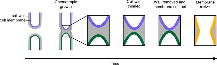
Fungi exhibit an enormous variety of morphologies, including yeast colonies, hyphal mycelia, and elaborate fruiting bodies. This diversity arises through a combination of polar growth, cell division, and cell fusion. Because fungal cells are nonmotile and surrounded by a protective cell wall that is essential for cell integrity, potential fusion partners must grow toward each other until they touch and then degrade the intervening cell walls without impacting cell integrity. Here, we review recent progress on understanding how fungi overcome these challenges. Extracellular chemoattractants, including small peptide pheromones, mediate communication between potential fusion partners, promoting the local activation of core cell polarity regulators to orient polar growth and cell wall degradation. However, in crowded environments, pheromone gradients can be complex and potentially confusing, raising the question of how cells can effectively find their partners. Recent findings suggest that the cell polarity circuit exhibits searching behavior that can respond to pheromone cues through a remarkably flexible and effective strategy called exploratory polarization.

Keywords: Cdc42; GPCR; anastomosis; cell polarity; fungi; mating; pheromone; yeast.


Conflict of interest statement

The authors declare no conflict of interest.

Figures

FIG 1
FIG 1
Examples of cell-cell fusion in fungi. Arrows indicate sites where fusion will occur. The color of the nuclei indicates mating type. A yellow outline indicates cells that have undergone fusion. (A) Mating yeast cells. (B) Mating monokaryotic hyphae (basidiomycetes). (C) Clamp connection formation in dikaryotic hyphae (basidiomycetes). To maintain a stable dikaryon, the tip (apical) cell grows a narrow protrusion that obtains a nucleus and septates to become a clamp cell, which homes toward and fuses with the neighboring subapical cell, allowing the nucleus to pass into the subapical cell. (D) Anastomosis (hyphal fusion) begins between germlings following spore germination. Later (not shown in figure), hyphae branch to form thinner fusion hyphae that similarly find each other and fuse to generate an interconnected mycelium. (E) Mating of trichogyne and conidium (filamentous ascomycetes). (F) Crozier formation (heterothallic filamentous ascomycetes). (G) Formation of adhesive loops and constricting rings in nematode-trapping fungi.
FIG 2
FIG 2
Challenges of cell-cell fusion. First, cells must use polar growth to come into contact with each other. Then, the cell wall between the two cells (and only that wall) must be removed to allow membrane contact and fusion.
FIG 3
FIG 3
The pheromone landscape. (A) Theoretical steady-state gradient formed by the diffusion of a chemoattractant from a single source. Plot of pheromone concentration as a function of distance from the source (R). Blue and orange lines indicate the change in concentration across a 10-μm detector placed 20 μm (blue) or 40 μm (orange) from the source. (B) Comparison of steady-state pheromone concentration (blue, in units of binding KD) and the resulting fraction of occupied receptors (orange) as a function of distance from the source (R). (C) Hyphal tips (green) can create multiple targets and a confusing net chemoattractant gradient for a searching hypha (purple). (D) Yeast cells often mate in environments where each cell has multiple potential fusion partners, creating a confusing net pheromone gradient. (E) Left: a computational model shows how the local secretion of a pheromone protease (Bar1) would reshape an imposed linear pheromone gradient (top color bar). Arrows indicate gradient direction sensed by each cell tip, leading to divergent growth. Right: When exposed to a linear pheromone gradient in a microfluidic device, cells secreting Bar1 exhibited self-avoidance (divergent growth), while bar1Δ cells did not. (Adapted from reference with permission from AAAS.)
FIG 4
FIG 4
Pheromone response in yeasts. (A) Cells of opposite mating type secrete unique prenylated (green) or unprenylated (purple) pheromones that are detected by G-protein-coupled receptors on cells of the opposite mating type. (B) Signaling from GPCR to MAPK for S. cerevisiae and S. pombe (dashed lines indicate where molecular links are not fully known). (C) Signaling from GPCR to Cdc42 for S. cerevisiae and S. pombe (dashed lines indicate where molecular links are not fully known). (D) Left: Conversion of Cdc42 between inactive and active forms, catalyzed by GEFs and GAPs. Active Cdc42 binds effectors that lead to local secretion. Right: Positive feedback loop leads to Cdc42 clustering; active Cdc42 can recruit effector-GEF complexes from the cytoplasm, thereby activating neighboring Cdc42. Locally depleted inactive Cdc42 is replenished by binding from the cytoplasm. (E) Pheromone receptor distribution is not uniform on the cell cortex. Left: Ste2-sfGFP in a cell in G1 (cortical signal is receptor; V, vacuolar signal from undegraded sfGFP). Right: Quantification of Ste2-sfGFP membrane distribution. Dark line is average from 71 cells, and shading represents standard deviation. (Adapted from reference .) (F) Mechanism for ratiometric sensing; pheromone-bound/active receptor promotes conversion of Gα-GDP to Gα-GTP. Inactive receptor binds Sst2, a GAP that converts Gα-GTP to Gα-GDP. Thus, Gα-GTP reports the ratio of active to inactive receptors.
FIG 5
FIG 5
Polarity site movement during yeast mating. (A) Vesicle delivery (marked by Spa2) trails the polarity site (marked by the Cdc42 effector Bem1) in S. cerevisiae. (Adapted from reference .) (B) Left: Polarity site movement illustrated for two touching cells of the opposite mating type. In the bottom cell, the polarity site movement in a 2-minute interval is illustrated by the arrow (t0 to t1). The optimal direction of movement (toward the partner cell’s polarity site) is indicated by the dashed line. The angle of polarity site movement (θ) relative to the optimal direction is shown. Top right: θ from cells with opposite mating type partners is biased toward small angles (i.e., toward the partner). Bottom right: θ from cells with same mating type partners shows no bias. (Adapted from reference .) (C) Pheromone receptor and Gβγ (marked by Ste4) surround and trail the site of vesicle delivery (Spa2). (Adapted from reference .) (D) Exploratory polarization model.
FIG 6
FIG 6
Self-signaling during hyphal anastomosis. Genetically identical conidial anastomosis tubes (CATs) in N. crassa grow toward each other alternating between states with either the scaffold SOFT (purple) or the MAPK MAK-2 (green) concentrating at the tip. According to the “ping-pong model,” an unknown chemoattractant (purple) is secreted from SOFT-enriched tips and received by MAPK-enriched tips, allowing chemotropic growth by alternating self-signaling between hyphae. Switches between the signaling and receiving states may involve feedback loops that render the system excitable.
FIG 7
FIG 7
Cell wall removal and membrane fusion. (A) The juxtaposition of cell walls between cells engaged in polar growth could enrich secreted hydrolases to promote enhanced degradation of the intervening wall. In the absence of cell-cell contact, secreted cell wall hydrolases escape rapidly via diffusion, acting only transiently. Upon cell-cell contact, diffusional escape paths are longer, allowing hydrolases more time to promote wall degradation. Gray lines indicate possible diffusional paths. (B) Focusing of secretion would alter the local ratio of hydrolases to synthases, enabling wall degradation. During vegetative growth, dispersed delivery of cell wall hydrolases and synthases to the growing tip maintains a balance between hydrolase and synthase activity. If actin cables become more tightly focused, it would concentrate the cell wall hydrolases, allowing them to outpace synthase activity to promote wall degradation. (C) Plasma membrane asymmetry during mating in S. pombe. One mating partner (green) has a taut, smooth plasma membrane that pushes into the slack, wavy membrane of the opposite partner (purple). The difference is correlated with a difference in the local abundance of exocytic and endocytic vesicles. An osmotic imbalance yielding higher hydrostatic pressure in the green cell may drive fusion.

References

    1. Bennett RJ, Turgeon BG. 2016. Fungal sex: the Ascomycota. Microbiol Spectr 4:4.5.20. 10.1128/microbiolspec.FUNK-0005-2016. - DOI - PubMed
    1. Coelho MA, Bakkeren G, Sun S, Hood ME, Giraud T. 2017. Fungal sex: the Basidiomycota. Microbiol Spectr 5:5.3.12. 10.1128/microbiolspec.FUNK-0046-2016. - DOI - PMC - PubMed
    1. Erental A, Dickman MB, Yarden O. 2008. Sclerotial development in Sclerotinia sclerotiorum: awakening molecular analysis of a “dormant” structure. Fungal Biol Rev 22:6–16. 10.1016/j.fbr.2007.10.001. - DOI
    1. Buller AHR. 1958. The formation of hyphal fusions in the mycelium of the higher fungi, p 27–32. In Researches on fungi: hyphal fusions and protoplasmic streaming in the higher fungi, together with an account of the production and liberation of spores in Sporobolomyces, Tilletia, and Sphaerobolus, vol 5. Hafner Publishing Co., New York, NY.
    1. Roca MG, Davide LC, Mendes-Costa MC, Wheals A. 2003. Conidial anastomosis tubes in Colletotrichum. Fungal Genet Biol 40:138–145. 10.1016/s1087-1845(03)00088-4. - DOI - PubMed

Publication types

Substances

LinkOut - more resources