

Human Polyomavirus 9-An Emerging Cutaneous and Pulmonary Pathogen in Solid Organ Transplant Recipients
- PMID: 35138364
- PMCID: PMC8829745
- DOI: 10.1001/jamadermatol.2021.5853
Human Polyomavirus 9-An Emerging Cutaneous and Pulmonary Pathogen in Solid Organ Transplant Recipients
Abstract
Importance: We describe the first report to our knowledge of cutaneous and systemic pathogenicity of human polyomavirus 9 in solid organ transplant recipients.
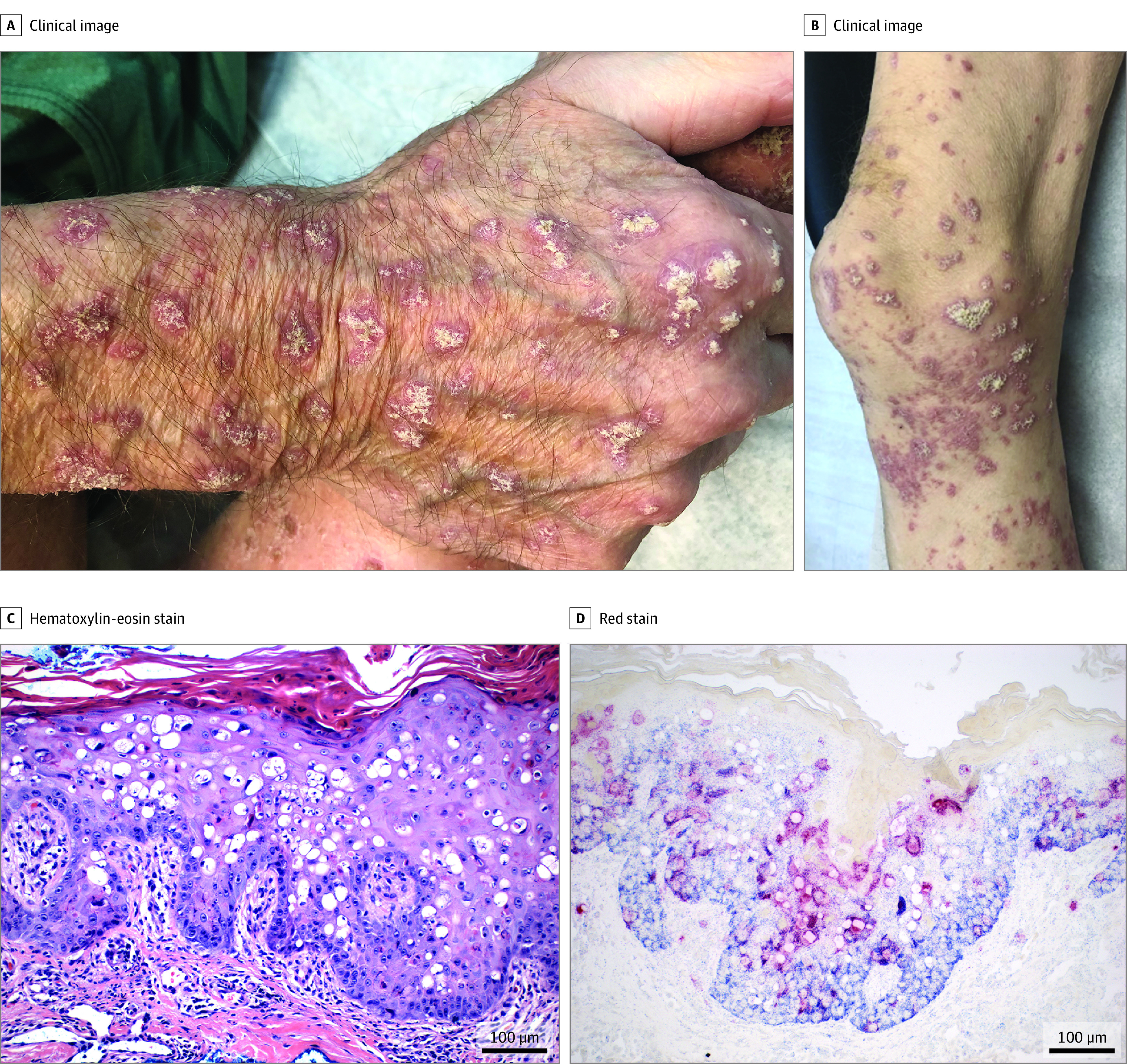
Objective: Three solid organ transplant recipients developed a widespread, progressive, violaceous, and hyperkeratotic skin eruption. All died from pulmonary and multiorgan failure around 1 year from onset of the rash. Routine clinical diagnostic testing could not identify any causative agent; therefore, samples and autopsies were investigated for novel pathogens using high-throughput sequencing.
Design, setting, and participants: This case series, including 3 solid organ transplant recipients who developed characteristic pink, violaceous, or brown hyperkeratotic papules and plaques throughout the body, was conducted at the Columbia University Medical Center. Lesional skin biopsies were collected from all 3 patients and subjected to high-throughput illumina sequencing for identification of microbial pathogens. Human polyomavirus 9 was identified in lesional skin biopsies. We subsequently collected ocular swabs, oral swabs, urine samples, and blood samples from patients, and organ tissues at autopsy in 1 patient. We investigated these samples for the presence of human polyomavirus 9 using in situ hybridization and quantitative polymerase chain reaction (PCR) assays.
Main outcomes and measures: A description of the clinical and pathologic findings of 3 patients.
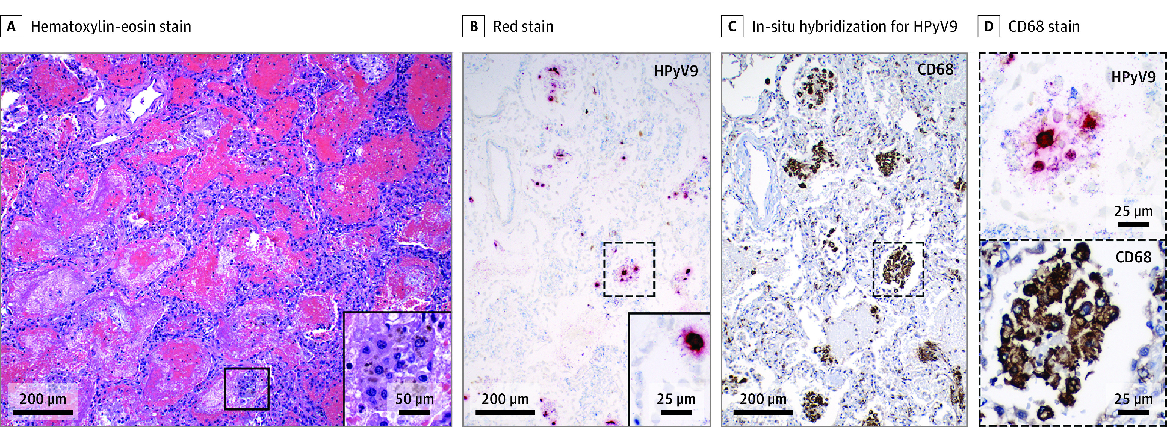
Results: This case series study found that human polyomavirus 9 was detected in the skin biopsies of all 3 patients by a capture-based high-throughput sequencing method platform (VirCapSeq-VERT). Human polyomavirus 9 was also detected in blood, oral, ocular swabs, and urine by real-time polymerase chain reaction (PCR) assay. In situ hybridization and quantitative PCR assays were performed on the skin biopsies from 3 patients and lung autopsy of 1 patient, which showed the presence of human polyomavirus 9 messenger RNA transcripts, indicating active viral replication and pathogenesis in the skin and lungs.
Conclusions and relevance: Human polyomavirus 9 was associated with the widespread cutaneous eruption. All 3 patients had progression of cutaneous disease, accompanied by clinical deterioration, pulmonary failure, and death. One patient underwent autopsy and human polyomavirus 9 was identified in the lungs and paratracheal soft tissue. These findings suggest that human polyomavirus 9 may be associated with cutaneous and possibly pulmonary infection and death in solid organ transplant recipients.
Conflict of interest statement
Figures
Similar articles
-
Human polyomavirus 7-associated pruritic rash and viremia in transplant recipients.J Infect Dis. 2015 May 15;211(10):1560-5. doi: 10.1093/infdis/jiu524. Epub 2014 Sep 17. J Infect Dis. 2015. PMID: 25231015 Free PMC article.
-
Prevalence of polyomavirus among United Arab Emirates kidney transplant recipients: results from a single center.Transplant Proc. 2015 May;47(4):1143-5. doi: 10.1016/j.transproceed.2014.11.060. Transplant Proc. 2015. PMID: 26036540
-
New findings about trichodysplasia spinulosa-associated polyomavirus (TSPyV)--novel qPCR detects TSPyV-DNA in blood samples.Diagn Microbiol Infect Dis. 2016 Feb;84(2):123-4. doi: 10.1016/j.diagmicrobio.2015.10.011. Epub 2015 Oct 21. Diagn Microbiol Infect Dis. 2016. PMID: 26602950
-
BK virus in solid organ transplant recipients: an emerging syndrome.Transplantation. 2001 Nov 27;72(10):1587-92. doi: 10.1097/00007890-200111270-00001. Transplantation. 2001. PMID: 11726814 Review.
-
Treatment of human polyomavirus-7-associated rash and pruritus with topical cidofovir in a lung transplant patient: Case report and literature review.Transpl Infect Dis. 2018 Feb;20(1). doi: 10.1111/tid.12793. Epub 2017 Nov 26. Transpl Infect Dis. 2018. PMID: 29064138 Review.
Cited by
-
[Human polyomavirus-associated skin diseases].Hautarzt. 2022 Jun;73(6):426-433. doi: 10.1007/s00105-022-04993-8. Epub 2022 Apr 28. Hautarzt. 2022. PMID: 35482045 Review. German.
-
A Review of Cutaneous Diseases Observed in Solid Organ Transplant Recipients.J Clin Aesthet Dermatol. 2022 Oct;15(10):21-31. J Clin Aesthet Dermatol. 2022. PMID: 36312823 Free PMC article. Review.
-
2025 American Society for Microbiology Awards and Prize Program: honorees from clinical microbiology.J Clin Microbiol. 2025 Mar 12;63(3):e0000125. doi: 10.1128/jcm.00001-25. Epub 2025 Feb 24. J Clin Microbiol. 2025. PMID: 40167424 Free PMC article. No abstract available.
References
Publication types
MeSH terms
Substances
Supplementary concepts
LinkOut - more resources
Full Text Sources
Medical
