Clinical Trial
. 2022 Jun;49(7):2174-2188.
doi: 10.1007/s00259-022-05712-0. Epub 2022 Feb 9.

Quantitative classification and radiomics of [18F]FDG-PET/CT in indeterminate thyroid nodules

Affiliations
Clinical Trial

Quantitative classification and radiomics of [18F]FDG-PET/CT in indeterminate thyroid nodules

Elizabeth J de Koster et al. Eur J Nucl Med Mol Imaging. 2022 Jun.

Abstract

Purpose: To evaluate whether quantitative [18F]FDG-PET/CT assessment, including radiomic analysis of [18F]FDG-positive thyroid nodules, improved the preoperative differentiation of indeterminate thyroid nodules of non-Hürthle cell and Hürthle cell cytology.

Methods: Prospectively included patients with a Bethesda III or IV thyroid nodule underwent [18F]FDG-PET/CT imaging. Receiver operating characteristic (ROC) curve analysis was performed for standardised uptake values (SUV) and SUV-ratios, including assessment of SUV cut-offs at which a malignant/borderline neoplasm was reliably ruled out (≥ 95% sensitivity). [18F]FDG-positive scans were included in radiomic analysis. After segmentation at 50% of SUVpeak, 107 radiomic features were extracted from [18F]FDG-PET and low-dose CT images. Elastic net regression classifiers were trained in a 20-times repeated random split. Dimensionality reduction was incorporated into the splits. Predictive performance of radiomics was presented as mean area under the ROC curve (AUC) across the test sets.

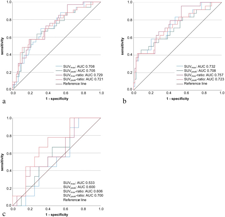
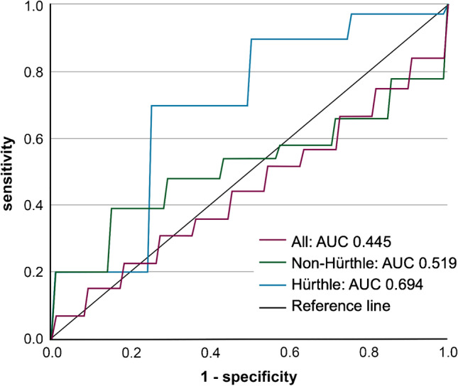
Results: Of 123 included patients, 84 (68%) index nodules were visually [18F]FDG-positive. The malignant/borderline rate was 27% (33/123). SUV-metrices showed AUCs ranging from 0.705 (95% CI, 0.601-0.810) to 0.729 (0.633-0.824), 0.708 (0.580-0.835) to 0.757 (0.650-0.864), and 0.533 (0.320-0.747) to 0.700 (0.502-0.898) in all (n = 123), non-Hürthle (n = 94), and Hürthle cell (n = 29) nodules, respectively. At SUVmax, SUVpeak, SUVmax-ratio, and SUVpeak-ratio cut-offs of 2.1 g/mL, 1.6 g/mL, 1.2, and 0.9, respectively, sensitivity of [18F]FDG-PET/CT was 95.8% (95% CI, 78.9-99.9%) in non-Hürthle cell nodules. In Hürthle cell nodules, cut-offs of 5.2 g/mL, 4.7 g/mL, 3.4, and 2.8, respectively, resulted in 100% sensitivity (95% CI, 66.4-100%). Radiomic analysis of 84 (68%) [18F]FDG-positive nodules showed a mean test set AUC of 0.445 (95% CI, 0.290-0.600) for the PET model.

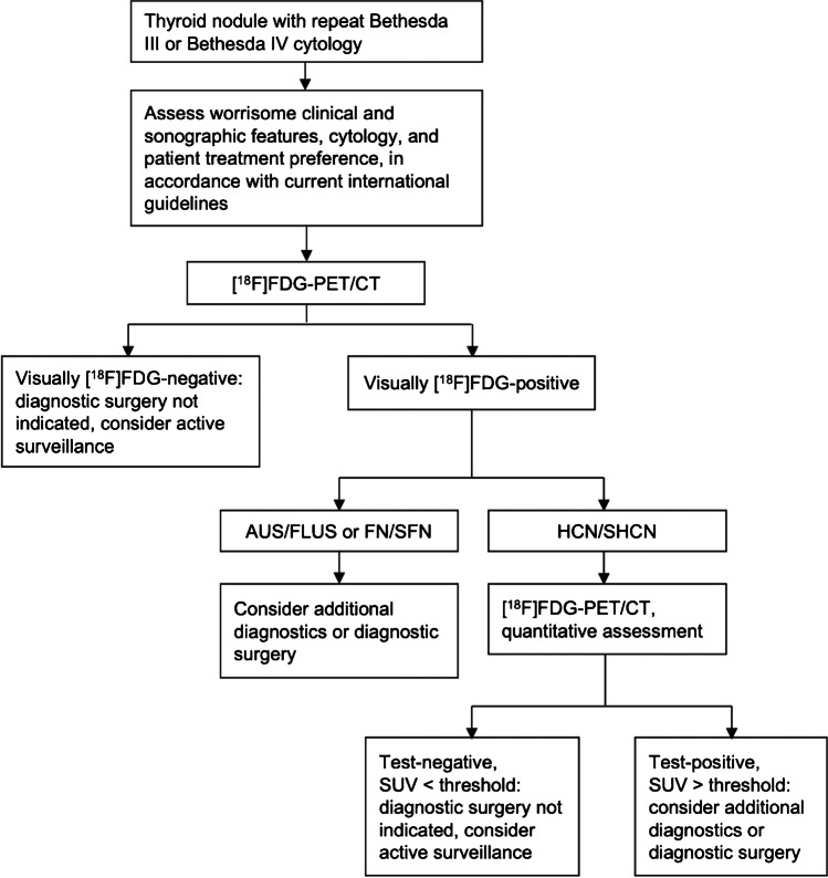
Conclusion: Quantitative [18F]FDG-PET/CT assessment ruled out malignancy in indeterminate thyroid nodules. Distinctive, higher SUV cut-offs should be applied in Hürthle cell nodules to optimize rule-out ability. Radiomic analysis did not contribute to the additional differentiation of [18F]FDG-positive nodules.

Trial registration number: This trial is registered with ClinicalTrials.gov: NCT02208544 (5 August 2014), https://clinicaltrials.gov/ct2/show/NCT02208544 .

Keywords: Indeterminate; Quantitative; Radiomics; Standardised uptake value; Thyroid carcinoma; Thyroid cytology; Thyroid nodule; [18F]FDG-PET/CT.


Conflict of interest statement

The authors declare no competing interests.

Figures

Fig. 1
Fig. 1
Study flowchart. aPatient screening for the original trial, including eligibility criteria, were previously published [4]. bBaseline characteristics of included patients (Table 1) were similar to those of excluded patients (Supplementary table 3). cnon-Hürthle cell nodules comprise nodules of AUS/FLUS (n = 55) and FN/SFN (n = 39) cytology. AUS/FLUS, atypia of undetermined significance or follicular lesion of undetermined significance. FN/SFN, cytology (suspicious for a) follicular neoplasm. FT-UMP, follicular tumour of uncertain malignant potential. HCN/SHCN, (suspicious for a) Hürthle cell neoplasm. NIFTP, non-invasive follicular thyroid neoplasm with papillary-like nuclear features
Fig. 2
Fig. 2
Quantitative [18F]FDG-PET/CT assessment and delineation of the VOI for radiomic analysis. Transverse and coronal [18F]FDG-PET/CT (a, b), maximum intensity projection (MIP) (c, d) and low-dose CT (e, f) images of a patient with a solitary, 30 mm Bethesda III thyroid nodule in the right lobe. Visual assessment (a) of the [18F]FDG-PET/CT showed an [18F]FDG-positive index nodule. Quantitative assessment (b) demonstrated a SUVmax of 9.7 g/mL and SUVpeak of 7.0 g/mL of the index nodule, and a SUVmax of 1.6 g/mL in the background of surrounding normal thyroid tissue. Consequently, the SUVmax-ratio and SUVpeak-ratio were 6.1 (9.7/1.6) and 4.4 (7.0/1.6), respectively. For radiomic analysis, VOIs were delineated on the [18F]FDG-PET scans using an isocontour that applies a threshold of 50% of the SUVpeak, corrected for local background (c, d) [29]. Boxing was applied to exclude [18F]FDG-positive tissue surrounding the index nodule and ldCT images were used as a visual reference (e, f). VOIs delineated on the PET images were resampled with a nearest neighbour algorithm to derive the ldCT VOIs
Fig. 3
Fig. 3
ROC curves of quantitative [18F]FDG-PET/CT analysis. ROC curves for SUVmax (blue line), SUVpeak (green), SUVmax-ratio (purple), and SUVpeak-ratio (red) in a all (n = 123), b non-Hürthle cell (n = 94), and c Hürthle cell (n = 29) nodules. a: In all nodules, the AUCs for the SUVmax, SUVpeak, SUVmax-ratio, and SUVpeak-ratio were 0.708 (95% CI, 0.609–0.807), 0.705 (0.601–0.810), 0.729 (0.633–0.824), and 0.721 (0.618–0.824), respectively. b: In non-Hürthle cell nodules, these AUCs were 0.732 (95% CI, 0.615–0.849), 0.708 (0.580–0.835), 0.757 (0.650–0.864), and 0.723 (0.601–0.844), respectively. c: In Hürthle cell nodules, these AUCs were 0.533 (95% CI, 0.320–0.747), 0.600 (0.392–0.808), 0.606 (0.388–0.823), and 0.700 (0.502–0.898), respectively
Fig. 4
Fig. 4
ROC curves of the PET model of the radiomic analysis. ROC curves for the PET/CT model of the radiomic analysis. The AUC was 0.445 (95% CI, 0.290–0.600) in all nodules (n = 84, purple line), 0.519 (95% CI, 0.298–0.740) in non-Hürthle cell nodules (n = 56, green), and 0.694 (95% CI, 0.461–0.926) in Hürthle cell nodules (n = 28, blue)
Fig. 5
Fig. 5
Proposed [18F]FDG-PET/CT-driven workup of Bethesda III/IV thyroid nodules

References

    1. Cibas ES, Ali SZ. The 2017 Bethesda system for reporting thyroid cytopathology. Thyroid. 2017;27:1341–1346. doi: 10.1089/thy.2017.0500. - DOI - PubMed
    1. Bongiovanni M, Spitale A, Faquin WC, Mazzucchelli L, Baloch ZW. The Bethesda system for reporting thyroid cytopathology: a meta-analysis. Acta Cytol. 2012;56:333–339. doi: 10.1159/000339959. - DOI - PubMed
    1. Durante C, Grani G, Lamartina L, Filetti S, Mandel SJ, Cooper DS. The diagnosis and management of thyroid nodules: a review. JAMA. 2018;319:914–924. doi: 10.1001/jama.2018.0898. - DOI - PubMed
    1. de Koster EJ, de Geus-Oei LF, Brouwers AH, van Dam EWCM, Dijkhorst-Oei LT, van Engen-van Grunsven ACH, et al. [18F]FDG-PET/CT to prevent futile surgery in indeterminate thyroid nodules: a blinded, randomised controlled multicentre trial. Eur J Nucl Med Mol Imaging 2022. 10.1007/s00259-021-05627-2. - PMC - PubMed
    1. de Geus-Oei LF, Pieters GF, Bonenkamp JJ, Mudde AH, Bleeker-Rovers CP, Corstens FH, et al. 18F-FDG PET reduces unnecessary hemithyroidectomies for thyroid nodules with inconclusive cytologic results. J Nucl Med. 2006;47:770–775. - PubMed

Publication types

MeSH terms

Substances

Associated data