. 2021:1349:195-223.
doi: 10.1007/978-981-16-4254-8_10.

Glial Chloride Channels in the Function of the Nervous System Across Species

Affiliations

Glial Chloride Channels in the Function of the Nervous System Across Species

Jesus Fernandez-Abascal et al. Adv Exp Med Biol. 2021.

Abstract

In the nervous system, the concentration of Cl- in neurons that express GABA receptors plays a key role in establishing whether these neurons are excitatory, mostly during early development, or inhibitory. Thus, much attention has been dedicated to understanding how neurons regulate their intracellular Cl- concentration. However, regulation of the extracellular Cl- concentration by other cells of the nervous system, including glia and microglia, is as important because it ultimately affects the Cl- equilibrium potential across the neuronal plasma membrane. Moreover, Cl- ions are transported in and out of the cell, via either passive or active transporter systems, as counter ions for K+ whose concentration in the extracellular environment of the nervous system is tightly regulated because it directly affects neuronal excitability. In this book chapter, we report on the Cl- channel types expressed in the various types of glial cells focusing on the role they play in the function of the nervous system in health and disease. Furthermore, we describe the types of stimuli that these channels are activated by, the other solutes that they may transport, and the involvement of these channels in processes such as pH regulation and Regulatory Volume Decrease (RVD). The picture that emerges is one of the glial cells expressing a variety of Cl- channels, encoded by members of different gene families, involved both in short- and long-term regulation of the nervous system function. Finally, we report data on invertebrate model organisms, such as C. elegans and Drosophila, that are revealing important and previously unsuspected functions of some of these channels in the context of living and behaving animals.

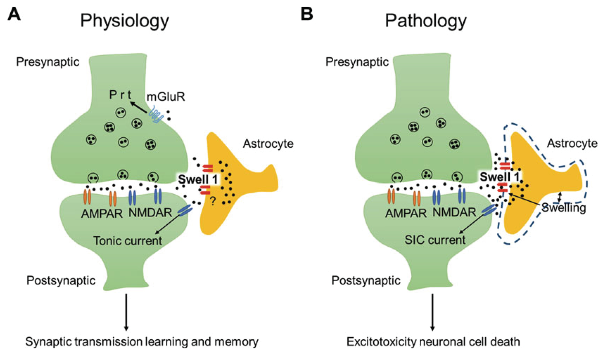
Keywords: Bestrophins; Channelopathies; ClC-2; Glial chloride channels; LRRC8; Maxi chloride channels; Nervous system development; Neuron; Pannexins; SWELL1; VRAC; glia interaction.


Figures

Fig. 10.1
Fig. 10.1
ClC-2 structure and function. (a) Membrane topology of a ClC channel. The 18 α-helices are labeled by letters (A through R). The amino acid sequences that contribute to the Cl selectivity are designated by the blue arrows. The two CBS motifs are shown in green. (b) Ribbon rendition of rat ClC-2 channel. The two subunits of the dimer are represented in red and blue, respectively. In both A and B, the light gray shaded area represents the plasma membrane. (c) Currents elicited by voltage steps from −160 mV to +60 in 20 mV increments from a holding potential of −30 mV in a oocyte expressing rat ClC-2 perfused with a solution in which the main anion was Cl. (d) Average current/voltage relationship from oocytes injected with rat ClC-2 and perfused with a Cl solution (filled circles) and with the Cl solution containing 2 mM CdCl2, a ClC-2 channel blocker (empty circles) (n = 8 for both, Sangaletti R., Johnson C.K., and Bianchi L., unpublished observations). (e) Perivascular astrocytes showcasing MLC1, Glialcam, and ClC2. All three proteins co-localize to the endfeet of perivascular astrocytes contacting blood vessels and astrocyte–astrocyte contacts [6, 7]. The localization and function of ClC-2 channels are controlled by the interaction with both GlialCAM and MLC1 [8]
Fig. 10.2
Fig. 10.2
ClC-2 type channels in Drosophila and C. elegans. (a and b) In Drosophila, ClC-a is expressed in the niche in cortex glia, which is associated with neurogenic tissues. Analysis of ClC-a mutant flies revealed that ClC-a controls both the wiring of the nervous system and the size of the fly brain. Indeed, in flies that are mutant for ClC-a, the brain is smaller and there are widespread wiring defects. Plazaola and colleagues proposed that ionic homeostasis mediated by glial ClC-a may nonautonomously affect neurogenesis and the assembly of neural circuits [37]. The photographs show representative confocal sections of adult optic lobes of wild type and ClC-a mutant photoreceptor arrays stained with anti-Chaoptin. Apparent is the axonal trajectory defect in the optic lobe of the ClC-a mutant fly. (c) Bright-field image of C. elegans glued on an agarose pad on a glass slide. The dashed arrows indicate the direction of the cuts made with a glass micropipette prior to pHlourin pH imaging experiments to expose the glia to the perfusing solution. (d) Fluorescent image of the same animal as in (c). The fluorescence is pHlourin expressed in an amphid sheath glial cell. Scale bars for C and D are 50 μm. (e) pHlourin-mediated pH imaging of amphid sheath glial cells in C. elegans perfused with an HCO3 buffer following baseline imaging perfusing with an HCO3 free and Cl free solution. Note the reduced alkalinization in the clh-1 mutant animal (red line), which is restored in the rescue animal (clh-1;pT02B11.3:clh-1, blue line), indicating that CLH-1 is important for HCO3 permeation into C. elegans glia. (f) Average alkalinization expressed as DF/F for wild type, clh-1 mutants, and clh-1 rescue C. elegans, n was 16, 8, and 8 respectively. (c–f) were adapted from Grant et al. [38]
Fig. 10.3
Fig. 10.3
LRRC8 structure and role in the regulation of volume decrease. (a) When cells sense hypotonic conditions they tend to swell causing VRAC/VSOAC/SWELL1 channels to open, allowing for an efflux of organic osmolytes and Cl. When the most substrate being transported is Cl then VRAC/VSOAC/SWELL1 causes efflux of K+ via the K+ channels to maintain electroneutrality. Water efflux across the plasma membrane is induced by the efflux of osmotically active substances. Water efflux is via the lipid bilayer or can be mediated by aquaporins. This mechanism allows the cellular volume to return to its original control level. (b) Schematic representation of the topology of an LRRC8 channel subunit. Transmembrane domains are labeled TMH1-4 (Transmembrane Helix 1–4), the extracellular loops are labeled EL1 and EL2 (Extracellular Loop 1 and 2), and the intracellular loops are labeled IL1 and IL2 (Intracellular Loop 1 and 2). The leucine-rich repeat domain (LRRD) is shown in purple. (c) Ribbon rendition of LRRC8A hexameric structure. Two subunits in the back are not shown for clarity (from [42]). Each subunit is shown in a different color. (d) Example of swelling activated currents in HCT166 cells coexpressing isoforms 8A and 8C of LRRC8. Currents were activated by voltage steps from −120 to +120 mV in 20 mV increments. (e) Current–voltage relationships of SWELL1 currents recorded in isotonic, hypertonic, and hypotonic solutions. (c, d) panels are adapted and reprinted with permission from Yamada and Strange [43]
Fig. 10.4
Fig. 10.4
Role of SWELL1 in learning and memory and in excitotoxicity. Astrocytes contribute to excitotoxicity and regulate synaptic transmission via glutamate release. The exact mechanisms by which astrocytic glutamate is released are not fully understood. VRAC/VSOAC/SWELL1 has been proposed to mediate non-vesicular glutamate release from astrocytes. Indeed, reduced ambient glutamate levels were observed in astrocyte-specific Swell1 knockout mice. These mutant mice exhibited impairment of learning and memory that was dependent on the hippocampus (a). Swell1 knockout mice were also protected from brain damage following an ischemic stroke due to reduced glutamate release from astrocytes (b) [62]
Fig. 10.5
Fig. 10.5
TMEM206 encodes an acid activated Cl channel across species. (a) Schematic topology of TMEM206. Human TMEM206 is 411 aa long, it is predicted to have two transmembrane domains (TM1 and TM2) and intracellular N and C termini. (b) Human TMEM206 and the corresponding homologs from the naked mole rat, chicken, green anole, and zebrafish expressed in TMEM206−/− HEK cells generate Cl currents activated by perfusion with a solution at pH 5 [77]
Fig. 10.6
Fig. 10.6
Pax1 encodes a Cl channel when activated by voltage. (a) Panx1 opens as a large conductance channel (500 pS) when exposed to high extracellular K+. The membrane patch was in the inside-out configuration and was clamped at −100 mV. The dashed and solid lines indicate subconductance and fully open and closed states, respectively. (b) Panx1 exhibits a low conductance state when activated by voltage. A membrane patch in the outside-out configuration was exposed to low K+ and clamped at +50 mV. Two small conductance channels, indicated by the red lines and by O1 and O2, are activated under these conditions. One of the channels is also shown on a 5 × scale. The scale shown in panel A applies also to panel B (10 pA). Modified from Wang et al. [107].
Fig. 10.7
Fig. 10.7
Bestrophins structure and function. (a) Schematic representation of the topology of a Bestrophin channel based on studies conducted on Best1 [132]. There are six hydrophobic domains, however, domains 3 and 4 are expected to be intracellular. (b) Best1 currents were recorded in retinal pigment epithelial cells differentiated from induced pluripotent stem cells of a wild-type donor using intracellular solutions containing 0, 0.6 μM, and 1.2 μM Ca2+. The voltage-clamp protocol is shown in the insert. The scale bar is 1 nA and 150 ms. (c) Ca2+ dose–response curve for Best1 currents similar to the ones shown in panel B. The number of cells tested was 5 or 6 for each data point. The dotted line represents the zero current level. Modified and reprinted with permission from Li et al. [133]
Fig. 10.8
Fig. 10.8
Best1 localization in hippocampal astrocytes and in Bergman glia. (a) Immunohistochemical staining of GFAP-GFP (green) and of Best1(red), and a merge of the two images demonstrating exclusive expression of Best1 channels in microdomains of hippocampal astrocytes. (b) Schematic representation of the subcellular localization of Best1 (red) in hippocampal astrocytes. (c) Representative current-voltage relationship of NPPB-sensitive currents showing anion conductance in hippocampal astrocytes. (d) Immunohistochemical staining of GFAP-GFP (green) and of Best1(red), and a merge of the two images showing exclusive localization of Best1 in the soma of Bergmann glia. (e) Schematic representation of the subcellular localization of Best1 in Bergman glia. (f) Representative current–voltage relationship of NPPB-sensitive currents in Bergman glia. Note that the experiments shown in c and f were conducted in isometric Cl predicting a reversal potential of 0 mV. The more negative reversal potential observed in hippocampal astrocytes is due to the space clamp error caused by the localization of Best1 at the end of the cellular processes in this cell type. From Park et al. [150]

Similar articles

Cited by

References

    1. Thiemann A, Grunder S, Pusch M, Jentsch TJ (1992) A chloride channel widely expressed in epithelial and non-epithelial cells. Nature 356:57–60 - PubMed
    1. Elorza-Vidal X, Gaitan-Penas H, Estevez R (2019) Chloride channels in astrocytes: structure, roles in brain homeostasis and implications in disease. Int J Mol Sci 20 - PMC - PubMed
    1. Hou X, Zhang R, Wang J, Li Y, Li F, Zhang Y, Zheng X, Shen Y, Wang Y, Zhou L (2018) CLC-2 is a positive modulator of oligodendrocyte precursor cell differentiation and myelination. Mol Med Rep 17:4515–4523 - PMC - PubMed
    1. Sirisi S, Elorza-Vidal X, Arnedo T, Armand-Ugon M, Callejo G, Capdevila-Nortes X, Lopez-Hernandez T, Schulte U, Barrallo-Gimeno A, Nunes V, Gasull X, Estevez R (2017) Depolarization causes the formation of a ternary complex between GlialCAM, MLC1 and ClC-2 in astrocytes: implications in megalencephalic leukoencephalopathy. Hum Mol Genet 26:2436–2450 - PubMed
    1. Walz W (2002) Chloride/anion channels in glial cell membranes. Glia 40:1–10 - PubMed

LinkOut - more resources