. 2021:1349:225-247.
doi: 10.1007/978-981-16-4254-8_11.

Physiological and Pathological Relevance of Selective and Nonselective Ca2+ Channels in Skeletal and Cardiac Muscle

Affiliations

Physiological and Pathological Relevance of Selective and Nonselective Ca2+ Channels in Skeletal and Cardiac Muscle

Jaime Balderas-Villalobos et al. Adv Exp Med Biol. 2021.

Abstract

Contraction of the striated muscle is fundamental for human existence. The action of voluntary skeletal muscle enables activities such as breathing, establishing body posture, and diverse body movements. Additionally, highly precise motion empowers communication, artistic expression, and other activities that define everyday human life. The involuntary contraction of striated muscle is the core function of the heart and is essential for blood flow. Several ion channels are important in the transduction of action potentials to cytosolic Ca2+ signals that enable muscle contraction; however, other ion channels are involved in the progression of muscle pathologies that can impair normal life or threaten it. This chapter describes types of selective and nonselective Ca2+ permeable ion channels expressed in the striated muscle, their participation in different aspects of muscle excitation and contraction, and their relevance to the progression of some pathological states.

Keywords: Calcium; Dystrophy; Hypertrophy; Malignant hyperthermia; Orai1; Resting Ca2+ entry; Ryanodine receptor; STIM1; Store-operated Ca2+ entry; TRPC; Voltage-gated calcium channel.


Figures

Figure 1.
Figure 1.
Cartoon showing the main Ca2+ fluxes in muscle cells. Primary and secondary active transport mechanisms in the plasma membrane, plasma membrane Ca2+ ATPase (PMCA) and Na+/Ca2+ exchanger (NCX), extrude Ca2+ from the cytosol against its electrochemical gradient and are coupled to ATP hydrolysis or the influx of Na+ down its electrochemical gradient, respectively. PMCA and NCX are important for keeping the resting Ca2+ concentration in quiescent cells or reestablishing resting Ca2+ levels after channel activation. Ca2+ permeability in the plasma membrane is low at rest (leak channels), but ion channels in the plasma membrane can open in response to stimuli, including voltage-gated channels and other channels such as store-operated channels, stretch-activated channels, and channels activated by signaling molecules (all grouped in “other channels”). When Ca2+ permeable channels open, Ca2+ enters the cells down its electrochemical gradient. Sarco-endoplasmic reticulum Ca2+ ATPase (SERCA) is expressed in the membrane of the SR; it is a Ca2+ ATPase that concentrates Ca2+ in the SR lumen. Intracellular ion channels expressed in the SR membrane such as the ryanodine receptor (RyR) can open enabling Ca2+ efflux from the SR lumen down its chemical gradient.
Figure 2.
Figure 2.
Structural organization of the membranous system in skeletal muscle and the disposition of the CaV1.1 and RyR1 channels in the t-tubule and junctional SR membranes, respectively. A. Drawing depicting the disposition of the membranous system with respect to the contractile machinery in amphibian muscle. Note that the t-tubule (TT) runs along the side of the Z-disk (ZD). Mammalian skeletal muscle has a similar disposition, but the t-tubule runs along between the A and I bands (AI) having two t-tubules per sarcomere. Drawing reproduced with publisher permission from Peachey, 1965 [35]. B. Thin section of a triad visualized using electron microscopy. The t-tubule (TT) is clearly observed as surrounded by two terminal cisternae (TC) of the SR. Two densities between the junctional SR membrane and the t-tubule membrane are identifiable (2 per junction, 4 in total). Each density is a “foot” that corresponds to the large cytoplasmic domain of the RyR1. It has been proposed that two rows of RyR1 run along each junctional SR. Image reproduced with publisher permission from Franzini-Armstrong, 1970 [55]. C. Freeze fracture and rotary shadowing experiments were used to visualize the CaV1.1 particles along the longitudinal axis of the t-tubule. A group of four CaV1.1 particles is a tetrad; the image depicts 4 tetrads. In addition, a hypothetical array of RyR1s was overlayed considering the dimensions from CryoEM reconstructions. The best approximation for CaV1.1/RyR1 disposition in vivo is shown. Image reproduced with publisher permission from Paolini, Protasi and Franzini-Armstrong 2004 [58].
Figure 3.
Figure 3.
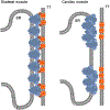
Comparison between skeletal and cardiac EC coupling. The current model for skeletal type EC coupling hypothesizes physical communication between the L-type Ca2+ channel (CaV1.1, orange) and the RyR1 (blue) expressed in the t-tubule (TT) and sarcoplasmic reticulum (SR) membranes, respectively. The effective communication between CaV1.1 and the RyR1 requires two essential accessory proteins β1a and Stac3. Other proteins are expressed in the junctional regions and may modulate skeletal EC coupling and some are relevant in skeletal muscle diseases (see [179]). In cardiac EC coupling, it is believed that CaV1.2 does not physically interact with the RyR2, but rather that clusters of these proteins are placed next to each other. Thus, a discrete group of CaV1.2 can activate a nearby cluster of RyR2 through a Ca2+-induced Ca2+ release mechanism.
Figure 4.
Figure 4.
Na+ and Ca2+ dysregulation can contribute to maladaptive changes in striated muscle pathologies. In several pathological models of striated muscle, upregulation or gain on function of some ion channels not activated by voltage (e.g., TRPC, STIM/Orai) can promote Ca2+ and Na+ dysregulation. Alterations in the homeostasis of these ions may influence downstream signaling pathways, metabolic changes, and oxidative stress that can contribute to the final pathological phenotype.

Similar articles

Cited by

References

    1. Berridge MJ. Elementary and global aspects of calcium signalling. J Exp Biol. 1997;200(Pt 2):315–9. - PubMed
    1. Aickin CC. Investigation of factors affecting the intracellular sodium activity in the smooth muscle of guinea-pig ureter. J Physiol. 1987;385:483–505. doi: 10.1113/jphysiol.1987.3522 - DOI - PMC - PubMed
    1. Powell T, Terrar DA, Twist VW. Electrical properties of individual cells isolated from adult rat ventricular myocardium. J Physiol. 1980;302:131–53. doi: 10.1113/jphysiol.1980.sp013234. - DOI - PMC - PubMed
    1. Lopez JR, Alamo L, Caputo C, DiPolo R, Vergara S. Determination of ionic calcium in frog skeletal muscle fibers. Biophys J. 1983;43(1):1–4. doi: 10.1016/S0006-3495(83)84316-1. - DOI - PMC - PubMed
    1. Lopez JR, Sanchez V, Lopez I, Ryan JF, Mendoza M, Sreter FA, et al. The effects of extracellular magnesium on myoplasmic [Ca2+] in malignant hyperthermia susceptible swine. Anesthesiology. 1990;73(1):109–17. doi: 10.1097/00000542-199007000-00016. - DOI - PubMed

LinkOut - more resources