. 2022 Mar;9(8):e2106058.
doi: 10.1002/advs.202106058. Epub 2022 Feb 9.

PIWI-Interacting RNA HAAPIR Regulates Cardiomyocyte Death After Myocardial Infarction by Promoting NAT10-Mediated ac4 C Acetylation of Tfec mRNA

Affiliations

PIWI-Interacting RNA HAAPIR Regulates Cardiomyocyte Death After Myocardial Infarction by Promoting NAT10-Mediated ac4 C Acetylation of Tfec mRNA

Kai Wang et al. Adv Sci (Weinh). 2022 Mar.

Abstract

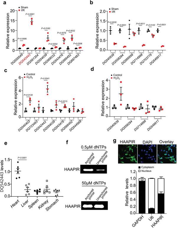
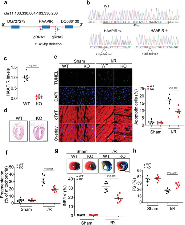
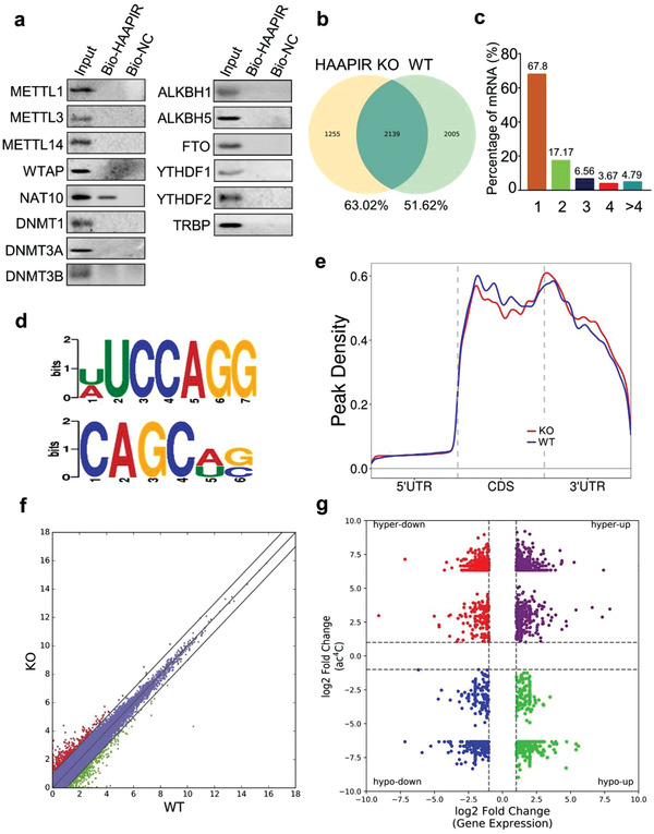
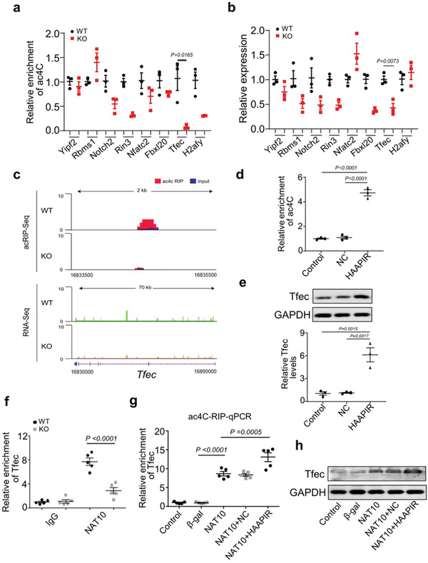
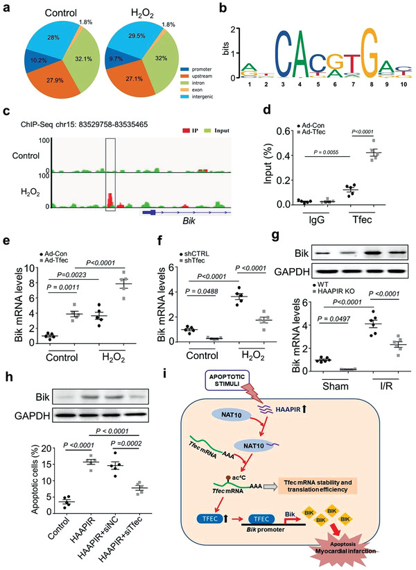
PIWI-interacting RNAs (piRNAs) are abundantly expressed in heart. However, their functions and molecular mechanisms during myocardial infarction remain unknown. Here, a heart-apoptosis-associated piRNA (HAAPIR), which regulates cardiomyocyte apoptosis by targeting N-acetyltransferase 10 (NAT10)-mediated N4-acetylcytidine (ac4 C) acetylation of transcription factor EC (Tfec) mRNA transcript, is identified. HAAPIR deletion attenuates ischemia/reperfusion induced myocardial infarction and ameliorate cardiac function compared to WT mice. Mechanistically, HAAPIR directly interacts with NAT10 and enhances ac4 C acetylation of Tfec mRNA transcript, which increases Tfec expression. TFEC can further upregulate the transcription of BCL2-interacting killer (Bik), a pro-apoptotic factor, which results in the accumulation of Bik and progression of cardiomyocyte apoptosis. The findings reveal that piRNA-mediated ac4 C acetylation mechanism is involved in the regulation of cardiomyocyte apoptosis. HAAPIR-NAT10-TFEC-BIK signaling axis can be potential target for the reduction of myocardial injury caused by cardiomyocyte apoptosis in ischemia heart diseases.

Keywords: ac4C acetylation; cardiomyocyte apoptosis; heart-apoptosis-associated piRNA (HAAPIR); piRNA; transcription factor EC (Tfec).


Conflict of interest statement

The authors declare no conflict of interest.

Figures

Figure 1
Figure 1
Identification of HAAPIR in cardiomyocytes. a,b) The expression levels of highly upregulated a) or downregulated b) piRNAs (selected from the Sham group in previous piRNA microarray data) in I/R injured mice hearts determined by qPCR (n = 3 independent experiments). c,d) qPCR analysis of highly upregulated c) or downregulated d) piRNAs in H2O2 treated cardiomyocytes selected from qPCR data in I/R injured mice hearts (n = 3 independent experiments). e) Relative expression level of DQ542443 in different tissues of normal adult mice as determined by qPCR (n = 6 independent experiments). f) Detection of 2′‐O‐methylation at the 3’ end of HAAPIR using RTL‐P approach. RT‐PCR reaction was performed with an unanchored or anchored RT primer at different concentrations of dNTPs. g) Representative images of fluorescence in situ hybridization with junction‐specific probes of HAAPIR indicates its subcellular localization (upper panel). Green represents HAAPIR and blue labels nuclei. Scale bar, 25 µm. The level of HAAPIR in the cytoplasmic or nuclear fractions of isolated cardiomyocytes as determined by qPCR. U6 and GAPDH used as internal controls (lower panel) (n = 3 independent experiments). Data are presented as Mean ± SEM. Two‐sided Student's t‐test a–d) or one‐way ANOVA test e).
Figure 2
Figure 2
Knockdown of HAAPIR attenuates H2O2‐induced cardiomyocyte apoptosis. a) Isolated neonatal mice cardiomyocytes were transfected with HAAPIR antagomir (anta) or its negative control (anta‐NC) for 24 h. Quantitative real‐time PCR (qPCR) analysis of the expression level of HAAPIR (n = 4 independent experiments). b–e) Isolated neonatal mice cardiomyocytes were transfected with HAAPIR antagomir (anta) or its negative control (anta‐NC) for 24 h and then cells were treated with H2O2 for an additional 24 h. b) qPCR analysis of the expression level of HAAPIR (n = 6 independent experiments). c) Apoptosis was determined by the TUNEL assay. DAPI indicates Nucleus. Bar = 50 µm. d) Quantitative analysis of the percentage of apoptotic cells (n = 5 independent experiments). e) Cardiomyocytes were staining with MitoTracker Red (red) /DAPI (blue). Representative images show the mitochondrial fission/fusion dynamics (Left panel). Bar = 10 µm. Quantitative analysis of the percentage of cells with fragmented mitochondria (Right panel, n = 5 independent experiments). f–h) Isolated neonatal mice cardiomyocytes were transfected with HAAPIR agomir (HAAPIR) or its negative control (NC) for 24 h. f) qPCR analysis of the expression level of HAAPIR (n = 6 independent experiments). g) Quantitative analysis of the percentage of apoptotic cells (n = 5 independent experiments). h) Quantitative analysis of the percentage of cells with fragmented mitochondria (n = 5 independent experiments). Data are presented as Mean ± SEM. All data were analyzed using one‐way ANOVA.
Figure 3
Figure 3
HAAPIR deficiency ameliorates ischaemia/reperfusion (I/R) induced heart injury. a) Schematic of HAAPIR location. CRISPR‐Cas9 gene editing system was used to knock out the genomic sequence of HAAPIR and generation of the mouse with mutated HAAPIR. b) HAAPIR knockout (KO) mice were genotyped by PCR and Sequencing. c) Quantitative real‐time PCR (qPCR) analysis of HAAPIR expression level in wild‐type (WT) and HAAPIR KO mice (n = 9 mice per group). d) Representative images of coronal sections of heart stained with hematoxylin and eosin (H&E) from WT and HAAPIR KO mice. Bar = 2 mm. e) Apoptosis was determined by the Terminal deoxynucleotidyl transferase (TdT) dUTP Nick‐End Labeling (TUNEL) assay in WT and HAAPIR KO mice heart (left panel). DAPI indicates Nucleus. Immunostaining of cTnT labels cardiomyocytes. Bar = 25 µm. Quantitative analysis of the percentage of apoptotic cardiomyocytes (right panel). n = 6 independent experiments. f) Quantitative analysis of the percentage of fragmented mitochondria (n = 6 independent experiments). g) HAAPIR KO mice exhibit reduced myocardial infarction upon I/R. WT and HAAPIR KO mice were exposed to I/R. The upper panels are representative photos of midventricular myocardial slices. The lower panel shows infarct sizes. left ventricle (LV), infarct area (INF). (n = 6 mice per group, Bar = 2 mm). h) Cardiac function was measured 24 h after cardiac I/R by left ventricle fractional shortening (FS) in WT and HAAPIR KO mice using echocardiography (n = 6 mice per group). Data are presented as Mean ± SEM. Two‐sided Student's t‐test c) or two‐way ANOVA test e–h).
Figure 4
Figure 4
HAAPIR binds to NAT10 and influences its N4‐acetylcytidine acetylation function. a) Cardiomyocytes were harvested and RNA pull‐down assay was performed using Bio‐ HAAPIR or Bio‐NC. Associated proteins were pulled down with streptavidin beads and bound levels of METTL1, METTL3, METTL14, NAT10, WTAP, DNMT1, DNMT3A, DNMT3B, ALKBH1, ALKBH5, FTO, YTHDF1, YTHDF2, and TRBP were analyzed by western blot. Representative image from three independent experiments. b–g) Ac4C acetylated RNA immunoprecipitation and sequencing (acRIP‐seq) was performed in HAAPIR knockout (KO) and wild‐type (WT) mice hearts. b) Numbers of ac4C peaks detected in HAAPIR KO (left circle) and WT (right circle) mice hearts. c) Percentage of mRNAs with different numbers of ac4C peaks. d) Sequence motifs enriched within ac4C peaks identified by acRIP‐seq. e) Metagene profile showing the distribution of ac4C peaks across the length of transcripts composed of three rescaled nonoverlapping segments 5′UTR, CDS, and 3′UTR in HAAPIR KO and WT mice hearts. f) Scatter plot of differential expression of mRNAs assessed from RNA‐seq data. Red dots denote up‐regulated genes and green dots denote down‐regulated genes. g) Correlation between the level of gene expression (overall transcript) and changes in ac4C level in HAAPIR KO mice hearts compared to WT.
Figure 5
Figure 5
HAAPIR promotes ac4C modification and expression of Tfec by targeting NAT10. a) Ac4C acetylated RNA immunoprecipitation and Quantitative real‐time PCR (acRIP‐qPCR) validation of ac4C modification levels of genes which selected from the results of acRIP‐seq and mRNA‐seq data in HAAPIR knockout (KO) and wild‐type (WT) mice hearts (n = 3 mice per group). b) qPCR validation of the expression levels of genes which selected from the results of acRIP‐seq and mRNA‐seq data in HAAPIR KO and WT mice hearts (n = 3 mice per group). c) Integrative Genomics Viewer (IGV) tracks displaying results of ac4C‐seq (upper panels) and RNA‐seq (lower panels) read distribution in Tfec mRNA of HAAPIR KO and WT mice hearts. d) acRIP‐qPCR analysis in isolated cardiomyocytes treated with or without HAAPIR agomir or agomir‐NC (NC) shows the ac4C modification level in Tfec mRNA. e) Expression levels of Tfec protein (upper panel) and mRNA (lower panel) in cardiomyocytes treated with HAAPIR agomir or NC (n = 3 independent experiments). f) RIP‐qPCR analysis in WT or HAAPIR KO mice hearts shows the level of Tfec mRNA binding to NAT10 (n = 5 mice per group). g) Ac4C enrichment level in Tfec mRNA was detected in cardiomyocytes infected with adenovirus harboring NAT10 or β‐gal and transfected with HAAPIR agomir or NC (n = 5 independent experiments). h) Western blot assay shows the protein level of TFEC in cardiomyocytes infected with adenovirus harboring NAT10 or β‐gal and transfected with HAAPIR agomir or NC. Data are presented as Mean ± SEM. Two‐sided Student's t‐test a,b), one‐way ANOVA test d,e,g) or two‐way ANOVA test f).
Figure 6
Figure 6
Inhibition of Tfec attenuates cardiomyocyte apoptosis in vitro and in vivo. a–d) Isolated neonatal mice cardiomyocytes were transfected with Tfec siRNA (si‐Tfec) or its negative control (si‐NC) for 24 h and then cells were treated with H2O2 for an additional 24 h. a) Representative western blot showing the expression of TFEC. b) The percentage of apoptotic cardiomyocytes was determined by TUNEL assay (n = 6 independent experiments). c) Cardiomyocytes were stained with MitoTracker Red (red) /DAPI (blue). Representative images show the mitochondrial fission/fusion dynamics. Bar = 10 µm. d) Quantitative analysis of the percentage of cells with fragmented mitochondria (n = 6 independent experiments). e–g) AAV9‐Tfec‐shRNA (shTfec) or AAV9‐control (shCTRL) were injected into mice and I/R induced heart injury was performed 3 weeks after the injection. e) The protein (upper panel) and mRNA (lower panel) levels of Tfec were detected by western blot assay and quantitative real‐time PCR (qPCR) assay (n = 6 mice per group). f) The percentage of apoptotic cardiomyocytes was determined by TUNEL assay (n = 6 mice per group). g) The infarct sizes after I/R induced heart injury were indicated by the ratio of infarct area (INF)/left ventricle (LV) (n = 6 mice per group). h,i) Cardiomyocytes were infected with adenovirus harboring NAT10 (NAT10) or its control (CTRL) and transfected with HAAPIR antagomir (anta) for 24 h, and then cells were treated with H2O2 for an additional 24 h. h) The mRNA levels of Tfec were detected by qPCR (n = 5 independent experiments). i) The percentage of apoptotic cardiomyocytes was determined by TUNEL assay (n = 5 independent experiments). Data are presented as Mean ± SEM. All data were analyzed using one‐way ANOVA.
Figure 7
Figure 7
Tfec regulates Bik expression during cardiomyocyte apoptosis. a–c) ChIP‐seq analysis was performed using flag‐tagged Tfec in cardiomyocytes treated with or without H2O2. a) Pie chart depicting the genomic distribution of Tfec‐enrichment. b) Motif analysis of the Tfec bound regions. c) Genome browser view showing Tfec ChIP‐seq signal around Bik. A Tfec peak in the promoter region on Bik was indicated. d) Isolated neonatal cardiomyocytes were infected with adenovirus harboring Tfec (Ad‐Tfec) or its negative control (Ad‐Con) for 24 h. CHIP‐qPCR assay was performed using antibodies against Tfec or IgG (n = 5 independent experiments). e,f) Cardiomyocytes were infected with Ad‐con or Ad‐Tfec e), control‐siRNA (sh‐CTRL) or Tfec‐siRNA (sh‐Tfec) f), and treated with H2O2 for 24 h. Bik mRNA levels were evaluated by qRT‐PCR (n = 5 independent experiments). g) HAAPIR knockout (KO) and wild‐type (WT) mice were exposed to I/R injury. The protein (upper panel) and mRNA (lower panel) levels of Bik were detected by western blot assay and qPCR, respectively (n = 6 mice per group). h) Cardiomyocytes were transfected with Tfec siRNA (siTfec) or its negative control (siNC) and transfected with HAAPIR agomir (HAAPIR) for 24 h. The protein levels of Bik were detected by western blot assay (upper panel). Quantitative analysis of the percentage of apoptotic cells were determined by TUNEL assay (n = 5 independent experiments). i) Model of HAAPIR function in apoptotic signaling. HAAPIR participates in the regulation of cardiac apoptosis through targeting NAT10/Tfec/Bik pathway. In our model, HAAPIR promotes NAT10‐mediated ac4C acetylation of Tfec mRNA transcript, which leads to an increase of Tfec expression, promotes Tfec‐induced Bik expression and thus leads to cardiomyocytes apoptosis. Data are presented as Mean ± SEM. All data were analyzed using one‐way ANOVA.

References

    1. Dorn L. E., Tual‐Chalot S., Stellos K., Accornero F., J. Mol. Cell Cardiol. 2019, 129, 272. - PMC - PubMed
    1. Stellos K., Cardiovasc. Res. 2017, 113, e2. - PubMed
    1. Gao X. Q., Zhang Y. H., Liu F., Ponnusamy M., Zhao X. M., Zhou L. Y., Zhai M., Liu C. Y., Li X. M., Wang M., Shan C., Shan P. P., Wang Y., Dong Y. H., Qian L. L., Yu T., Ju J., Wang T., Wang K., Chen X. Z., Wang Y. H., Zhang J., Li P. F., Wang K., Nat. Cell Biol. 2020, 22, 1319. - PubMed
    1. Song H., Feng X., Zhang H., Luo Y., Huang J., Lin M., Jin J., Ding X., Wu S., Huang H., Yu T., Zhang M., Hong H., Yao S., Zhao Y., Zhang Z., Autophagy 2019, 15, 1419. - PMC - PubMed
    1. Mathiyalagan P., Adamiak M., Mayourian J., Sassi Y., Liang Y., Agarwal N., Jha D., Zhang S., Kohlbrenner E., Chepurko E., Chen J., Trivieri M. G., Singh R., Bouchareb R., Fish K., Ishikawa K., Lebeche D., Hajjar R. J., Sahoo S., Circulation 2019, 139, 518. - PMC - PubMed

Publication types

LinkOut - more resources