

Brain MR Atlas Construction Using Symmetric Deep Neural Inpainting
- PMID: 35139030
- PMCID: PMC9250592
- DOI: 10.1109/JBHI.2022.3149754
Brain MR Atlas Construction Using Symmetric Deep Neural Inpainting
Abstract
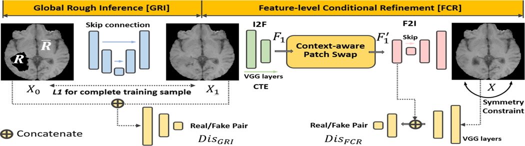
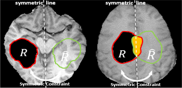
Modeling statistical properties of anatomical structures using magnetic resonance imaging is essential for revealing common information of a target population and unique properties of specific subjects. In brain imaging, a statistical brain atlas is often constructed using a number of healthy subjects. When tumors are present, however, it is difficult to either provide a common space for various subjects or align their imaging data due to the unpredictable distribution of lesions. Here we propose a deep learning-based image inpainting method to replace the tumor regions with normal tissue intensities using only a patient population. Our framework has three major innovations: 1) incompletely distributed datasets with random tumor locations can be used for training; 2) irregularly-shaped tumor regions are properly learned, identified, and corrected; and 3) a symmetry constraint between the two brain hemispheres is applied to regularize inpainted regions. Henceforth, regular atlas construction and image registration methods can be applied using inpainted data to obtain tissue deformation, thereby achieving group-specific statistical atlases and patient-to-atlas registration. Our framework was tested using the public database from the Multimodal Brain Tumor Segmentation challenge. Results showed increased similarity scores as well as reduced reconstruction errors compared with three existing image inpainting methods. Patient-to-atlas registration also yielded better results with improved normalized cross-correlation and mutual information and a reduced amount of deformation over the tumor regions.
Figures


References
-
- Liang Z-P and Lauterbur PC, Principles of magnetic resonance imaging: a signal processing perspective. SPIE OE Press, 2000. 1
-
- Orrison WW, Lewine J, Sanders J, and Hartshorne MF, Functional brain imaging. Elsevier Health Sciences, 2017. 1
-
- Murphy DG, DeCarli C, Schapiro MB et al. , “Age-related differences in volumes of subcortical nuclei, brain matter, and cerebrospinal fluid in healthy men as measured with magnetic resonance imaging,” Arc. Neurology, vol. 49, no. 8, pp. 839–845, 1992. 1 - PubMed
-
- Evans AC, Collins DL, Mills S, Brown E, Kelly R, and Peters TM, “3d statistical neuroanatomical models from 305 mri volumes,” in 1993 IEEE NSS MIC. IEEE, 1993, pp. 1813–1817. 1
Publication types
MeSH terms
Grants and funding
LinkOut - more resources
Full Text Sources
Medical
