. 2022 Feb 9;18(2):e1009986.
doi: 10.1371/journal.ppat.1009986. eCollection 2022 Feb.

Keap1 recognizes EIAV early accessory protein Rev to promote antiviral defense

Affiliations

Keap1 recognizes EIAV early accessory protein Rev to promote antiviral defense

Yan Wang et al. PLoS Pathog. .

Abstract

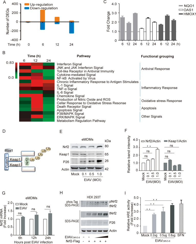
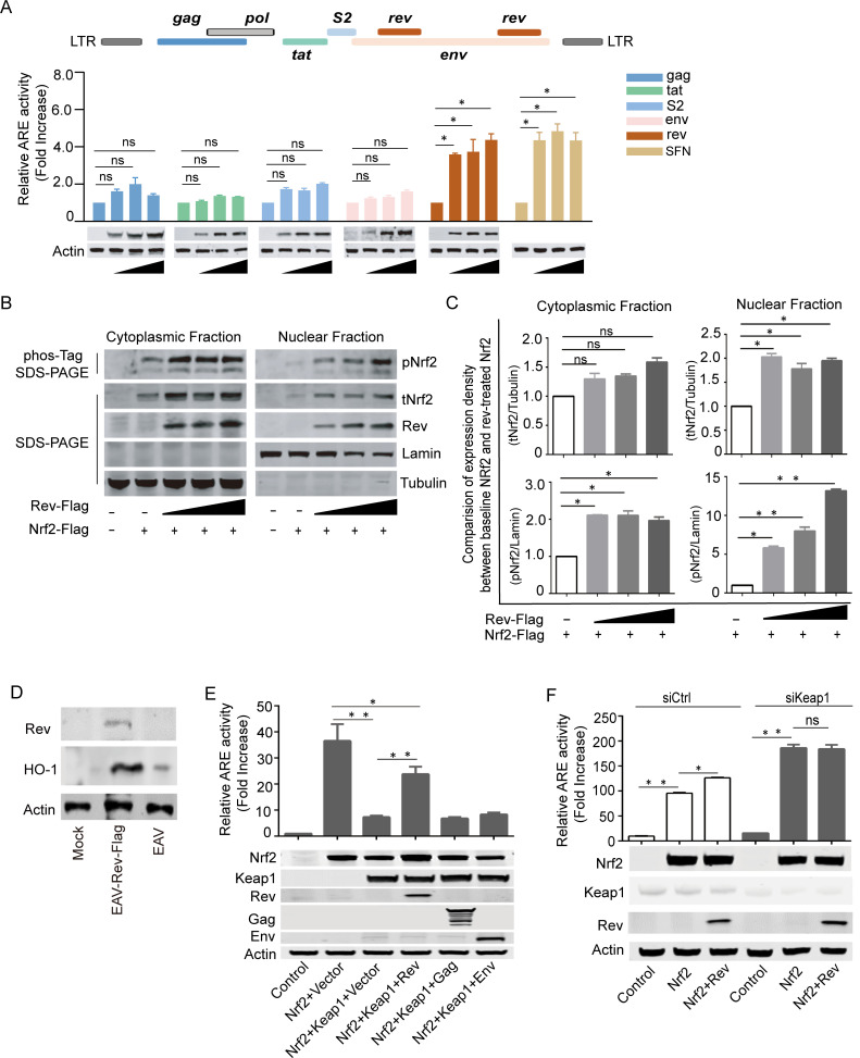
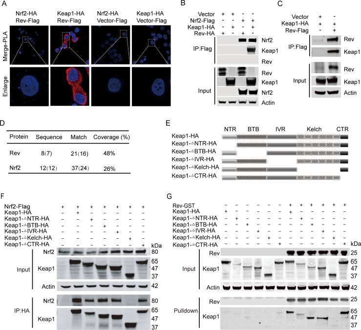
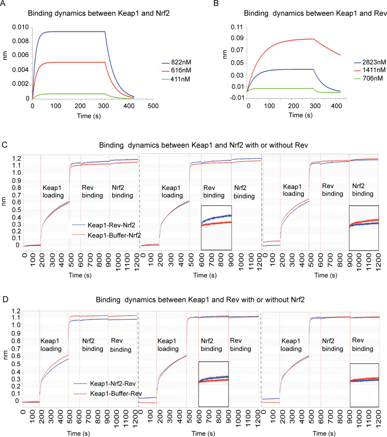
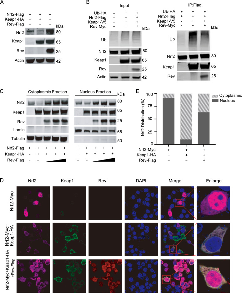
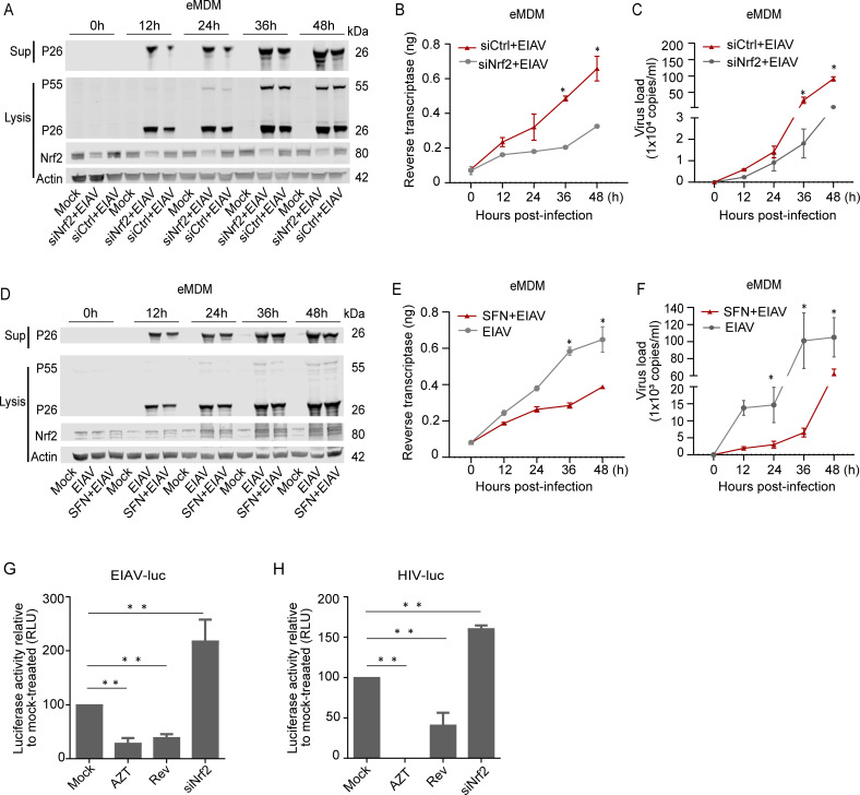
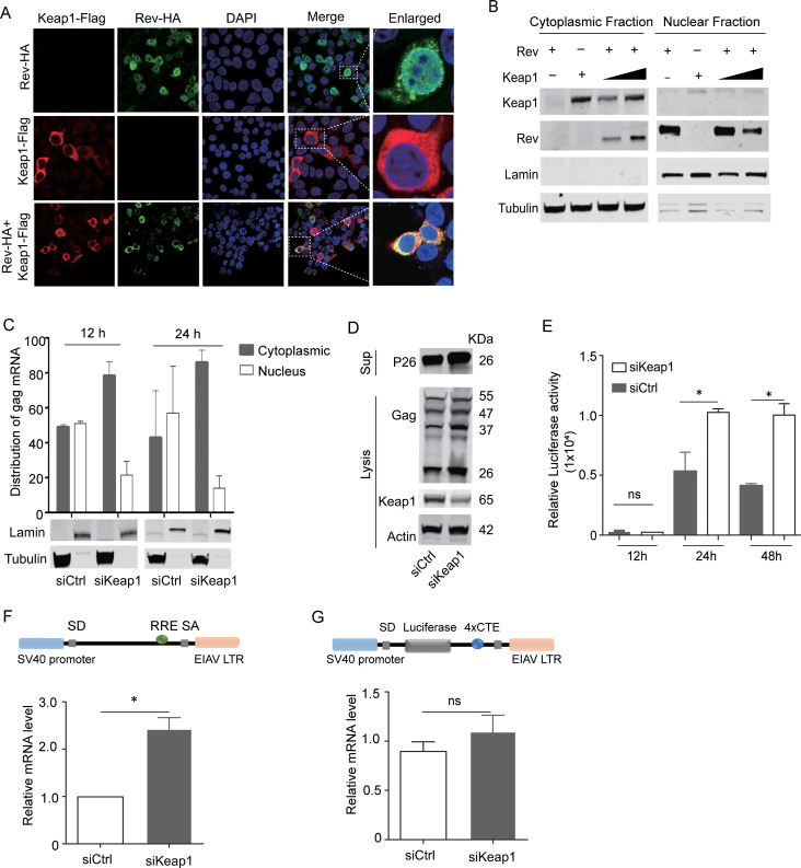
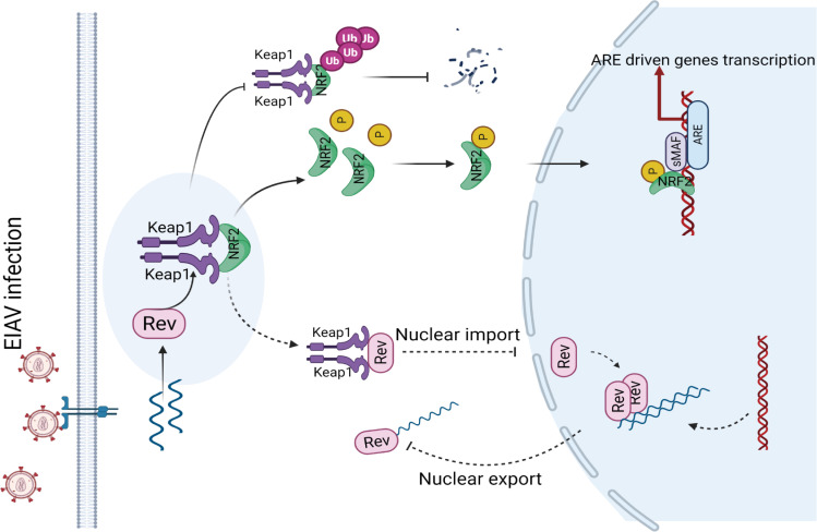
The Nrf2/Keap1 axis plays a complex role in viral susceptibility, virus-associated inflammation and immune regulation in host cells. However, whether or how the Nrf2/Keap1 axis is involved in the interactions between equine lentiviruses and their hosts remains unclear. Here, we demonstrate that the Nrf2/Keap1 axis was activated during EIAV infection. Mechanistically, EIAV-Rev competitively binds to Keap1 and releases Nrf2 from Keap1-mediated repression, leading to the accumulation of Nrf2 in the nucleus and promoting Nrf2 responsive genes transcription. Subsequently, we demonstrated that the Nrf2/Keap1 axis represses EIAV replication via two independent molecular mechanisms: directly increasing antioxidant enzymes to promote effective cellular resistance against EIAV infection, and repression of Rev-mediated RNA transport through direct interaction between Keap1 and Rev. Together, these data suggest that activation of the Nrf2/Keap1 axis mediates a passive defensive response to combat EIAV infection. The Nrf2/Keap1 axis could be a potential target for developing strategies for combating EIAV infection.


Conflict of interest statement

The authors have declared that no competing interests exist.

Figures

Fig 1
Fig 1. The Nrf2-Keap1 axis is activated by EIAV infection.
(A) Waterfall plot representing the total number of up- and down-regulated genes at each time point following transcriptome analysis (DEGs, differentially expressed genes; selected based on fold change>1.5, P value<0.05). (B) Heat map showing statistically significant canonical pathways commonly regulated at 6 h, 12 h and 24 h post EIAV infection, compared to the control. Heat map colors represent the ratio of regulated genes after EIAV infection (red and green correspond to over- and under-regulated genes, respectively). (C) Real-time PCR analysis of NQO1, OAS1 and HMOX1 mRNA in equine macrophages infected with EIAV at the indicated times (6 h, 12 h and 24 h) post infection. (D) Schematic representation of Nrf2-Keap1 interaction. (E) Quantification of Nrf2 and Keap1 expression in equine macrophages infected with EIAV at varied infection dose. (F) Densitometric analyses of pNrf2 and tNrf2 band intensity shown after normalization to actin. (G) Nrf2 mRNA was quantified using real-time PCR as above. (H) The effects of EIAV infection on the phosphorylation of Nrf2 were analyzed using a Phos-tag assay. Immunoblots for total Nrf2, p26 and actin were performed on normal SDS-PAGE gels as previously described. (I) The activation of Nrf2/Keap1 axis triggered by EIAV infection was analyzed using the ARE reporter gene assay. 293T cells were co-transfected together with the ARE luciferase reporter plasmid, as well as pcDNA3.1 (empty vector) or increasing concentrations of EIAVCMV3-8. Twenty-four hours later, cells were lysed and firefly luciferase activities was assayed. Data are representative of two (A-B) or three (C-I) independent experiments.
Fig 2
Fig 2. EIAV-Rev Induces Nrf2/Keap1 axis Activation.
(A) The ability of EIAV-coded proteins inducing Nrf2/Keap1 axis activation were evaluated using the ARE gene reporter. The assay protocol was the same as that shown in Fig 1I but cells were transfected with EIAV-env, EIAV-gag, EIAV-rev, EIAV-S2 or EIAV-Tat separately. SFN was chosen as a positive control. Western blot depicting the expression of each of the transfected constructs, including the loading control, actin. (B-C) Rev triggered Nrf2/Keap1 axis activation. 293T cells were transfected with Flag-tagged-rev, and the cytoplasmic and nuclear proteins were fractionated and then immunoblotted for pNrf2 and tNrf2. Densitometric analyses of pNrf2 and tNrf2 band intensity shown after normalization to Tubulin (cytoplasmic purity control) or Lamin (nuclear purity control). (D) Immunoblot analysis for HO-1 expression of extracts from equine macrophage cells infected with either the EAV infectious clone or EAV-Rev-Flag. (E) The ARE gene reporter was assayed for Nrf2 activation in the presence of Rev. 293T cells were transfected with the indicated plasmids. After 24 h, the luciferase activities were assessed (upper) and exogenous expression of proteins was measured using western blotting (lower). (F) Same assay protocol as D but 293T cells were pre-treated with siCtrl or siKeap1 and then transfected with the indicated plasmids. Data shown represent three independent experiments. Data are the mean values ± SDs, ns (non-significant), P > 0.05; *P < 0.05; **P < 0.01 (Student’s t test).
Fig 3
Fig 3. EIAV-Rev interacts with Keap1 but not Nrf2.
(A) PLA assay was used to analyze the interactions between Rev and Keap1 or Nrf2. The red spots represent interacting complexes of the examined proteins. The nuclei were stained with DAPI (blue). Cells co-transfected with Keap1 (Nrf2) and vector were used as negative controls. (B) and (C) Reciprocal immunoprecipitations of Rev and Keap1 or Nrf2 were performed on 293T cells co-transfected with plasmids for Nrf2-Flag and rev-HA, Nrf2-Flag and Keap1-HA or with Keap1-HA and Flag-rev. 24 hours post-transfection, cells were collected and subjected to pull down with Flag beads. (D) Mass spectrometry analysis of Rev and Nrf2 peptides after Keap1 pull-down. (E) Schematic diagram of Keap1 domain structure and Keap1 deletion mutants used in (E and F). All Keap1 deletion mutants were HA tagged at the amino terminal end. (F) The interactions between Keap1 mutants and Nrf2 were screened with Co-IP with the selected antibodies. (G) as (F) with the addition of GST-tagged-rev instead of Nrf2-Flag.
Fig 4
Fig 4. Rev-competitive inhibition antagonizes Nrf2 binding to Keap1.
(A-B) Biolayer interferometry graphs showing association and dissociation steps using different concentrations of Nrf2 (A) or Rev (B) to Keap1 immobilized on the anti-streptavidin biosensors. (C-D) BLI profiles showing the effect of Rev on the affinity of the Keap1-Nrf2 interaction. Rev incubated with Keap1 pre-coated with Nrf2 on the biosensors (C). Nrf2 (blue line) incubated with Keap1 pre-coated with Rev (D). The data are expressed as means and SD for at least three independent replicates.
Fig 5
Fig 5. EAIV-Rev reduces Keap1-mediated Nrf2 ubiquitination and facilitates Nrf2 translocation into the nuclei.
(A) Rev prevents Keap1-mediated Nrf2 degradation. Keap1 and rev were expressed in 293T cells alone or in combination as indicated. Cells were analyzed using western blotting at 24 h p.t. (B) Rev reduces Keap1-mediated Nrf2 ubiquitination. 293T cells were transfected with the indicated plasmids. At 24 h post-transfection, whole-cell lysates were immunoprecipitated using Flag beads and then ubiquitination was analyzed by blotting with HA-K48 ubiquitin antibody. (C) Protocol as in (A), with the addition of a step separating the cytoplasmic and nuclear proteins. Western blotting was performed as in Fig 3B. (D) The distribution of Nrf2 was visualized with or without Rev using confocal imaging. The plasmids encoding Nrf2 (purple) and Keap1 (green) were expressed alone or in combination with rev (red) in 293T cells. Twenty-four hours later, cells were fixed using acetone/methanol and subjected to immunofluorescence analysis using the indicated antibodies. Nuclei were visualized by staining with DAPI. Quantifications are given in (E). In each transfection experiment, at least 100 cells were scored, and the gray and stripe bars represent cytosolic and nuclear localization, respectively. Each of these experiments was repeated at least twice, and consistent results were obtained.
Fig 6
Fig 6. Nrf2 blocks EIAV replication in equine macrophages.
(A-C) eMDMs were pre-treated with Nrf2-specific siRNA or scramble siRNA. Six-hours after treatment, cells were either mock infected or infected with EIAV at 1×10⁵ TCID50. 24 hours post infection, cell extracts were analyzed using western blotting. Viral replication was determined using real-time RT PCR and reverse transcriptase activity assays. (D-F) same procedure as in A-C, except that eMDMs were treated with media supplemented with 20 μM SFN. (G-H) 293T cells were transfected with rev or Nrf2-specific siRNA. After treatment for 12h, cells were inoculated with VSV-G-pseudotyped EIAV encoding firefly luciferase (K) and VSV-G-pseudotyped HIV-1 (L). Twenty-four hours later, luciferase activity was measured using photon emission. AZT (Zidovudine), a specific reverse transcription inhibitor, served as a positive control for viral inhibition. The data are expressed as means and SD for at least three independent replicates. *P <0.05, * *P<0.01.
Fig 7
Fig 7. Keap1 hijacks rev in the cytoplasm and limits rev-mediated RNA transport.
(A) Distribution of Rev was screened with or without Keap1 using confocal imaging. Scale bars 10μm. Images are representative of 3 independent experiments. (B) 293T cells were co-transfected with HA-tagged-rev and Keap1. The cytoplasmic and nuclear proteins were fractionated as in Fig 2B and then immunoblotted for Keap1 and Rev. (C) Distribution of cytoplasmic and nuclear unspliced gag RNAs in siKeap1 and control cells was analyzed using real-time PCR with primers specific for gag mRNA. Lamin and tubulin were used as nuclear and cytoplasmic controls, respectively. (D) 293T cells were treated with siRNA targeting Keap1 (siKeap1) or scrambled siRNA (siCtrl) and then transfected with EIAVCMV3-8. Cells were lysed and samples were analyzed for expression of Gag, Keap1, and Actin using western blotting. (E) EIAV pseudovirions were generated separately in siKeap1 or siCtrl cells and then used to infect 293T cells. The luciferase activity in the supernatant was assayed at the indicated timepoints (12 h, 24 h and 48 h). (F) Rev/RRE RNA export reporter plasmids were transfected into siKeap1 and control cells and viral mRNA synthesis was calculated using real-time RT PCR. (G) The protocol is as (F), but with the Rev-independent RNA export reporter plasmid (4×CTE). The graph represents three independent experiments; error bars represent results from SEM.
Fig 8
Fig 8. A proposed model for the cellular Nrf2/Keap1 axis manipulation of EIAV replication.
Nrf2/Keap1 plays a crucial role in the management of oxidative stress. Under normal conditions, Keap1 interacts with Nrf2 in the cytoplasm, targeting it for proteasomal degradation to maintain redox homeostasis. Under EIAV infection, EIAV-Rev interacts with Keap1, disrupting the interaction of Keap1-Nrf2, leading to the enhancement of pNrf2 and nuclear translocation which results in the activation of Nrf2. The activation of the Nrf2 signal in turn inhibits EIAV replication via increasing expression of several antioxidant enzymes and also by limiting rev-mediated RNA transport.

Similar articles

Cited by

References

    1. Craigo JK, Barnes S, Cook SJ, Issel CJ, Montelaro RC. Divergence, not diversity of an attenuated equine lentivirus vaccine strain correlates with protection from disease. Vaccine. 2010;28(51):8095–104. doi: 10.1016/j.vaccine.2010.10.003 ; PubMed Central PMCID: PMC2997116. - DOI - PMC - PubMed
    1. Haas L. [Equine infectious anemia—a review]. Berl Munch Tierarztl Wochenschr. 2014;127(7–8):297–300. . - PubMed
    1. Zimmerli U, Thur B. [Equine Infectious Anaemia—a review from an official veterinary perspective]. Schweiz Arch Tierheilkd. 2019;161(11):725–38. Epub 2019/11/07. doi: 10.17236/sat00232 . - DOI - PubMed
    1. Cursino AE, Vilela APP, Franco-Luiz APM, de Oliveira JG, Nogueira MF, Junior JPA, et al.. Equine infectious anemia virus in naturally infected horses from the Brazilian Pantanal. Arch Virol. 2018;163(9):2385–94. Epub 2018/05/13. doi: 10.1007/s00705-018-3877-8 . - DOI - PubMed
    1. Wang XF, Lin YZ, Li Q, Liu Q, Zhao WW, Du C, et al.. Genetic Evolution during the development of an attenuated EIAV vaccine. Retrovirology. 2016;13:9. Epub 2016/02/05. doi: 10.1186/s12977-016-0240-6 ; PubMed Central PMCID: PMC4738788. - DOI - PMC - PubMed

Publication types

MeSH terms