. 2022 Feb 23;70(7):2389-2400.
doi: 10.1021/acs.jafc.1c07225. Epub 2022 Feb 9.

Structure, Immunogenicity, and IgE Cross-Reactivity among Walnut and Peanut Vicilin-Buried Peptides

Affiliations

Structure, Immunogenicity, and IgE Cross-Reactivity among Walnut and Peanut Vicilin-Buried Peptides

Alexander C Y Foo et al. J Agric Food Chem. .

Abstract

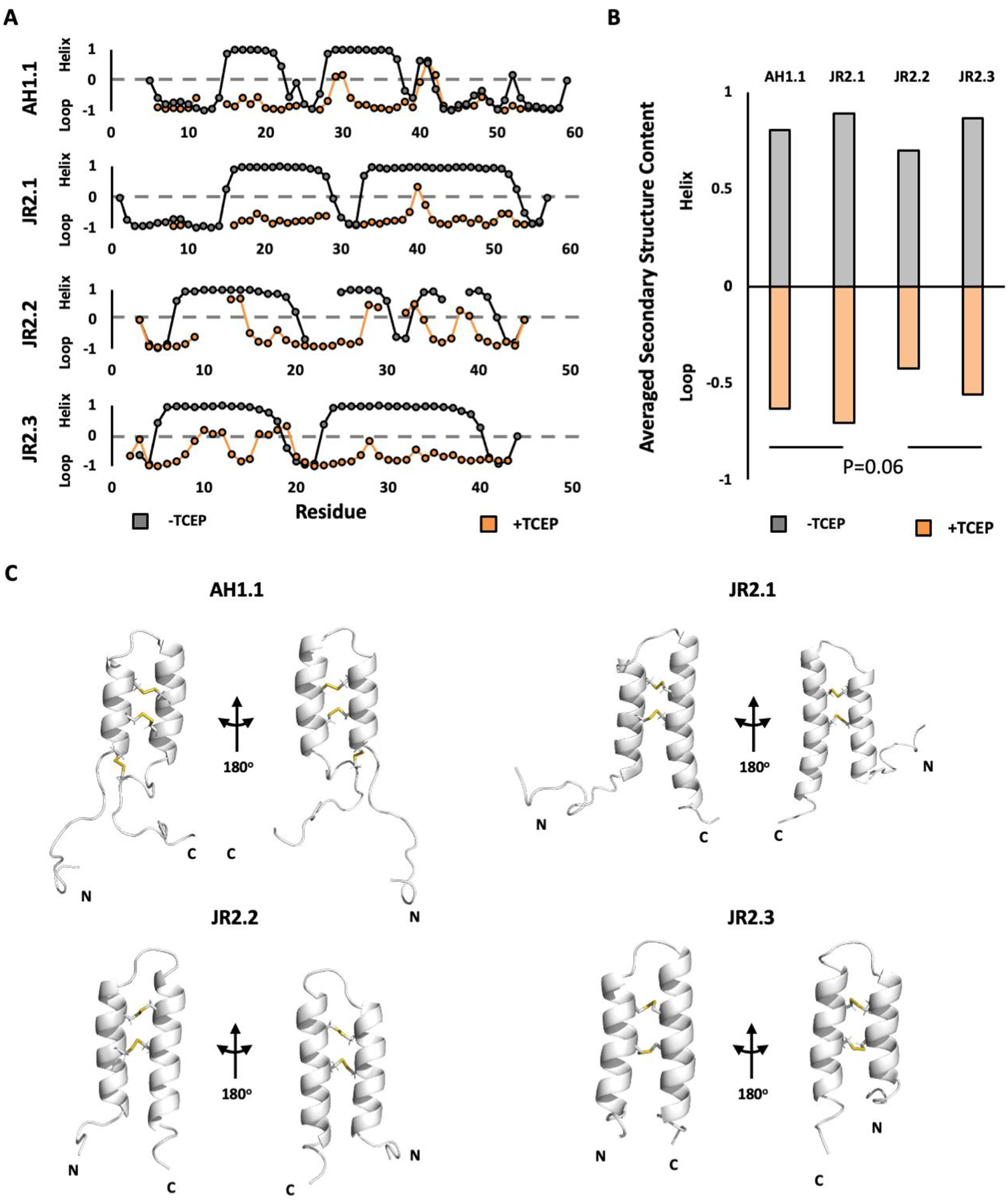
Vicilin-buried peptides (VBPs) from edible plants are derived from the N-terminal leader sequences (LSs) of seed storage proteins. VBPs are defined by a common α-hairpin fold mediated by conserved CxxxCx(10-14)CxxxC motifs. Here, peanut and walnut VBPs were characterized as potential mediators of both peanut/walnut allergenicity and cross-reactivity despite their low (∼17%) sequence identity. The structures of one peanut (AH1.1) and 3 walnut (JR2.1, JR2.2, JR2.3) VBPs were solved using solution NMR, revealing similar α-hairpin structures stabilized by disulfide bonds with high levels of surface similarity. Peptide microarrays identified several peptide sequences primarily on AH1.1 and JR2.1, which were recognized by peanut-, walnut-, and dual-allergic patient IgE, establishing these peanut and walnut VBPs as potential mediators of allergenicity and cross-reactivity. JR2.2 and JR2.3 displayed extreme resilience against endosomal digestion, potentially hindering epitope generation and likely contributing to their reduced allergic potential.

Keywords: IgE epitopes; cross-reactivity; vicilin-buried peptide; α-hairpinin.


Figures

Figure 1:
Figure 1:
Amino acid sequence of Peanut/Walnut VBPs. A) N-terminal leader sequence (LS) from Ara h 1 and Jug r 2. Individual VBP sequences are highlighted in orange. Residues present in the NMR structures, but not the native sequence are denoted in parentheses. Conserved cysteine forming the characteristic CxxxC motif highlighted in red B) sequence identity matrix comparing the various Peanut/Walnut VBP sequences.
Figure 2:
Figure 2:
Peanut and walnut VBPs adopt a common α-hairpin structure mediated by conserved disulfide bonds. A) Secondary structure plot for the peanut/walnut VBPs under oxidizing (black) and reducing (orange) conditions, as calculated based on the backbone and side chain NMR chemical shifts using the TALOS algorithm. Average secondary structure content for all α-hairpin residues are shown in B. C) NMR structures of AH1.1, JR2.1, JR2.2, and JR2.3. Cartoon figures depict representative conformations of each structure. Inter-helix disulfides shown as sticks and highlighted in yellow. Complete NMR data tables are available in Figures S1.
Figure 3:
Figure 3:
Loss of VBP structure under reducing conditions. A) Representative circular-dichroism spectra of peanut/walnut VBPs in the absence (black) and presence (orange) of the reducing agent tris(2-carbocyethyl)phosphine (TCEP). The local minima at 220/210 nm and 205 nm are indicative of α-helical and random-coil secondary structure respectively. The relative loss of α-helical structure upon addition of TCEP is quantified in B. Data values and error bars represent the mean and standard deviation obtained from at least three trials from two biological replicates.
Figure 4:
Figure 4:
Quantifying structural similarity across peanut and walnut VBPs A) Structure of AH1.1 colored by surface similarity (ΔSIM) values when compared against JR2.1, JR2.2, and JR2.3 as calculated using the SPADE surface comparison algorithm., Cyan and purple represent regions of low and high surface similarity respectively. Total ΔSIM (Σ[ΔSIM]) for all residues is indicated. B) Structure of WN VBPs colored by surface similarity (ΔSIM) values against AH1.1 as calculated using the SPADE surface comparison algorithm. Total ΔSIM (Σ[ΔSIM]) for all residues is indicated. Additional SPADE comparisons were performed on VBP homologues from tomato (VBP-8) and buckwheat (BWI-2c) (Figure S2).
Figure 5:
Figure 5:
Potential IgE-reactive and cross reactive regions on peanut and walnut VBPs. A) Sequence of peanut/walnut VBPs used in the peptide microarray analysis. Residues depicted in the solution-NMR structures are shown in capital letters. Residues from the expression system used to generate the NMR samples but are not present in the peptide microarrays are denoted with brackets. Residues highlighted in blue represent alpha-helices identified in the available NMR structures. Epitopes identified in previous works by Maleki et al. and Burks et al. are indicated in grey rectangles, whereas cross-reactive peptides identified in this present work are indicated by colored rectangles. B) IgE-reactive peptides mapped onto the structure of peanut/walnut VBPs. Peptides color-coded as in (A).
Figure 6:
Figure 6:
Conformational stability of peanut and walnut VBPs contribute to proteolytic stability. Half-life of peanut and walnut VBPs subjected to simulated gastric (A) and duodenal (B) digestion under reducing and oxidizing conditions. C) Half-life of peanut and walnut VBPs subjected to simulated endosomal digestion (reducing conditions only). Faded bars represent conditions under which no appreciable (<10%) digestion was detected. Data values and error bars represent the mean and standard deviation obtained from at least three trials from two biological replicates.
Figure 7:
Figure 7:
Proposed model of VBP allergenicity. Following uptake by antigen presenting cells, potential allergens are subjected to endosomal degradation to generate T-cell epitopes. Proteins with an intermediate stability are primarily digested in the late endosome, resulting in efficient MHCII loading and a tolerogenic (IgG/IgG4) immune response as reviewed by Foo et al. Proteins with above-average stability experience peak epitope generation in the lysosome, increasing the probability of an allergic (IgE) response due to the scarcity of MHCII. Hyper-stabilized proteins are completely resilient to endosomal digestion and yield only minimal levels of epitope fragments over the endosomal lifecycle, resulting in a diminished immune response. The results reported in this work suggest that extremely immunogenic VBPs such as AH1.1 and JR2.1 belong to the former, while hyper-stabilized homologues such as JR2.2 and JR2.3 fall into the latter category, contributing to their reduced sensitizing potential.

References

    1. Burney P; Summers C; Chinn S; Hooper R; Van Ree R; Lidholm J Prevalence and Distribution of Sensitization to Foods in the European Community Respiratory Health Survey: A EuroPrevall Analysis. Allergy Eur. J. Allergy Clin. Immunol 2010, 65 (9), 1182–1188. 10.1111/j.1398-9995.2010.02346.x. - DOI - PubMed
    1. Chong KW; Ruiz-Garcia M; Patel N; Boyle RJ; Turner PJ Reaction Phenotypes in IgE-Mediated Food Allergy and Anaphylaxis. Ann. Allergy, Asthma Immunol 2020, 124 (5), 473–478. 10.1016/j.anai.2019.12.023. - DOI - PMC - PubMed
    1. Fischer D; Vander Leek TK; Ellis AK; Kim H Anaphylaxis. Allergy, Asthma Clin. Immunol 2018, 14 (Suppl 2). 10.1186/s13223-018-0283-4. - DOI - PMC - PubMed
    1. Weinberger T; Sicherer S Current Perspectives on Tree Nut Allergy: A Review. J. Asthma Allergy 2018, 11, 41–51. 10.2147/JAA.S141636. - DOI - PMC - PubMed
    1. Maloney JM; Rudengren M; Ahlstedt S; Bock SA; Sampson HA The Use of Serum-Specific IgE Measurements for the Diagnosis of Peanut, Tree Nut, and Seed Allergy. J. Allergy Clin. Immunol 2008, 122 (1), 145–151. 10.1016/j.jaci.2008.04.014. - DOI - PubMed