. 2022 Mar;13(3):4674-4687.
doi: 10.1080/21655979.2022.2027066.

Improving the repair mechanism and miRNA expression profile of tibial defect in rats based on silent information regulator 7 protein analysis of mesenchymal stem cells

Affiliations

Improving the repair mechanism and miRNA expression profile of tibial defect in rats based on silent information regulator 7 protein analysis of mesenchymal stem cells

Rui Chen et al. Bioengineered. 2022 Mar.

Abstract

The aim of this study was to verify the role of Silent Information Regulator 7 (SIRT7) in improving the repair mechanism of bone marrow mesenchymal stem cells (BMMSCs) and the expression of microribonucleic acid (miRNA). Human BMMSCs were extracted from patients with femoral fractures, and the proliferation activity of human BMMSCs before and after knockout SIRT7 and the expression levels of bone-related genes and proteins were compared. Thirty-two 8-week-old male Sprague-Dawley (SD) rats were randomly divided into a blank group, a chitosan scaffold group, a control group, and a silence information regulator knockout group 7 (n = 8). In addition to the blank group, the chitosan scaffold, the green fluorescent protein (GFP) transfected stem cell composite chitosan scaffold, and the SIRT7 knockout stem cell composite chitosan scaffold were implanted in the other three groups, respectively. The X-rays and small animal in vivo three-dimensional tomography (Micro-CT) were adopted to quantitatively analyze the volume fraction, the number of trabeculae, and the connection density. Compared with the other three groups, the bone defect was formed more in the medullary mesenchymal stem cell knockout group, and the bone volume fraction, number of trabeculae and connection density were significantly increased (P < 0.05). MiR-98-5p can significantly promote the formation of bone molecules and bone structure in rats (P < 0.05). Human BMMSCs combined with chitosan scaffold can accelerate the repair of tibial defects. MiR-98-5p targeting and regulating bone formation gene (CKIP-1) could significantly improve the process of osteogenesis in rats.

Keywords: BMMSCs; SIRT7 protein; miRNA; tibial defect; tibial defect model.


Conflict of interest statement

No potential conflict of interest was reported by the author(s).

Figures

Figure 1.
Figure 1.
Endogenous expression of human BMMSCs (a: mRNA expression changes of human BMMSCs in osteogenic differentiation; b: quantitative analysis of protein expression. (*: compared with 0d, P < 0.05)).
Figure 2.
Figure 2.
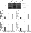
SIRT7 knockout effect of BMMSCs in the 3rd and 9th generation (a: Western blot electrophoresis; b, c: mRNA and protein levels of SIRT7 in the 3rd generation after transfection; d, e: mRNA and protein levels of SIRT7 in the 9th generation after transfection. Lenti-SIRT7: SIRT7 knockout group; Lenti-Con: control group; Mock-Treated: simulation group (not added with any lentivirus but added with a lentivirus transfection agent). (*: compared with control group, P < 0.05)).
Figure 3.
Figure 3.
The value-added activity detection of human BMMSCs (Lenti-SIRT7: SIRT7 knockout group; Lenti-Con: control group. (*: compared with control group, P < 0.05)).
Figure 4.
Figure 4.
The mRNA expression levels of various genes in osteoblasts (Lenti-SIRT7: SIRT7 knockout group; Lenti-con: control group; Mock-Treated: simulation Group (not added with any lentivirus but added with a lentivirus transfer agent). a. RUNX2 gene; b. OSX gene; c. OPN gene; d. COL1A1 gene (*: compared with the control group, P < 0.05)).
Figure 5.
Figure 5.
Changes of ALP activity and calcium nodule level in osteoblasts (Lenti-SIRT7: SIRT7 knockout group; Lenti-con: control group; Mock-Treated: simulation Group (not added with any lentivirus but added with a lentivirus transfer agent); a. ALP activity; b. calcium nodule level (*: compared with the control group, P < 0.05)).
Figure 6.
Figure 6.
X-ray during 6 weeks after tibial defect in rats (Blank: blank group; CS-Only: chitosan scaffold group; Lenti-con: control group; Lenti-SIRT7: knockout SIRT7 group. The circle showed the degree of bone defect.).
Figure 7.
Figure 7.
Missing surface and reconstructed image of Micro-CTD (Blank: blank group; CS-Only: chitosan scaffold group; Lenti-con: control group; Lenti-SIRT7: knockout SIRT7 group. The circle shows the degree of bone defect.).
Figure 8.
Figure 8.
Quantitative analysis of Micro-CT (Blank: blank group; CS-Only: chitosan scaffold group; Lenti-con: control group; Lenti-SIRT7: knockout SIRT7 group. a. Bone volume fraction; b. trabecular number; c. connectivity density; (*: compared with Black, P < 0.05)).
Figure 9.
Figure 9.
Expression changes of different miRNAs in osteogenic differentiation (a. different groups; b. different induced differentiation time; (*: P < 0.05 compared with the control group; #: P < 0.05 compared with that before osteogenic differentiation (0d)).
Figure 10.
Figure 10.
Eosin-hematoxylin staining results of tissue sections of tibial defects 6 weeks postoperatively (a. blank control group; b. chitosan scaffold group; c. control group; d. knockout SIRT7 group).

Similar articles

References

    1. Sagi HC, Young ML, Gerstenfeld L, et al. Qualitative and quantitative differences between bone graft obtained from the medullary canal (with a reamer/ irrigator/ aspirator) and the iliac crest of the same patient. J Bone Joint Surg Am. 2012;94(23):2128–2135. - PubMed
    1. Wubneh A, Tsekoura EK, Ayranci C, et al. Current state of fabrication technologies and materials for bone tissue engineering. Acta Biomater. 2018;80:1–30. - PubMed
    1. Bartold PM, Gronthos S, Haynes D, et al. Mesenchymal stem cells and biologic factors ioading to bone formation. J Clin Periodontol. 2019;46(21):12–32. 4 6. - PubMed
    1. Li D, Cheng P, Jiang H, et al. Vascularization converts the lineage fate of bone mesenchymal stem cells to endothelial cells cells in tissue-engineered bone grafts by modulating FGF2-RhoA/ ROCK signaling. Cell Death Dis. 2018;9(10):959. - PMC - PubMed
    1. Camorani S, Hill BS, Fontanella R, et al. Inhibition of bone marrow-derived mesenchymal stem cells homing towards triple-negative breast cancer microenvironment using an anti-PDGFRbeta aptamer. Theranostics. 2017;7(14):3595–3607. - PMC - PubMed

LinkOut - more resources