. 2022 Feb;13(2):4428-4440.
doi: 10.1080/21655979.2022.2034584.

Acetylshikonin suppresses diffuse large B-Cell Lymphoma cell growth by targeting the T-lymphokine-activated killer cell-originated protein kinase signalling pathway

Affiliations

Acetylshikonin suppresses diffuse large B-Cell Lymphoma cell growth by targeting the T-lymphokine-activated killer cell-originated protein kinase signalling pathway

Jieke Cui et al. Bioengineered. 2022 Feb.

Abstract

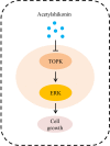
Diffuse large B-cell lymphoma (DLBCL) is one of the most common causes of cancer death worldwide, and responds poorly to the existing treatments. Thus, identifying novel therapeutic targets of DLBCL is urgently needed. In this study, we found that T-lymphokine-activated killer cell-originated protein kinase (TOPK) was highly expressed in DLBCL cells and tissues. Data from the GEPIA database also indicated that TOPK was highly expressed in DLBCL tissues. The high expression levels of proteins were identified via Western blots and immunohistochemistry (IHC). TOPK knockdown inhibited cell growth and induced apoptosis of DLBCL cells with 3-(4,5-dimethylthiazol-2-yl)-5-(3-carboxymethoxyphenyl)-2-(4-sulfophenyl)-2 H-tetrazolium (MTS) and flow cytometry. Further experiments demonstrated that acetylshikonin, a compound that targeted TOPK, could attenuate cell growth and aggravate cell apoptosis through TOPK/extracellular signal-regulated kinase (ERK)-1/2 signaling, as shown by MTS, flow cytometry and Western blots. In addition, we demonstrated that TOPK modulated the effect of acetylshikonin on cell proliferation and apoptosis in U2932 and OCI-LY8 cells using MTS, flow cytometry and Western blots. Taken together, the present study suggests that acetylshikonin suppresses the growth of DLBCL cells by attenuating TOPK signaling, and the targeted inhibition of TOPK by acetylshikonin may be a promising approach for the treatment of DLBCL.

Keywords: DLBCL; ERK; TOPK; acetylshikonin; cell growth.


Conflict of interest statement

No potential conflict of interest was reported by the author(s).

Figures
None
Graphical abstract
Figure 1.
Figure 1.
TOPK is highly expressed in human DLBCL. (a) The GEPIA database demonstrated the overexpression of TOPK in DLBCL tumor tissues compared to normal tissues (*p < 0.05). (b) IHC staining revealed nuclear staining of TOPK in the DLBCL tissue array, control group (20 samples), and DLBCL group (40 samples). The values were normalized to control tissue expression, and the values are represented as the mean ± S.D. (***p < 0.001). (c) TOPK protein levels were measured in DLBCL cell lines (U2932, OCI-LY8, SUDHL-6), WIL2S cell lines and PBMCs using Western blots. The data represent the mean ± S.D. for three individual experiments (ns: no significant difference, **p < 0.01, ***p < 0.001, n = 3).
Figure 2.
Figure 2.
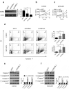
TOPK knockdown inhibits cell growth and induces cell apoptosis in U2932 and OCI-LY8 cell lines. (a) OCI-LY8 cells were infected with shTOPK.1, shTOPK.2 or shNT, and the TOPK knockdown efficiency was detected by Western blots. The data represent the mean ± S.D. for three individual experiments (**p < 0.01, ***p < 0.001, n = 3). (b and c) U2932 and OCI-LY8 cells were infected with shTOPK.1, shTOPK.2 or shNT, and cell proliferation was detected at 24, 48, and 72 h with the MTS assay. The data represent the mean ± S.D. for three individual experiments (*p < 0.05, **p < 0.01, ***p < 0.001, n = 3). (d) U2932 and OCI-LY8 cells were infected with shTOPK.1, shTOPK.2 or shNT for 72 h, and cell apoptosis was detected by staining with annexin V and PI (Q1 indicates dead cells, Q2 indicates late apoptotic cells, Q3 indicates early apoptotic cells, Q4 indicates viable cells). The data represent the mean ± S.D. for three individual experiments (**p < 0.01, ***p < 0.001, n = 3). (e and f) U2932 and OCI-LY8 cells were infected with shTOPK.2 or shNT, and the cleaved caspase 3 and caspase 7 protein levels were assessed by Western blots. The data are the mean ± S.D. for three individual experiments (***p < 0.001, n = 3).
Figure 3.
Figure 3.
TOPK inhibitors suppress cell proliferation in DLBCL cells. (a-b) WIL2S cells were treated with different concentrations (5 µM, 10 µM, 20 µM, 40 µM) of acetylshikonin or 3-DSC, and cell proliferation was assessed at 24, 48, and 72 h by the MTS assay. The data represent the mean ± S.D. for three individual experiments (*p < 0.05, **p < 0.01, n = 3). (c-f) U2932 and OCI-LY8 cells were treated with different concentrations (5 µM, 10 µM, 20 µM, 40 µM) of acetylshikonin or 3-DSC, and cell proliferation was assessed at 24, 48, and 72 h by the MTS assay. The data are the mean ± S.D. for three individual experiments (*p < 0.05, **p < 0.01, ***p < 0.001, n = 3).
Figure 4.
Figure 4.
Acetylshikonin induces DLBCL cell apoptosis and reduces the expression of proteins involved in TOPK signaling. (a) U2932 and OCI-LY8 cells were treated with 10 µM acetylshikonin for 24 h, and cell apoptosis was detected by staining with annexin V and PI (Q1 indicates dead cells, Q2 indicates late apoptotic cells, Q3 indicates early apoptotic cells, Q4 indicates viable cells). The data represent the mean ± S.D. for three individual experiments (***p < 0.001, n = 3). (b) U2932 cells were treated with 10 µM acetylshikonin for 24 h, and the protein levels of pTOPK, total TOPK, pERK, total ERK, pRSK, total RSK, pc-Jun, total c-Jun were detected by Western blots. The data represent the mean ± S.D. for three individual experiments (***p < 0.001, n = 3).
Figure 5.
Figure 5.
Acetylshikonin inhibits DLBCL cell growth in vivo. (a-b) The effect of acetylshikonin on the size of tumors was assessed. Vehicle or acetylshikonin (120 mg.kg−1 body weight) was administered by gavage. Data are shown as mean values ± SD, from n = 6 in each group. (c) Tumor weight was measured after treatment on the last day of the study. Data are shown as mean values ± SD (***p < 0.001, n = 6).
Figure 6.
Figure 6.
TOPK overexpression attenuates the effect of acetylshikonin. (a-b) U2932 and OCI-LY8 cells were infected with a lentivirus carrying TOPK cDNA or treated with 10 µM acetylshikonin, and cell growth was determined at 72 h using the MTS assay. The data represent the mean ± S.D. for three individual experiments (*p < 0.05, **p < 0.01, n = 3). (c) U2932 and OCI-LY8 cells were infected with a lentivirus carrying TOPK cDNA or treated with 10 µM acetylshikonin, and cell apoptosis was detected by staining with annexin V and PI (Q1 indicates dead cells, Q2 indicates late apoptotic cells, Q3 indicates early apoptotic cells, Q4 indicates viable cells). The data represent the mean ± S.D. for three individual experiments (*p < 0.05, ***p < 0.001, n = 3). (d) U2932 cells were infected with a lentivirus carrying TOPK cDNA or treated with 10 µM acetylshikonin, and the cleaved caspase 3 and caspase 7 protein levels were assessed by Western blots. The data represent the mean ± S.D. for three individual experiments (**p < 0.01, ***p < 0.001, n = 3).

References

    1. Tilly H, Gomes da Silva M, Vitolo U, et al. Diffuse large B-cell lymphoma (DLBCL): ESMO clinical practice guidelines for diagnosis, treatment and follow-up. Ann Oncol. 2015;26(Suppl 5):v116–25. - PubMed
    1. Wang Y, Zhang M, Xu H, et al. Discovery and validation of the tumor-suppressive function of long noncoding RNA PANDA in human diffuse large B-cell lymphoma through the inactivation of MAPK/ERK signaling pathway. Oncotarget. 2017;8(42):72182–72196. - PMC - PubMed
    1. Feugier P, Van Hoof A, Sebban C, et al. Long-term results of the R-CHOP study in the treatment of elderly patients with diffuse large B-cell lymphoma: a study by the Groupe d’Etude des Lymphomes de l’Adulte. J Clin Oncol. 2005;23(18):4117–4126. - PubMed
    1. Vercellino L, Cottereau AS, Casasnovas O, et al. High total metabolic tumor volume at baseline predicts survival independent of response to therapy. Blood. 2020;135(16):1396–1405. - PMC - PubMed
    1. Sehn LH, Gascoyne RD.. Diffuse large B-cell lymphoma: optimizing outcome in the context of clinical and biologic heterogeneity. Blood. 2015;125:22–32. - PubMed

Publication types

MeSH terms

Substances