

Therapeutic effect of adipose stromal vascular fraction spheroids for partial bladder outlet obstruction induced underactive bladder
- PMID: 35139904
- PMCID: PMC8826668
- DOI: 10.1186/s13287-022-02739-w
Therapeutic effect of adipose stromal vascular fraction spheroids for partial bladder outlet obstruction induced underactive bladder
Abstract
Background: Underactive bladder (UAB) is a common clinical problem but related research is rarely explored. As there are currently no effective therapies, the administration of adipose stromal vascular fraction (ad-SVF) provides a new potential method to treat underactive bladder.
Methods: Male Sprague-Dawley rats were induced by partial bladder outlet obstruction (PBOO) for four weeks and randomly divided into three groups: rats treated with PBS (Sham group); rats administrated with ad-SVF (ad-SVF group) and rats performed with ad-SVF spheroids (ad-SVFsp group). After four weeks, urodynamic studies were performed to evaluate bladder functions and all rats were sacrificed for further studies.
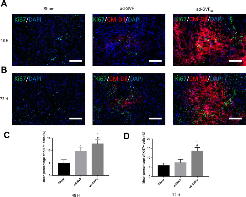
Results: We observed that the bladder functions and symptoms of UAB were significantly improved in the ad-SVFsp group than that in the Sham group and ad-SVF group. Meanwhile, our data showed that ad-SVF spheroids could remarkably promote angiogenesis, suppress cell apoptosis and stimulate cell proliferation in bladder tissue than that in the other two groups. Moreover, ad-SVF spheroids increased the expression levels of bFGF, HGF and VEGF-A than ad-SVF. IVIS Spectrum small-animal in vivo imaging system revealed that ad-SVF spheroids could increase the retention rate of transplanted cells in bladder tissue.
Conclusions: Ad-SVF spheroids improved functions and symptoms of bladder induced by PBOO, which contributes to promote angiogenesis, suppress cell apoptosis and stimulate cell proliferation. Ad-SVF spheroids provide a potential treatment for the future patients with UAB.
Keywords: Bladder outlet obstruction; Spheroid; Stromal vascular fraction; Underactive bladder.
© 2022. The Author(s).
Conflict of interest statement
The authors declare that they have no competing interests.
Figures


Similar articles
-
Adipose Derived Stromal Vascular Fraction and Mesenchymal Stem Cells Improve Angiogenesis in a Rat Hindlimb Ischaemia Model.Eur J Vasc Endovasc Surg. 2024 May;67(5):828-837. doi: 10.1016/j.ejvs.2023.11.036. Epub 2023 Nov 22. Eur J Vasc Endovasc Surg. 2024. PMID: 37995961
-
Improved bladder contractility after transplantation of human mesenchymal stem cells overexpressing hepatocyte growth factor into underactive bladder from bladder outlet obstruction models of rats.PLoS One. 2021 Dec 22;16(12):e0261402. doi: 10.1371/journal.pone.0261402. eCollection 2021. PLoS One. 2021. PMID: 34936660 Free PMC article.
-
Preischemic Administration of Nonexpanded Adipose Stromal Vascular Fraction Attenuates Acute Renal Ischemia/Reperfusion Injury and Fibrosis.Stem Cells Transl Med. 2016 Sep;5(9):1277-88. doi: 10.5966/sctm.2015-0223. Epub 2016 Jun 30. Stem Cells Transl Med. 2016. PMID: 27365485 Free PMC article.
-
Autologous Adipose-Derived Tissue Stromal Vascular Fraction (AD-tSVF) for Knee Osteoarthritis.Int J Mol Sci. 2022 Nov 4;23(21):13517. doi: 10.3390/ijms232113517. Int J Mol Sci. 2022. PMID: 36362308 Free PMC article. Review.
-
Underactive Bladder Versus Bladder Outlet Obstruction: Don't Get Tricked!Eur Urol Focus. 2022 Mar;8(2):388-390. doi: 10.1016/j.euf.2022.03.015. Epub 2022 Mar 31. Eur Urol Focus. 2022. PMID: 35370120 Review.
Cited by
-
Lesson on obesity and anatomy of adipose tissue: new models of study in the era of clinical and translational research.J Transl Med. 2024 Aug 14;22(1):764. doi: 10.1186/s12967-024-05547-3. J Transl Med. 2024. PMID: 39143643 Free PMC article. Review.
-
Efficacy of autologous micrografts technology: a promising approach for chronic wound healing and tissue regeneration-a pilot study.Front Med (Lausanne). 2024 Jul 26;11:1417920. doi: 10.3389/fmed.2024.1417920. eCollection 2024. Front Med (Lausanne). 2024. PMID: 39131083 Free PMC article.
References
-
- Osman NI, Esperto F, Chapple CR. Detrusor underactivity and the underactive bladder: a systematic review of preclinical and clinical studies. Eur Urol. 2018;74(5):633–643. - PubMed
-
- Abrams P, Cardozo L, Fall M, Griffiths D, Rosier P, Ulmsten U, et al. The standardisation of terminology of lower urinary tract function: report from the Standardisation Sub-committee of the International Continence Society. Am J Obstet Gynecol. 2002;187(1):116–126. - PubMed
-
- De Nunzio C, Lombardo R, Cicione A, Trucchi A, Carter S, Tema G, et al. The role of bladder wall thickness in the evaluation of detrusor underactivity: development of a clinical nomogram. Neurourol Urodyn. 2020;39(4):1115–1123. - PubMed
Publication types
MeSH terms
LinkOut - more resources
Full Text Sources
Research Materials
