. 2022 Mar;41(12):1821-1834.
doi: 10.1038/s41388-022-02212-1. Epub 2022 Feb 9.

DDR1 promotes hepatocellular carcinoma metastasis through recruiting PSD4 to ARF6

Affiliations

DDR1 promotes hepatocellular carcinoma metastasis through recruiting PSD4 to ARF6

Xiaochao Zhang et al. Oncogene. 2022 Mar.

Abstract

Discoidin domain receptor 1 (DDR1) is a member of the receptor tyrosine kinase family, and its ligand is collagen. Previous studies demonstrated that DDR1 is highly expressed in many tumors. However, its role in hepatocellular carcinoma (HCC) remains obscure. In this study, we found that DDR1 was upregulated in HCC tissues, and the expression of DDR1 in TNM stage II-IV was higher than that in TNM stage I in HCC tissues, and high DDR1 expression was associated with poor prognosis. Gene expression analysis showed that DDR1 target genes were functionally involved in HCC metastasis. DDR1 positively regulated the migration and invasion of HCC cells and promoted lung metastasis. Human Phospho-Kinase Array showed that DDR1 activated ERK/MAPK signaling pathway. Mechanically, DDR1 interacted with ARF6 and activated ARF6 through recruiting PSD4. The kinase activity of DDR1 was required for ARF6 activation and its role in metastasis. High expression of PSD4 was associated with poor prognosis in HCC. In summary, our findings indicate that DDR1 promotes HCC metastasis through collagen induced DDR1 signaling mediated PSD4/ARF6 signaling, suggesting that DDR1 and ARF6 may serve as novel prognostic biomarkers and therapeutic targets for metastatic HCC.


Conflict of interest statement

The authors declare no competing interests.

Figures

Fig. 1
Fig. 1. DDR1 plays a prometastatic role in HCC in vitro and in vivo.
A Immunohistochemical staining (IHC) and expression scoring of DDR1 was performed in 169 HCC tissues. Representative pictures were shown (scale bar: 200 μm). B, C Kaplan–Meier analysis was used to illustrate the correlation between DDR1 expression and overall survival or disease-free survival of HCC patients. The cutoff for determining low or high DDR1 expression was the median value. D, E Indicated cells were treated with (+) or without (−) collagen I for 3 h, and immunoprecipitation with anti-DDR1. The blots were probed with the indicated antibodies. F, G Trans-well migration and invasion assays, wound healing assays were performed in indicated cells. H, J Lung metastasis from nude mice injected with SK-Hep1 and HLF cells by tail veins from both groups killed at 8 weeks, was measured by bioluminescent imaging (BLI), representative images of lung tissue sections, and number of lung metastatic foci in both groups (n = 9). I, K Representative hematoxylin and eosin staining of lung tissue sections, and incidence of lung metastasis in both groups of BALB/c (nu/nu) mice (n = 9). *P < 0.05, **P < 0.01, ***P < 0.001. #P < 0.05, ##P < 0.01, ###P < 0.001: the scramble group compared with the shDDR1#3 group.
Fig. 2
Fig. 2. DDR1 physically interacts and colocalized with ARF6.
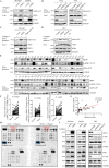
A Cellular extracts from SK-Hep1 and HLF cells transfected with FLAG-tagged DDR1 or FLAG-tagged vector were subsequently immunoprecipitated with anti-FLAG antibody. Eluted proteins were separated on SDS-PAGE and visualized by Coomassie blue staining. Eluted proteins were identified by mass spectrometry analysis. B Selective genes from mass spectrometry analysis were co-immunoprecipitated with Myc-tagged DDR1 (up). Detection of ARF6 by mass spectrometry (bottom). C 293 T cells were transiently co-transfected with indicated plasmids and co-immunoprecipitation assays were performed. D Immunoblots of co-immunoprecipitated (IP) endogenous DDR1 and endogenous ARF6 in HLE cell extracts. Immunoglobulin G (IgG) is negative control. E Confocal assays were shown to observe the co-localization of exogenously expressed DDR1 and ARF6 in 293 T cells (Scale bar: 15 μm). F Confocal assays were shown to observe the co-localization of endogenous DDR1 and ARF6 in HLF and HLE cells (Scale bar: 30 μm). G Schematic diagram of FLAG-tagged full-length or deletion constructs of ARF6 used in this study (left panel). Co-precipitation of HA-tagged DDR1 with FLAG-tagged ARF6 or its mutants, analyzed by anti-FLAG immunoprecipitation and anti-FLAG/ HA immunoblots (right panel).
Fig. 3
Fig. 3. DDR1 promotes ARF6 activation in a kinase activity-independent manner, and promotes MAPK signaling pathway activation.
A Analysis of the activation level of ARF6 in SK-Hep1 and Hep3B cell lines with collagen I. B Analysis of the activation level of ARF6 in HLE and HLF cell lines stably knocked down of DDR1, compared with the scramble groups. C Analysis of the activation level of ARF6 in SK-Hep1 cell lines stably overexpressed DDR1, DDR1-K618A, compared with the vector groups with collagen I. D Analysis of the activation level of ARF6 in HLE cell lines, treated with or without imatinib, 7rh, and collagen I. E The protein or phosphorylation level of DDR1 and ARF6-GTP was analyzed in 50 paired HCC tissues (tumor, T) with corresponding adjacent non-cancerous tissues (normal, N) by western blot. Representative western blot results were shown. F Statistical analysis showed DDR1, phosphorylated DDR1 and ARF6-GTP upregulated in HCC tissues, compared with that in normal liver, where GAPDH was used as a control. G Spearman correlation analysis between ARF6-GTP and DDR1 expression (N = 50). H Representative images of human phosphor-kinase array analysis in indicated SK-Hep1 cells with collagen I. The boxed dots are reference protein (blue) and phosphorylated ERK (red). I Western blot analysis of the phosphorylation level of P38, ERK and c-jun in indicated HCC cells.
Fig. 4
Fig. 4. Active ARF6 plays a prometastatic role in HCC progress in vitro and in vivo, and promotes MAPK signaling pathway activation.
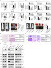
A, B Trans-well migration and invasion assays, wound healing assays were performed in indicated cells. C, E Lung metastasis from nude mice injected with SK-Hep1 and HLF cells by tail veins from both groups killed at eight weeks, was measured by bioluminescent imaging (BLI), representative images of lung tissue sections, and number of lung metastatic foci in both groups (n = 8). D, F Representative hematoxylin and eosin staining of lung tissue sections, and incidence of lung metastasis in both groups of BALB/c (nu/nu) mice (n = 8). G Western blot analysis of the phosphorylation level of P38, ERK and c-jun in indicated HCC cells. *P < 0.05, **P < 0.01, ***P < 0.001. #P < 0.05, ##P < 0.01, ###P < 0.001: the scramble group compared with the shARF6#2 group.
Fig. 5
Fig. 5. DDR1 promotes the migration, invasion and metastasis of HCC cells through ARF6.
DDR1-overexpressing SK-Hep1 (A) and Hep3B (B) cells were stably transfected with scramble or shARF6 (shARF6#1 and shARF6#2) plasmids to generate control (vector+scramble), DDR1-overexpressing (DDR1 + scramble), and DDR1-overexpressing but ARF6-knockdown (DDR1 + shARF6#1 and DDR1 + shARF6#2) stable cells. A, B, E Trans-well migration and invasion assays, wound healing assays were performed in indicated cells. C Lung metastasis from nude mice injected with SK-Hep1 and HLF cells by tail veins from both groups killed at 8 weeks, was measured by bioluminescent imaging (BLI), representative images of lung tissue sections, and number of lung metastatic foci in both groups (n = 9). D Representative hematoxylin and eosin staining of lung tissue sections, and incidence of lung metastasis in both groups of BALB/c (nu/nu) mice (n = 9).
Fig. 6
Fig. 6. DDR1 recruits PSD4 to activating ARF6.
A 293 T cells were transiently co-transfected with FLAG-tagged ARF-GEFs and HA-tagged DDR1. Co-immunoprecipitation of DDR1 by ARF-GEFs was shown. B 293 T cells were transiently co-transfected with FLAG-tagged PSD4 and HA-tagged DDR1 and co-immunoprecipitation assays were performed. C 293 T cells were transiently co-transfected with FLAG-tagged PSD4 and HA-tagged ARF6 and co-immunoprecipitation assays were performed. D Confocal assays were shown to observe the co-localization of exogenously expressed PSD4 and DDR1 or ARF6 in 293 T cells (Scale bar 15 μm). E 293 T cells were transiently co-transfected with FLAG-tagged PSD4 and HA-tagged ARF6 with or without MYC-tagged DDR1. Co-immunoprecipitation of ARF6 by PSD4 was shown.
Fig. 7
Fig. 7. PSD4 plays a prometastatic role in HCC progress in vitro and in vivo.
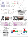
A Immunohistochemical staining (IHC) and expression scoring of PSD4 was performed in 169 HCC tissues. Representative pictures were shown (scale bar: 200 μm). B Spearman correlation analysis between PSD4 and DDR1 expression. N = 169 (right panel). C, D Kaplan–Meier analysis was used to illustrate the correlation between PSD4 expression and overall survival or disease-free survival of HCC patients. The cutoff for determining low or high PSD4 expression was the median value. E Analysis of the activation level of ARF6 in indicated SK-Hep1 and HLF cells. F, H Lung metastasis from nude mice injected with SK-Hep1 and HLF cells by tail veins from both groups killed at 8 weeks, was measured by bioluminescent imaging (BLI), representative images of lung tissue sections, and number of lung metastatic foci in both groups. G, I Representative hematoxylin and eosin staining of lung tissue sections, and incidence of lung metastasis in both groups of BALB/c (nu/nu) mice. J Analysis of the activation level of ARF6 in indicated Hep3B cells with collagen I. K Proposed oncogenic DDR1/PSD4/ARF6 signaling pathway in HCC metastasis. *P < 0.05, **P < 0.01, ***P < 0.001; #P < 0.05, ##P < 0.01, ###P < 0.001: the scramble group compared with the shPSD4#3 group.

References

    1. Villanueva A. Hepatocellular Carcinoma. N Engl J Med. 2019;380:1450–62. - PubMed
    1. Yang JD, Hainaut P, Gores GJ, Amadou A, Plymoth A, Roberts LR. A global view of hepatocellular carcinoma: trends, risk, prevention and management. Nat Rev Gastroenterol Hepatol. 2019;16:589–604. - PMC - PubMed
    1. Tsochatzis EA, Meyer T, Burroughs AK. Hepatocellular carcinoma. N Engl J Med 366. 2012;92:92–3. - PubMed
    1. Agarwal G, Mihai C, Iscru DF. Interaction of discoidin domain receptor 1 with collagen type 1. J Mol Biol. 2007;367:443–55. - PubMed
    1. Weiner HL, Huang H, Zagzag D, Boyce H, Lichtenbaum R, Ziff EB. Consistent and selective expression of the discoidin domain receptor-1 tyrosine kinase in human brain tumors. Neurosurgery. 2000;47:1400–9. - PubMed

MeSH terms

Substances