. 2022 Mar;15(3):471-479.
doi: 10.1038/s41385-022-00490-2. Epub 2022 Feb 9.

Infection with the enteric pathogen C. rodentium promotes islet-specific autoimmunity by activating a lymphatic route from the gut to pancreatic lymph node

Affiliations

Infection with the enteric pathogen C. rodentium promotes islet-specific autoimmunity by activating a lymphatic route from the gut to pancreatic lymph node

Sakari Pöysti et al. Mucosal Immunol. 2022 Mar.

Abstract

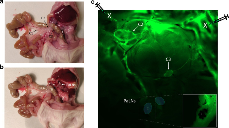
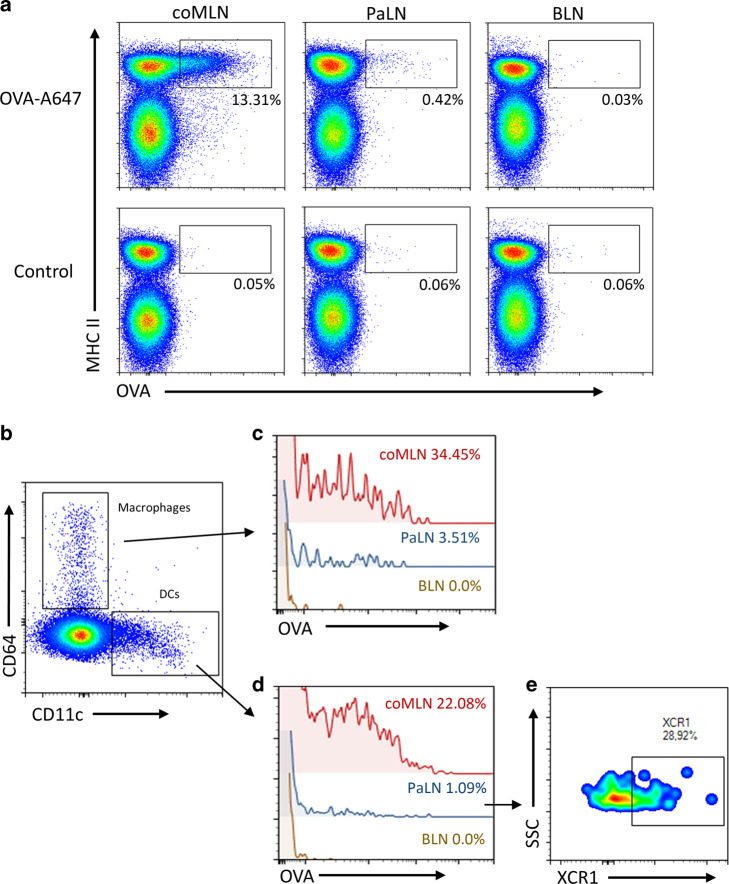
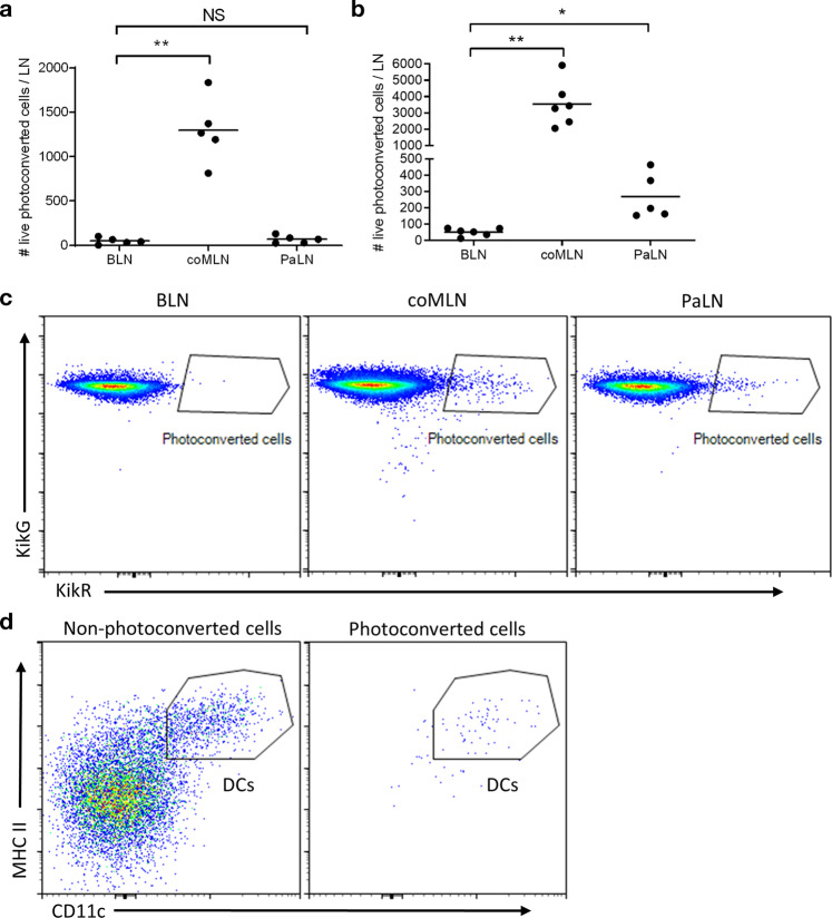
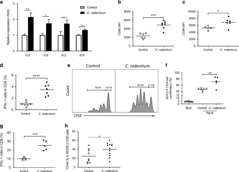
In nonobese diabetic (NOD) mice, C. rodentium promotes priming of islet-specific T-cells in pancreatic lymph nodes (PaLN), which is a critical step in initiation and perpetuation of islet-autoimmunity. To investigate mechanisms by which C. rodentium promotes T-cell priming in PaLN, we used fluorescent imaging of lymphatic vasculature emanating from colon, followed dendritic cell (DC) migration from colon using photoconvertible-reporter mice, and evaluated the translocation of bacteria to lymph nodes with GFP-C. rodentium and in situ hybridization of bacterial DNA. Fluorescent dextran injected in the colon wall accumulated under subcapsular sinus of PaLN indicating the existence of a lymphatic route from colon to PaLN. Infection with C. rodentium induced DC migration from colon to PaLN and bacterial DNA was detected in medullary sinus and inner cortex of PaLN. Following infection with GFP-C. rodentium, fluorescence appeared in macrophages and gut-derived (CD103+) and resident (CD103-/XCR1+) DC, indicating transportation of bacteria from colon to PaLN both by DC and by lymph itself. This induced proinflammatory cytokine transcripts, activation of DC and islet-specific T-cells in PaLN of NOD mice. Our findings demonstrate the existence of a direct, enteric pathogen-activated route for lymph, cells, and bacteria from colon, which promotes activation of islet-specific T-cells in PaLN.


Conflict of interest statement

The authors declare no competing interests.

Figures

Fig. 1
Fig. 1. FITC-dextran injected in colon wall accumulates in colon-draining and in pancreatic lymph nodes.
a A photograph of a dissected mouse showing colon-draining (coMLN) lymph nodes C1-C3 located in the mesenterium; and b pancreatic (PaLN) lymph nodes located in the rear of peritoneal cavity. c A photomicrograph showing diffusion of FITC dextran injected into colon wall (injection sites marked by an X). FITC-dextran accumulates under subcapsular sinus of two colon-draining lymph nodes (C2, C3, arrows) and under subcapsular sinus of a separate lymph node located at the rear of peritoneal cavity (PaLN). Due to their location close to vertebral column, PaLN were visible only after further preparation (inset). During dissection, PaLN block was marked with black ink (I in the insert) to help its identification.
Fig. 2
Fig. 2. Soluble antigen injected subserosally in colon wall is endocytosed by macrophages and DC in coMLN and PaLN.
a Injection of OVA-A647 (upper panels) leads to a fluorescent signal in MHCII+ cells in coMLN and PaLN but not in BLN (brachial lymph node) as compared to PBS injection (“control”, lower panels). b MHCII+ cells divided into macrophages (MHCII+/CD64+) and DCs (MHCII+/CD11c+). c OVA-A647 positivity in CD64+MHCII+ macrophages in coMLN and PaLN. d Also CD11c+MHCII+ DC show OVA-A647 positivity in coMLN and PaLN. e XCR1 expression in OVA-A647 positive DCs. Lymph nodes were analyzed 1 h after injection of OVA-A647. In (c and d) background was reduced from the frequencies of positive cells indicated in histograms. Single live cells were gated as CD45+/CD19/TCRβ cells before further population identification. In figure a percentages represents portions from MHCII+ cells. Regions were gated according to FMO stainings. Data are representative of 4 mice/group and two independent experiments.
Fig. 3
Fig. 3. DC migrate from colon to PaLN only during dysbiosis.
a Under steady-state conditions, photoconverted cells are found in coMLNs but not in PaLN or BLN. (b, c) Following photoconversion of colon wall after C. rodentium infection, photoconverted (KikRHI) cells are found also in PaLN. b Phenotyping of photoconverted cells in PaLN identifies them as MHCIIHI CD11cHI DC. **p ≤ 0.01, ***p ≤ 0.001. Single live cells were gated as CD45+/CD19/TCRβ before further population identification. Data are from four independent experiments and each data point represents one mouse. One-way ANOVA and Dunnett’s T3 multiple comparisons test was used to compare groups.
Fig. 4
Fig. 4. Dysbiosis increases translocation of bacterial DNA to coMLN and PaLN.
a Photomicrographs of coMLN and PaLN sections (20x magnification) of NOD mice following in situ hybridization for bacterial 16S RNA gene. b Bacterial DNA is increased significantly in both coMLN and PaLN sections in NOD mice infected with C. rodentium. c NOD mice have significantly more bacterial DNA in PaLN also during steady state when compared to C57BL/6 mice. GFP signal in medullary sinus of PaLN after infecting mice with GFP-C. rodentium (e) compared to control mice (d). GFP is co-localized with F4/80+ cells (20x magnification). f Increased GFP positivity in DCs (MHCII+/CD11c+ cells), and in CD103+ DCs and CD103/XCR1+ DCs after oral inoculation of GFP-C.rodentium. g Representative flow cytometry dot plots of GFP signal in MHCII+/CD11c+ DCs. For flow cytometry, live single cells were gated as CD45+/CD19/TCRβ before further population identification. *p ≤ 0.05, **p ≤ 0.01 (unpaired Student’s t test). Bacterial counts (b, c) were quantified using ImageJ software and are expressed as qty/AU (quantity/arbitrary units). Data in each panel are from three independent experiments. Each data point represents an individual mouse.
Fig. 5
Fig. 5. C. rodentium infection promotes DC activation and T cell response in PaLN of NOD mice.
a Relative expressions of cytokine transcripts in PaLN of control and C. rodentium -infected mice (n = 6, data represent mean + SD). (b, c) CD80 and CD86 levels on CD11c+MHC II+ DC. d IFNγ production in CD4 T cells in PaLN. e Representative dilution of CFSE label among successive cell divisions in adoptively transferred islet-specific BDC2.5 T cells in PaLN of uninfected and C. rodentium infected mice. f C. rodentium infection increases BDC2.5 cell proliferation in PaLN. g IFNγ production in CD8 T cells in PaLN. h CD44 in IGRP-specific T cells in PaLN of 5 week old NOD.8.3 mice 6 days after C. rodentium infection. BLN, brachial lymph node. *p < 0.05; **p < 0.01; ***p < 0.001; ****p < 0.0001 (unpaired Student’s t-test). T cells were gated as single live cells and CD45+/CD19/TCRβ+ before further population identification. Data are from 3 independent experiments. Each data point represents an individual mouse.

References

    1. Atarashi K, et al. Induction of colonic regulatory T cells by indigenous Clostridium species. Science. 2011;331:337–341. doi: 10.1126/science.1198469. - DOI - PMC - PubMed
    1. Hooper LV, Littman DR, Macpherson AJ. Interactions between the microbiota and the immune system. Science. 2012;336:1268–1273. doi: 10.1126/science.1223490. - DOI - PMC - PubMed
    1. Clemente JC, Manasson J, Scher JU. The role of the gut microbiome in systemic inflammatory disease. BMJ. 2018;360:j5145. doi: 10.1136/bmj.j5145. - DOI - PMC - PubMed
    1. Levy M, Kolodziejczyk AA, Thaiss CA, Elinav E. Dysbiosis and the immune system. Nat. Rev. Immunol. 2017;17:219–232. doi: 10.1038/nri.2017.7. - DOI - PubMed
    1. Meng X, et al. Gut dysbacteriosis and intestinal disease: mechanism and treatment. J. Appl Microbiol. 2020;129:787–805. doi: 10.1111/jam.14661. - DOI - PMC - PubMed

Publication types