. 2022 Feb 2:15:635-648.
doi: 10.2147/JIR.S338598. eCollection 2022.

IL-17 is a Potential Therapeutic Target in a Rodent Model of Otitis Media with Effusion

Affiliations

IL-17 is a Potential Therapeutic Target in a Rodent Model of Otitis Media with Effusion

Nanfeng Zhang et al. J Inflamm Res. .

Abstract

Background: Otitis media with effusion (OME) is a non-suppurative inflammation of the middle ear that is characterized by middle ear effusion and hearing loss. However, the mechanisms of OME are not fully understood. The aim of this study was to determine the function and the mechanism of the IL-17 cytokine in the pathogenesis of OME and to investigate IL-17 as a potential strategy for the treatment of OME.

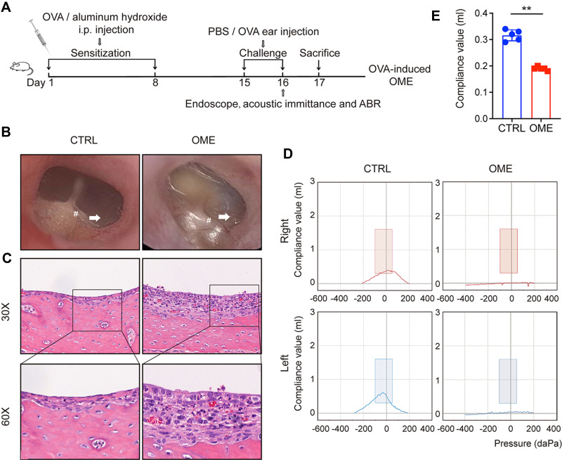
Methods: In this study, the OME rat model was induced by ovalbumin (OVA) as previously described. The severity of OME was determined with an oto-endoscope, by histochemical analysis, and by acoustic immittance. Kyoto Encyclopedia of Genes and Genomes (KEGG) analysis of RNA-sequencing (RNA-seq) data was carried out to analyze the signaling pathways related to the pathogenesis of OME, which indicated that IL-17 is involved in OME. The anti-IL-17A monoclonal antibody was administrated by nasal drip to block IL-17 to treat OME in the rat model. The rats were finally injected intraperitoneally with the inhibitor of Notch signaling pathway to study the mechanisms of IL-17-induced inflammation. Serum and lavage fluid were collected for the detection of related cytokines, and middle ear tissue was collected for Western blot, quantitative real-time PCR (qRT-PCR), and immunohistochemical and immunofluorescence analysis.

Results: KEGG analysis of RNA-seq data suggested that the IL-17 signaling pathway might be involved in the onset of OME. IL-17 expression was confirmed to be increased in both the serum and the middle ear of the rat model. The monoclonal antibody against IL-17 neutralized IL-17, inhibited the inflammation in the middle ear, and reduced the overall severity of OME in vivo. Furthermore, the Notch signaling pathway was activated upon IL-17 upregulation in OME and was suppressed by IL-17 blockage. However, there was no change in IL-17 expression after Notch inhibitor treatment, which reduced the severity of OME in the rat middle ear.

Conclusion: IL-17 plays a key role in the pathogenesis of the OVA-induced OME rat model. IL-17 induced inflammatory responses via the Notch signaling pathway and targeting IL-17 might be an effective approach for OME therapy.

Keywords: IL-17; anti-IL-17A monoclonal antibody; notch; otitis media with effusion.


Conflict of interest statement

The authors declare that no conflict of interest exists.

Figures

Figure 1
Figure 1
Evaluation of the OVA-induced OME rat model. (A) The scheme of the experimental OVA-stimulated OME establishment. The rats were sensitized with 1.2 mg ovalbumin on day 1 and day 8 and then challenged by intra-aural injection of OVA or PBS as a control. (B) The morphology of the tympanic membrane viewed through an endoscope showing the secretions (arrow) and the radial vascularity (pound sign). (C) The severity of the middle ear injury was evaluated by HE staining at 30× and 60× magnification. (D) The acoustic immittance confirmed the existence of OME in the OME rats. In the control group, both the right ear and left ear were type A or As, which meant that the control group had normal hearing and no otitis media in the ears. In the OME group both the right ear and left ear were type B, which meant that the rats had hearing loss and OME. (E) The statistical analysis of the acoustic immittance according to the compliance values. The error bars represent the s.e.m. of 5 rats per group. **P < 0.01.
Figure 2
Figure 2
The IL-17 signaling pathway was activated in OME. (A) the KEGG database was used to analyze the pathways using the RNA-seq data. The dot size represents the gene count. The KEGG pathway analysis showed the top 20 enrichments related to OME. (B) The expression levels of genes related to the IL-17 signaling pathway are shown as log2 (FPKM + 1). (C) qPCR and Western blot were used to measure the mRNA and protein expression of Il-17 in the rat middle ear tissue. The error bars represent the s.e.m. of three pairs of ears per group. (D) IL-17 levels in the serum and MEE of the OME rats were measured by ELISA. The error bars represent the s.e.m. of 5 rats per group. *P < 0.05, **P < 0.01, ***P < 0.001. Data are representative of three independent experiments.
Figure 3
Figure 3
The blockage of IL-17 signaling reduced the overall severity of OME in rats. (A) The outline of the experimental OVA-stimulated OME. The rats in the WT group received no treatment. The other rats were sensitized with 1.2 mg OVA on day 1 and 8 and then challenged on days 15 and 16 with OVA or PBS as a control. The OME+IL-17 mAb group inhaled IL-17 mAb (diluted to 80 µL/mL with PBS) into the nostril 1 h before injection of OVA into the middle ear. The rats in the OME+Saline group inhaled PBS as the control. (B) The morphology of the tympanic membrane as viewed through an endoscope showing the secretions (arrow) and the radial vascularity (pound sign) (C) The acoustic immittance confirmed the existence of otitis media in the OME group of rats and little otitis media in the OME+IL-17 mAb group. (D) The statistical analysis of the acoustic immittance according to the compliance values, and ABR measurement was conducted in the rats. The error bars represent the s.e.m. of 5 rats per group. *P < 0.05, ##P < 0.01. Data are representative of three independent experiments.
Figure 4
Figure 4
IL-17 mAb ameliorated OME in rats. (A) The severity of the middle ear injury was evaluated by HE staining at 1×, 30×, and 60× magnification (TC, tympanic cavity). (B) Representative photos are the PAS-stained middle ear sections in different groups. The goblet cells (arrows) and the secretion of mucin was evaluated by PAS staining at 30× and 60× magnification. The positivity of PAS staining was purple-red. The statistical analysis of all data from three rats is presented (C). **P < 0.01, ##P < 0.01. Data are representative of three independent experiments.
Figure 5
Figure 5
Anti-IL17 mAb neutralized IL-17 and attenuated IL-17-induced inflammation. (A) IL-17 levels in the serum and MEE were detected by ELISA. The error bars represent the s.e.m. of five rats per group. (B and C) The expression of IL-17 in the middle ear (ME) tissues was measured by qPCR, Western blot. The error bars represent the s.e.m. of three pairs of ears per group. (D) IL-17 expression (brown) was determined by the immunohistochemistry at 1×, 30×, 60× magnification. (E) IL-17 expression (brown) was determined by the immunofluorescence analysis at 1×, 40×, 60× magnification. IL-17 (red), DAPI (blue). (F) qPCR was used to measure IL-17 expression in the immune cells of the peripheral blood. The error bars represent the s.e.m. of three rats per group. (G) Flow cytometry was used to assess the IL-17 expression of CD4+ T-cells in the peripheral blood mononuclear cell (PBMC), lymph node (LN), and spleen, and the IL-17 expressing cells were gated from CD4+ T cell subset as shown in Figure S1. The error bars represent the s.e.m. of three rats per group. *P < 0.05, **P < 0.01, ##P < 0.01. Data are representative of three independent experiments.
Figure 6
Figure 6
IL-17 induced inflammatory responses in the middle ear via the Notch signaling pathway. (A) Pathway analysis using the Reactome database showed the top 20 pathways related to OME. (BF) qPCR and Western blot were used to detect the mRNA expression and protein expression of Notch1, Jagged1, and IL-17 in the rat middle ear tissue. The error bars represent the s.e.m. of three pairs of ears per group. (G) The acoustic immittance confirmed little otitis media in the OME+DAPT group. The error bars represent the s.e.m. of five ears per group. *P < 0.05, **P < 0.01, #P < 0.05, ##P < 0.01. Data are representative of three independent experiments.

References

    1. Schilder AG, Chonmaitree T, Cripps AW, et al. Otitis media. Nat Rev Dis Primers. 2016;2:16063. doi: 10.1038/nrdp.2016.63 - DOI - PMC - PubMed
    1. Monasta L, Ronfani L, Marchetti F, et al. Burden of disease caused by otitis media: systematic review and global estimates. PLoS One. 2012;7:e36226. doi: 10.1371/journal.pone.0036226 - DOI - PMC - PubMed
    1. Pichichero ME. Helping children with hearing loss from otitis media with effusion. Lancet. 2018;392:533–534. doi: 10.1016/S0140-6736(18)31862-2 - DOI - PubMed
    1. Rosenfeld RM, Shin JJ, Schwartz SR, et al. Clinical practice guideline: otitis media with effusion (update). Otolaryngol Head Neck Surg. 2016;154:S1–S41. doi: 10.1177/0194599815623467 - DOI - PubMed
    1. Rovers MM, Schilder AG, Zielhuis GA, Rosenfeld RM. Otitis media. Lancet. 2004;363:465–473. doi: 10.1016/S0140-6736(04)15495-0 - DOI - PubMed