

Loss of physical contact in space alters the dopamine system in C. elegans
- PMID: 35141505
- PMCID: PMC8810405
- DOI: 10.1016/j.isci.2022.103762
Loss of physical contact in space alters the dopamine system in C. elegans
Abstract
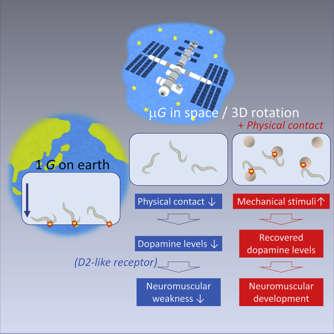
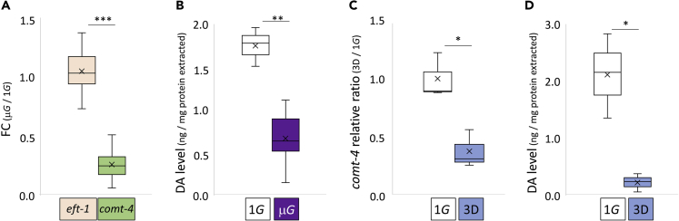
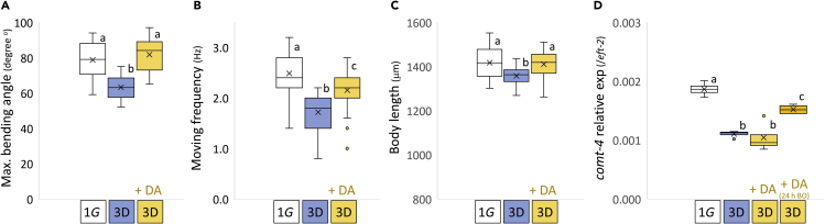
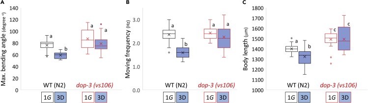
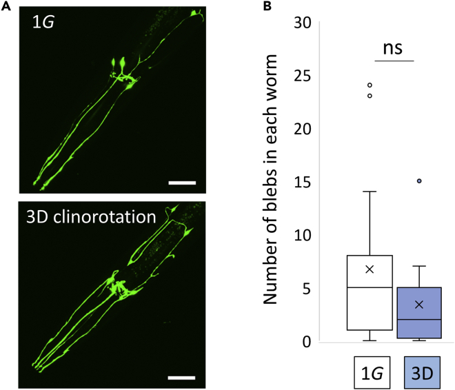
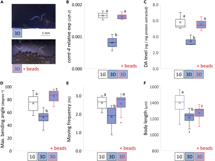
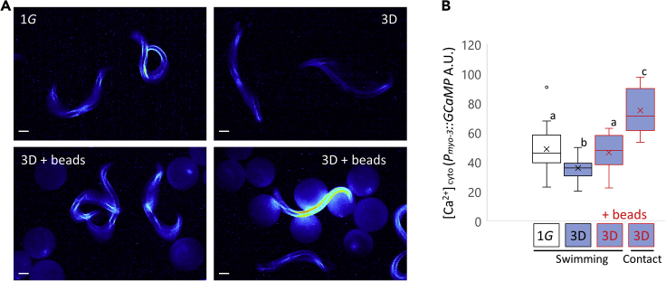
Progressive neuromuscular decline in microgravity is a prominent health concern preventing interplanetary human habitation. We establish functional dopamine-mediated impairments as a consistent feature across multiple spaceflight exposures and during simulated microgravity in C. elegans. Animals grown continuously in these conditions display reduced movement and body length. Loss of mechanical contact stimuli in microgravity elicits decreased endogenous dopamine and comt-4 (catechol-O-methyl transferase) expression levels. The application of exogenous dopamine reverses the movement and body length defects caused by simulated microgravity. In addition, increased physical contact made comt-4 and dopamine levels rise. It also increased muscular cytoplasmic Ca2+ firing. In dop-3 (D2-like receptor) mutants, neither decrease in movement nor in body length were observed during simulated microgravity growth. These results strongly suggest that targeting the dopamine system through manipulation of the external environment (contact stimuli) prevents muscular changes and is a realistic and viable treatment strategy to promote safe human deep-space travel.
Keywords: Aerospace Engineering; Space medicine.
© 2022 The Author(s).
Conflict of interest statement
Authors declare that they have no competing interests.
Figures
Similar articles
-
Integrated spaceflight transcriptomic analyses and simulated space experiments reveal key molecular features and functional changes driven by space stressors in space-flown C. elegans.Life Sci Space Res (Amst). 2025 Feb;44:10-22. doi: 10.1016/j.lssr.2024.11.004. Epub 2024 Nov 22. Life Sci Space Res (Amst). 2025. PMID: 39864902
-
Microgravity alters the expressions of DNA repair genes and their regulatory miRNAs in space-flown Caenorhabditis elegans.Life Sci Space Res (Amst). 2023 May;37:25-38. doi: 10.1016/j.lssr.2023.02.002. Epub 2023 Feb 17. Life Sci Space Res (Amst). 2023. PMID: 37087176
-
Microgravity elicits reproducible alterations in cytoskeletal and metabolic gene and protein expression in space-flown Caenorhabditis elegans.NPJ Microgravity. 2016 Jan 21;2:15022. doi: 10.1038/npjmgrav.2015.22. eCollection 2016. NPJ Microgravity. 2016. PMID: 28725720 Free PMC article.
-
Microgravity and Human Body: Unraveling the Potential Role of Heat-Shock Proteins in Spaceflight and Future Space Missions.Biology (Basel). 2024 Nov 13;13(11):921. doi: 10.3390/biology13110921. Biology (Basel). 2024. PMID: 39596876 Free PMC article. Review.
-
The Lungs in Space: A Review of Current Knowledge and Methodologies.Cells. 2024 Jul 6;13(13):1154. doi: 10.3390/cells13131154. Cells. 2024. PMID: 38995005 Free PMC article. Review.
Cited by
-
Caenorhabditis elegans in microgravity: An omics perspective.iScience. 2023 Jun 20;26(7):107189. doi: 10.1016/j.isci.2023.107189. eCollection 2023 Jul 21. iScience. 2023. PMID: 37456835 Free PMC article. Review.
-
The Protective Role of Neurogenetic Components in Reducing Stress-Related Effects during Spaceflights: Evidence from the Age-Related Positive Memory Approach.Life (Basel). 2022 Aug 2;12(8):1176. doi: 10.3390/life12081176. Life (Basel). 2022. PMID: 36013355 Free PMC article. Review.
-
Neural mechanisms of dopamine function in learning and memory in Caenorhabditis elegans.Neuronal Signal. 2024 Jan 18;8(1):NS20230057. doi: 10.1042/NS20230057. eCollection 2024 Jan. Neuronal Signal. 2024. PMID: 38572143 Free PMC article. Review.
-
Deficiency in RCAT-1 Function Causes Dopamine Metabolism Related Behavioral Disorders in Caenorhabditis elegans.Int J Mol Sci. 2022 Feb 21;23(4):2393. doi: 10.3390/ijms23042393. Int J Mol Sci. 2022. PMID: 35216508 Free PMC article.
-
A Compact Imaging Platform for Conducting C. elegans Phenotypic Assays on Earth and in Spaceflight.Life (Basel). 2023 Jan 10;13(1):200. doi: 10.3390/life13010200. Life (Basel). 2023. PMID: 36676149 Free PMC article.
References
Grants and funding
LinkOut - more resources
Full Text Sources
Molecular Biology Databases
Miscellaneous
