. 2022 Sep;136(5):1391-1406.
doi: 10.1007/s00414-022-02789-y. Epub 2022 Feb 9.

A multi-method assessment of 3D printed micromorphological osteological features

Affiliations

A multi-method assessment of 3D printed micromorphological osteological features

Rachael M Carew et al. Int J Legal Med. 2022 Sep.

Abstract

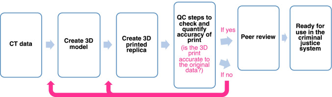
The evaluation of 3D printed osteological materials has highlighted the difficulties associated with accurately representing fine surface details on printed bones. Moreover, there is an increasing need for reconstructions to be demonstrably accurate and reliable for use in the criminal justice system. The aim of this study was to assess the surface quality of 3D prints (n = 9) that presented with micromorphological alterations from trauma, taphonomy and pathology processes. The archaeological bones were imaged using micro-CT scanning and 3D printed with selective laser sintering (SLS) printing. A multi-method experimental approach subsequently identified: (1) the 3D printed bones to be metrically accurate to within 1.0 mm; (2) good representation of micromorphological surface features overall, albeit with some loss of intricate details, depths, and fine textures that can be important for visual processing; (3) five of the nine 3D printed bones were quantitatively scored as accurate using the visual comparison method; and, (4) low mesh comparison distances (± 0.2 mm) between the original models and the digitised 3D print models. The findings offer empirical data that can be used to underpin 3D printed reconstructions of exhibits for use in courts of law. In addition, an adaptable pathway was presented that can be used to assess 3D print accuracy in future reconstructions.

Keywords: 3D imaging; 3D modelling; 3D printing; Evidence reconstruction; Forensic anthropology; Trauma.


Conflict of interest statement

The authors declare no competing interests.

Figures

Fig. 1
Fig. 1
A X-TEK Benchtop micro-CT, main unit; B bones mounted on platform using Blu Tack, with platform removed from micro-CT scanner for positioning (left; sample G), and with platform in position in micro-CT scanner (right; sample A); C sample C in position on rotating platform in the micro-CT scanner (x-ray tube upper left), with illustration of the field of view of the bone captured by the radiation as the orange triangular ‘beam’; D micro-CT workstation, with ‘Optimise’ parameter selected under Sample Setup; E STL model of sample H viewed in Avizo; F deleting floating part from sample F using Blender
Fig. 2
Fig. 2
Photographs of the archaeological bones (left column) including section imaged (pink boxes), with screenshots of the corresponding virtual 3D models viewed in 3D Slicer (central column), and photographs of the SLS 3D prints (right column). Two views provided each for samples H and I. Note, 3D models and prints approximate to scale provided
Fig. 3
Fig. 3
Colour map showing mesh comparison distances when compared with the ‘Optimised’ setting model, for models O1–O6 (sample A)
Fig. 4
Fig. 4
Comparisons of 3D prints to original bone samples: Sample A, fracture line visible in yellow rectangular box; Sample B, exposed trabecular bone in larger red circle, toothmarks in smaller blue circle; Sample C, (left image) depressed fracture in smaller blue circle and (right image) toothmarks in larger red circle; Sample D, butchery/chop mark in yellow rectangular box; Sample E, (left image) macroporosity in larger blue circle and smaller orange circle, and (right image) pathological remodelling features in larger blue circle; Sample F, endocranial root etchings in large red circle; Sample G, exposed trabecular bone in smaller pink circle, osteophytes in larger red circle, and eburnation and grooves in yellow rectangular box; Sample H, transverse fractures in blue circle; Sample I, detail on external surface (left image) and features on internal surface (right image). Scales in cm
Fig. 5
Fig. 5
Stacked bar chart illustrating the quantitative scores obtained with samples A–I (dashed line representing the cut-off total score value for determining the prints as accurate). The adjusted scoring method without the fracture pattern element resulted in accurate scores for six of nine 3D prints (B and D to H), where each print scored greater than the adjusted cut-off value of 12
Fig. 6
Fig. 6
Histograms for each mesh comparison for samples A through I, illustrating the distribution of the mesh distance values (C2M = cloud to mesh). Scales are model specific in each histogram
Fig. 7
Fig. 7
Example of a standard operating procedure incorporating the presented quality control (QC) steps. A 3D print will be re-modelled or re-printed if deemed to be inaccurate depending upon the issue

References

    1. Blau S, et al. Evaluating the impact of different formats in the presentation of trauma evidence in court: a pilot study. Aust J Forensic Sci. 2018;51(6):695–704. doi: 10.1080/00450618.2018.1457717. - DOI
    1. Errickson D, et al. The effect of different imaging techniques for the visualisation of evidence in court on jury comprehension. Int J Legal Med. 2019 doi: 10.1007/s00414-019-02221-y. - DOI - PMC - PubMed
    1. Baier W et al (2020) Forensic 3D printing from micro-CT for court use—process validation. Forensic Sci Int 110560. 10.1016/j.forsciint.2020.110560. - PubMed
    1. Carew RM, Morgan RM, Rando C. A preliminary investigation into the accuracy of 3D modeling and 3D printing in forensic anthropology evidence reconstruction. J Forensic Sci. 2019;64(2):342–352. doi: 10.1111/1556-4029.13917. - DOI - PMC - PubMed
    1. Baier W, et al. Introducing 3D printed models as demonstrative evidence at criminal trials. J Forensic Sci. 2018;63(4):1298–1302. doi: 10.1111/1556-4029.13700. - DOI - PubMed

LinkOut - more resources