

In vivo development and single-cell transcriptome profiling of human brain organoids
- PMID: 35141969
- PMCID: PMC8891563
- DOI: 10.1111/cpr.13201
In vivo development and single-cell transcriptome profiling of human brain organoids
Abstract
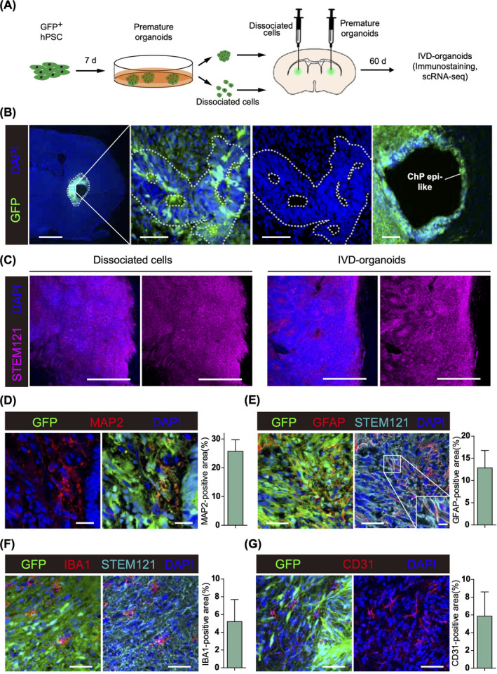
Objectives: Human brain organoids can provide not only promising models for physiological and pathological neurogenesis but also potential therapies in neurological diseases. However, technical issues such as surgical lesions due to transplantation still limit their applications.
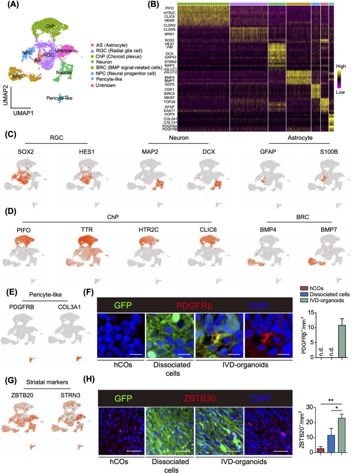
Materials and methods: Instead of applying mature organoids, we innovatively developed human brain organoids in vivo by injecting small premature organoids into corpus striatum of adult SCID mice. Two months after injection, single-cell transcriptome analysis was performed on 6131 GFP-labeled human cells from transplanted mouse brains.
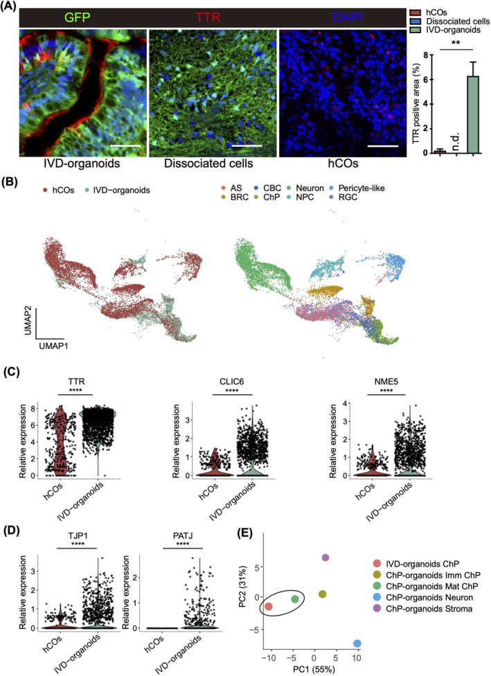
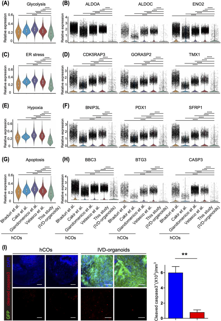
Results: Eight subsets of cells (including neuronal cells expressing striatal markers) were identified in these in vivo developed organoids (IVD-organoids) by unbiased clustering. Compared with in vitro cultured human cortical organoids, we found that IVD-organoids developed more supporting cells including pericyte-like and choroid plexus cells, which are important for maintaining organoid homeostasis. Furthermore, IVD-organoids showed lower levels of cellular stress and apoptosis.
Conclusions: Our study thus provides a novel method to generate human brain organoids, which is promising in various applications of disease models and therapies.
© 2022 The Authors. Cell Proliferation Published by John Wiley & Sons Ltd.
Conflict of interest statement
The authors declare no conflict of interest.
Figures
Similar articles
-
Robust production of uniform human cerebral organoids from pluripotent stem cells.Life Sci Alliance. 2020 Apr 17;3(5):e202000707. doi: 10.26508/lsa.202000707. Print 2020 May. Life Sci Alliance. 2020. PMID: 32303588 Free PMC article.
-
Dynamic Characterization of Structural, Molecular, and Electrophysiological Phenotypes of Human-Induced Pluripotent Stem Cell-Derived Cerebral Organoids, and Comparison with Fetal and Adult Gene Profiles.Cells. 2020 May 23;9(5):1301. doi: 10.3390/cells9051301. Cells. 2020. PMID: 32456176 Free PMC article.
-
Brain organoids: an ensemble of bioassays to investigate human neurodevelopment and disease.Cell Death Differ. 2021 Jan;28(1):52-67. doi: 10.1038/s41418-020-0566-4. Epub 2020 Jun 1. Cell Death Differ. 2021. PMID: 32483384 Free PMC article. Review.
-
Cellular complexity in brain organoids: Current progress and unsolved issues.Semin Cell Dev Biol. 2021 Mar;111:32-39. doi: 10.1016/j.semcdb.2020.05.013. Epub 2020 Jun 1. Semin Cell Dev Biol. 2021. PMID: 32499191 Review.
-
Bioengineering tissue morphogenesis and function in human neural organoids.Semin Cell Dev Biol. 2021 Mar;111:52-59. doi: 10.1016/j.semcdb.2020.05.025. Epub 2020 Jun 12. Semin Cell Dev Biol. 2021. PMID: 32540123 Free PMC article. Review.
Cited by
-
Organoids in Haematologic Research: Advances and Future Directions.Cell Prolif. 2025 Jun;58(6):e13806. doi: 10.1111/cpr.13806. Epub 2025 Jan 26. Cell Prolif. 2025. PMID: 40538372 Free PMC article. Review.
-
The Evolution of Single-Cell RNA Sequencing Technology and Application: Progress and Perspectives.Int J Mol Sci. 2023 Feb 2;24(3):2943. doi: 10.3390/ijms24032943. Int J Mol Sci. 2023. PMID: 36769267 Free PMC article. Review.
-
Transplantation Strategies to Enhance Maturity and Cellular Complexity in Brain Organoids.Biol Psychiatry. 2023 Apr 1;93(7):616-621. doi: 10.1016/j.biopsych.2023.01.004. Epub 2023 Jan 13. Biol Psychiatry. 2023. PMID: 36739209 Free PMC article. Review.
-
Brain Organoid Transplantation: A Comprehensive Guide to the Latest Advances and Practical Applications-A Systematic Review.Cells. 2025 Jul 14;14(14):1074. doi: 10.3390/cells14141074. Cells. 2025. PMID: 40710327 Free PMC article. Review.
-
Functional recovery through the plastic adaptation of organoid grafts in cortical tissue.Cell Mol Life Sci. 2025 Jun 9;82(1):227. doi: 10.1007/s00018-025-05767-w. Cell Mol Life Sci. 2025. PMID: 40490613 Free PMC article. Review.
References
MeSH terms
Grants and funding
LinkOut - more resources
Full Text Sources
Molecular Biology Databases
