. 2022 Mar 15;135(6):jcs259297.
doi: 10.1242/jcs.259297. Epub 2022 Mar 17.

Hedgehog pathway modulation by glypican 3-conjugated heparan sulfate

Affiliations

Hedgehog pathway modulation by glypican 3-conjugated heparan sulfate

Yulu Cherry Liu et al. J Cell Sci. .

Abstract

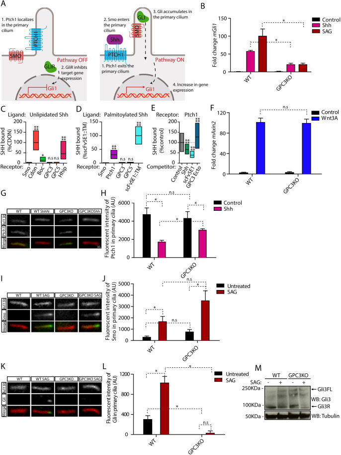
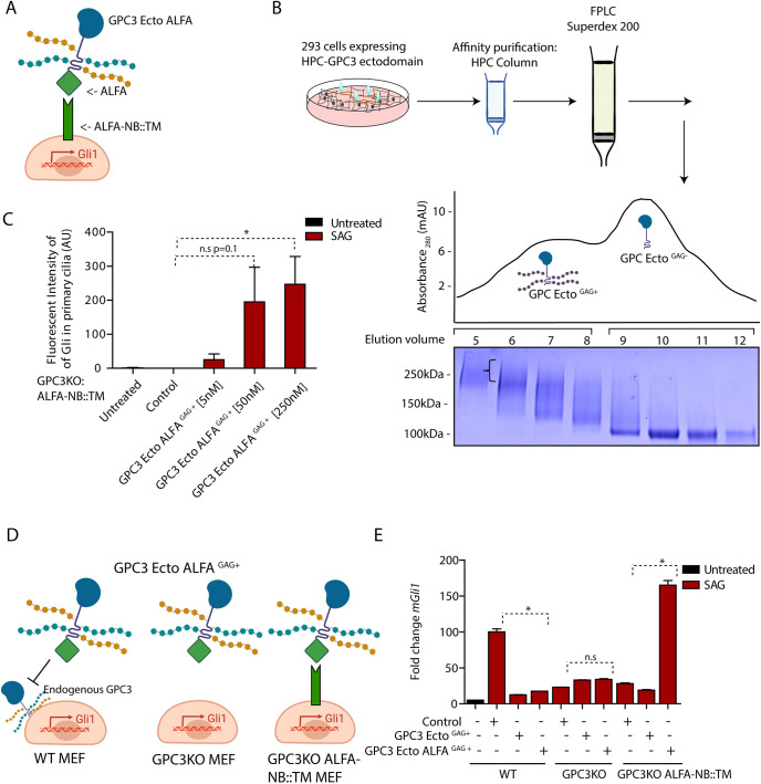
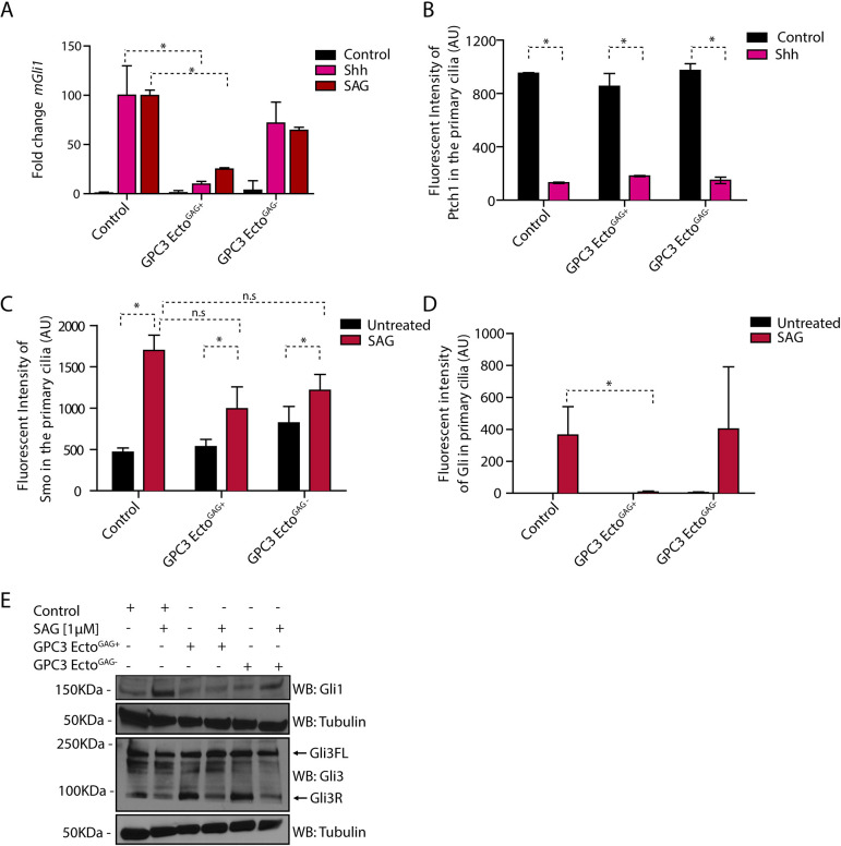
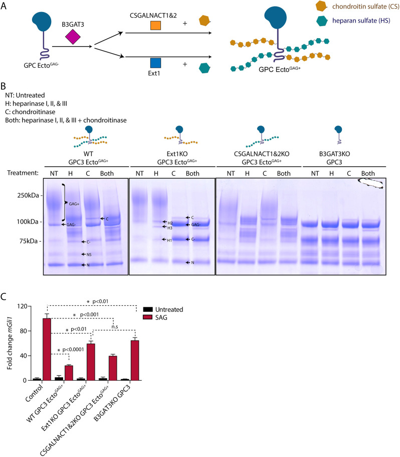
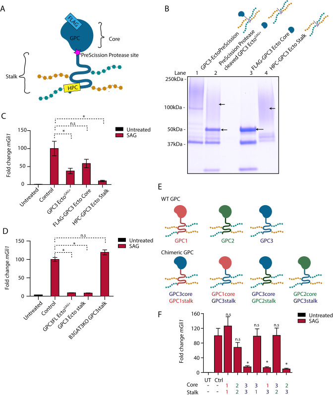
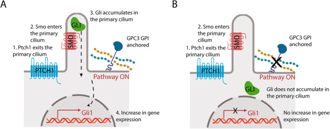
Glypicans are a family of cell surface heparan sulfate proteoglycans that play critical roles in multiple cell signaling pathways. Glypicans consist of a globular core, an unstructured stalk modified with sulfated glycosaminoglycan chains, and a glycosylphosphatidylinositol anchor. Though these structural features are conserved, their individual contribution to glypican function remains obscure. Here, we investigate how glypican 3 (GPC3), which is mutated in Simpson-Golabi-Behmel tissue overgrowth syndrome, regulates Hedgehog signaling. We find that GPC3 is necessary for the Hedgehog response, surprisingly controlling a downstream signal transduction step. Purified GPC3 ectodomain rescues signaling when artificially recruited to the surface of GPC3-deficient cells but has dominant-negative activity when unattached. Strikingly, the purified stalk, modified with heparan sulfate but not chondroitin sulfate, is necessary and sufficient for activity. Our results demonstrate a novel function for GPC3-associated heparan sulfate and provide a framework for the functional dissection of glycosaminoglycans by in vivo biochemical complementation. This article has an associated First Person interview with the first author of the paper.

Keywords: Glycosaminoglycans; Glypican; Hedgehog; Heparan sulfate; Signaling.


Conflict of interest statement

Competing interests The authors declare no competing or financial interests.

Figures

Fig. 1.
Fig. 1.
Endogenous GPC3 is necessary for Hh pathway activation. (A) Schematic of the Hh pathway. Left: in the absence of signaling, Ptch1 in primary cilia inhibits Smo activation and localization to cilia. Right: upon pathway activation by Shh, Ptch1 is inhibited and exits from cilia, whereas Smo becomes active and accumulates in cilia. Smo activates Gli proteins, which accumulate at ciliary tips. Active Gli proteins turn on the transcriptional targets of the pathway, including Gli1. (B) Wild-type (WT) or Gpc3KO MEFs were incubated with Shh, SAG (1 µM) or control medium for 24 h, and Hh signaling was measured by qRT-PCR for Gli1. Data are mean±s.e.m. of three replicates and were normalized from 0% (untreated) to 100% activation of the Hh pathway by saturating SAG. Hh pathway activation by Shh and SAG was significantly decreased in the absence of GPC3. (C) Fluorescently labeled unlipidated Shh was incubated with HEK293T cells transiently transfected with eGFP-tagged GPC3, GPC5 or control receptors, and bound ligand was quantified by fluorescence microscopy. Cdon, Boc and Hhip (left), as well as Ptch1 and a single-chain variant of the anti-Shh monoclonal antibody (scFv5E1) (Wierbowski et al., 2020) (right), were used as positive controls, and Smo was used as negative control. Data are normalized between binding to the negative control (0%) and the highest bound signal (100%). Boxplots represent the median and the first and third quartiles of binding. At least 200 cells were measured per condition. Neither GPC3 nor GPC5 binds unlipidated Shh. (D) As in C but using fluorescently labeled palmitoylated Shh. At least 1000 cells were measured per condition. Neither GPC3 nor GPC5 binds palmitoylated Shh. (E) As in C, except binding of palmitoylated Shh to Ptch1 was measured after preincubation of Shh with purified GPC3 ectodomain (GPC3-Ecto) or control competitors. Data are normalized between binding to Smo (negative control, 0%) and binding to Ptch1 in the presence of control competitor (100%). At least 1000 cells were measured per condition. GPC3-Ecto does not compete with Ptch1 for Shh binding. Shh binding was competed by scFv5E1 or by excess unlipidated Shh. (F) Wild-type or Gpc3KO MEFs were incubated with Wnt3A-conditioned medium or control medium for 24 h, and Wnt signaling was measured by qRT-PCR for Axin2. Wnt pathway activation by Wnt3A was normal in the absence of GPC3. Data are mean±s.e.m. of three replicates. (G) Wild-type or Gpc3KO MEFs stably expressing Ptch1-eGFP were incubated with Shh as in B, and ciliary intensity of Ptch1 was measured by immunofluorescence microscopy. Cilia were detected by staining for endogenous Arl13B. Ptch1 is green, Arl13B is red. In both wild-type and Gpc3KO cells, Shh induces Ptch1 exit from cilia. (H) Quantification of the experiment in G. Data are mean±s.d. (300-400 cilia were measured per condition). (I) As in G but measuring ciliary levels of endogenous Smo following treatment with SAG (1 µM). Smo is green, Arl13B is red. In both wild-type and Gpc3KO cells, SAG causes normal ciliary accumulation of Smo. (J) As in H but quantifying the experiment in I (300-400 cilia were measured per condition). (K) As in G but measuring ciliary intensity of endogenous Gli proteins. The anti-Gli antibody recognizes both full-length Gli2 and Gli3 (Tukachinsky et al., 2010). Gli is green, Arl13B is red. In the absence of GPC3, Gli proteins did not localize to the tips of cilia and their ciliary recruitment in response to SAG was greatly reduced. (L) As in H but quantifying the experiment in K (300-400 cilia were measured per condition). (M) As in B but cells were analyzed by immunoblotting for Gli3. Blotting for tubulin served as loading control. In the absence of GPC3, Gli3R was cleared normally by Hh pathway stimulation. Blots shown are representative of three experiments. *P<0.05; ****P<0.0001; n.s., not significant (two-tailed paired t-test). AU, arbitrary units. Scale bar: 1 μm (shown in G, also applies to I and K).
Fig. 2.
Fig. 2.
Purified GPC3-GAG (+) recruited to the cell surface rescues loss of endogenous GPC3. (A) Cartoon illustration of Gpc3KO MEFs expressing the membrane-bound ALFA nanobody (NB) construct ALFA-NB::TM, used to recruit GPC3-Ecto-ALFA to the cell surface. (B) Affinity-purified GPC3-Ecto fused to HaloTag was separated by size-exclusion chromatography to isolate GAG-modified and GAG-unmodified fractions. The indicated fractions were analyzed by SDS-PAGE and Coomassie staining. The bracket indicates the high molecular weight imparted by GAG modification of GPC3-Ecto. Gel shown is representative of three experiments. The y axis shows absorbance at 280 nm, expressed in mAU. (C) Gpc3KO MEFs stably expressing ALFA-NB::TM were incubated with SAG (1 µM) for 6 h in the presence of the indicated concentrations of purified GPC3-Ecto-ALFAGAG+. Ciliary intensity of endogenous Gli proteins was measured by immunofluorescence microscopy. Recruitment of GPC3-Ecto-ALFAGAG+ to the cell surface rescued signal-dependent accumulation of Gli at ciliary tips in a dose-dependent manner. Data are mean±s.d. (300-400 cilia were measured per condition). (D) Cartoon illustration of the experiment in E. (E) Wild-type (WT) MEFs, Gpc3KO MEFs or Gpc3KO MEFs stably expressing ALFA-NB::TM were incubated with SAG (1 µM) for 24 h in the presence or absence of 50 nM purified GPC3-Ecto-ALFAGAG+ or GPC3-EctoGAG+. Hh pathway output was measured by qRT-PCR for Gli1. Data are mean±s.e.m. of three replicates. Hh pathway activation is rescued when GPC3-Ecto-ALFAGAG+ is recruited to the cell surface. *P<0.05; n.s., not significant (two-tailed paired t-test). AU, arbitrary units.
Fig. 3.
Fig. 3.
Purified GPC3 ectodomain inhibits Hh signaling as dominant negative. (A) Wild-type MEFs treated with GPC3-EctoGAG+ or GPC3-EctoGAG− were incubated with SAG (1 µM) or Shh for 24 h, and Hh pathway output was measured by qRT-PCR for Gli1. GPC3-EctoGAG+ inhibits Hh signaling in wild-type MEF cells. Data are mean±s.e.m. of three replicates. (B) Wild-type MEFs stably expressing Ptch1-eGFP were incubated with Shh for 24 h in the presence or absence of 1 µM of purified GPC3-EctoGAG+ or GPC3-EctoGAG−, and ciliary intensity of Ptch1 was measured by immunofluorescence microscopy. Cilia were detected by staining for endogenous Arl13B. GPC3-EctoGAG+ and GPC3-EctoGAG− had no effect on Shh-induced Ptch1 exit from cilia. Data are mean±s.d. (300-400 cilia were measured per condition). (C) As in B but treating wild-type MEFs with SAG (1 µM) and measuring the intensity of endogenous Smo in primary cilia. GPC3-EctoGAG+ and GPC3-EctoGAG− had no effect on SAG-induced ciliary accumulation of Smo. Data are mean±s.d. (300-400 cilia were measured per condition). (D) As in C but measuring the intensity of endogenous Gli proteins in primary cilia. GPC3-EctoGAG+ abolishes Gli recruitment to ciliary tips by Hh pathway activation, whereas GPC3-EctoGAG− has no effect. Data are mean±s.d. (300-400 cilia were measured per condition). (E) As in D but cells were analyzed by immunoblotting for Gli1 and Gli3. Blotting for tubulin served as a loading control. GPC3-EctoGAG+ did not block the reduction in Gli3R levels caused by Hh pathway activation but blocks the accumulation of Gli1. Blots shown are representative of three experiments. *P<0.05; n.s., not significant (two-tailed paired t-test). AU, arbitrary units.
Fig. 4.
Fig. 4.
Heparan sulfate modification is required for the effect of GPC3 on Hh signaling. (A) Schematic of the pathways for glypican modification with heparan sulfate (teal) or chondroitin sulfate (brown). B3GAT3 functions in the synthesis of the tetrasaccharide linker shared between chondroitin sulfate and heparan sulfate. CSGALNACT1 and 2 function in CS biosynthesis, whereas EXT1 functions in HS biosynthesis. (B) GPC3-Ecto was expressed and purified from HEK293T cells of the indicated genetic backgrounds [wild-type (WT), EXT1KO, CSGALNACT1,2KO, or B3GAT3KO]. The proteins were then digested with heparinase I, II and III (H), chondroitinase (C), or both, for 4 h at room temperature. The reactions were analyzed by SDS-PAGE and Coomassie staining. In wild-type cells, GPC3-Ecto was modified with both heparan sulfate and chondroitin sulfate chains. The choice of expression host cells allowed modification of GPC3-Ecto with heparan sulfate, chondroitin sulfate or both. GPC3-Ecto also underwent incomplete Furin-mediated proteolytic cleavage into two fragments (38 and 22 kDa; see Fig. S4F). Band notation is as follows: GAG+, GAG-modified full-length ectodomain and C-terminal cleavage product; GAG−, full-length ectodomain, uncleaved; N, N-terminal Furin cleavage product; C, chondroitinase; H1, heparinase I; H2, heparinase II; H3, heparinase III; C−, GAG− C-terminal Furin cleavage product; NS, nonspecific degradation product. See Fig. S4F for schematic. Gels shown are representative of three experiments. (C) Wild-type MEFs were incubated with SAG (1 µM) or control medium for 24 h in the absence or presence of the indicated differentially GAG-modified purified GPC3-Ecto (1 µM). Hh signaling was measured by qRT-PCR for Gli1. GPC3-Ecto required heparan sulfate chains to inhibit Hh signaling. Data are mean±s.e.m. of three replicates. *P<0.05; n.s., not significant (two-tailed paired t-test).
Fig. 5.
Fig. 5.
The GAG-modified GPC3 stalk is necessary and sufficient for Hh pathway antagonism. (A) Schematic of the GPC3-Ecto variant with an engineered PreScission protease cleavage site between the core and stalk domains (GPC3-EctoPreScission). The fusion is FLAG tagged on the N terminus (to isolate the core domain after cleavage, FLAG-GPC3core) and HPC tagged on the C terminus (to isolate the stalk region, HPC-GPC3 stalk). (B) Affinity-purified GPC3-EctoPreScission (lane 1) was cleaved with PreScission protease (lane 2), followed by purification of the core domain (lane 3) and stalk (lane 4) by FLAG and HPC affinity, respectively. Proteins were analyzed by SDS-PAGE and Coomassie staining. Following protease cleavage, the globular core collapsed to defined bands (lane 3), whereas the stalk region migrated as a high molecular weight smear (lane 4) due to GAG modification. Gel shown is representative of three experiments. (C) Wild-type (WT) MEFs were incubated with SAG (1 µM) in the absence or presence of the indicated purified proteins (1 µM) for 24 h. Hh signaling was measured by qRT-PCR for Gli1. The GAG-modified GPC3 stalk potently inhibited Hh signaling, in contrast to the unmodified globular core. Untreated is no treatment. Cells were kept in DMEM. (D) As in C but with incubation with GPC3 stalk expressed and purified from B3GAT3KO cells. The unmodified GPC3 stalk did not inhibit Hh signaling. (E) Schematic of GPCs GPC1, GPC2 and GPC3, and the chimeric GPCs generated by domain swapping. (F) As in C but with incubation with purified chimeric GPC ectodomains. Hh signaling antagonism required the presence of the GPC3 stalk.; Ctrl, cells treated with the buffer for the purified protein; UT, untreated. Data are mean±s.e.m. of three replicates. *P<0.05; n.s., not significant (two-tailed paired t-test).
Fig. 6.
Fig. 6.
Model of GPC3 function in Hh signaling. (A) Endogenous GPI-anchored GPC3 promotes Hh signaling at the level of Gli proteins. (B) Loss of GPC3 impairs Hh signaling.

Similar articles

Cited by

References

    1. Ahn, J., Lüdecke, H.-J., Lindow, S., Horton, W. A., Lee, B., Wagner, M. J., Horsthemke, B. and Wells, D. E. (1995). Cloning of the putative tumour suppressor gene for hereditary multiple exostoses (EXT1). Nat. Genet. 11, 137-143. 10.1038/ng1095-137 - DOI - PubMed
    1. Aza-Blanc, P., Lin, H. Y., Ruiz i Altaba, A. and Kornberg, T. B. (2000). Expression of the vertebrate Gli proteins in Drosophila reveals a distribution of activator and repressor activities. Development 127, 4293-4301. 10.1242/dev.127.19.4293 - DOI - PubMed
    1. Baeg, G. H., Lin, X., Khare, N., Baumgartner, S. and Perrimon, N. (2001). Heparan sulfate proteoglycans are critical for the organization of the extracellular distribution of Wingless. Development 128, 87-94. 10.1242/dev.128.1.87 - DOI - PubMed
    1. Bai, C. B. and Joyner, A. L. (2001). Gli1 can rescue the in vivo function of Gli2. Development 128, 5161-5172. 10.1242/dev.128.24.5161 - DOI - PubMed
    1. Bangs, F. and Anderson, K. V. (2017). Primary cilia and mammalian Hedgehog signaling. Cold Spring Harb. Perspect. Biol. 9, a028175. 10.1101/cshperspect.a028175 - DOI - PMC - PubMed

Supplementary concepts