. 2022 Mar;36(3):e22177.
doi: 10.1096/fj.202101323R.

Exercise improves angiogenic function of circulating exosomes in type 2 diabetes: Role of exosomal SOD3

Affiliations

Exercise improves angiogenic function of circulating exosomes in type 2 diabetes: Role of exosomal SOD3

Kareem Abdelsaid et al. FASEB J. 2022 Mar.

Abstract

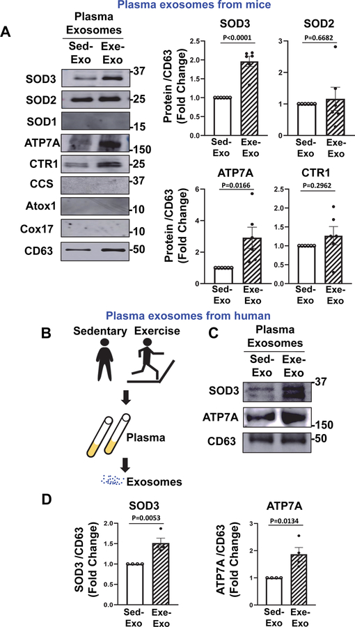
Exosomes, key mediators of cell-cell communication, derived from type 2 diabetes mellitus (T2DM) exhibit detrimental effects. Exercise improves endothelial function in part via the secretion of exosomes into circulation. Extracellular superoxide dismutase (SOD3) is a major secretory copper (Cu) antioxidant enzyme that catalyzes the dismutation of O2•- to H2 O2 whose activity requires the Cu transporter ATP7A. However, the role of SOD3 in exercise-induced angiogenic effects of circulating plasma exosomes on endothelial cells (ECs) in T2DM remains unknown. Here, we show that both SOD3 and ATP7A proteins were present in plasma exosomes in mice, which was significantly increased after two weeks of volunteer wheel exercise. A single bout of exercise in humans also showed a significant increase in SOD3 and ATP7A protein expression in plasma exosomes. Plasma exosomes from T2DM mice significantly reduced angiogenic responses in human ECs or mouse skin wound healing models, which was associated with a decrease in ATP7A, but not SOD3 expression in exosomes. Exercise training in T2DM mice restored the angiogenic effects of T2DM exosomes in ECs by increasing ATP7A in exosomes, which was not observed in exercised T2DM/SOD3-/- mice. Furthermore, exosomes overexpressing SOD3 significantly enhanced angiogenesis in ECs by increasing local H2 O2 levels in a heparin-binding domain-dependent manner as well as restored defective wound healing and angiogenesis in T2DM or SOD3-/- mice. In conclusion, exercise improves the angiogenic potential of circulating exosomes in T2DM in a SOD3-dependent manner. Exosomal SOD3 may provide an exercise mimetic therapy that supports neovascularization and wound repair in cardiometabolic disease.

Keywords: SOD3; exercise; exosome; type 2 diabetes.


Conflict of interest statement

DISCLOSURES

The authors declare no competing financial interests.

Figures

Figure 1:
Figure 1:. Characterization of plasma exosomes isolated from sedentary and voluntary wheel running exercised mice.
(A) Image of voluntary wheel exercise model. (B) Tracing of wheel activity of exercised mice (Interval distance). (C) Representative western blots for phospho-eNOS and total eNOS in mesenteric arteries at day 0,7,14,28 after exercise. (D)Transmission electron microscopy (TEM) image from sedentary and two weeks exercised mice (scale bar=100nm). (E) Exosome size and number of particles characterization from sedentary and two weeks exercised mice by Zetaview analysis (n=7). (F) Nanosight tracking of plasma exosomes. (G) Plasma exosomes concentration in sedentary and two weeks exercised mice (n=7) (H) Lysates from equal number of plasma exosomes from sedentary and exercised mice were immunoblotted (IB) with exosome markers (Tsg101 and CD63) antibodies (n=3). Results are presented as mean ± SEM.
Figure 2:
Figure 2:. Exercise enhances the angiogenic effects of plasma exosomes.
(A) Left: Representative fluorescence images to show the dose-dependent uptake of PKH67-labeled plasma exosomes (green) isolated from mice by HUVEC. Nuclei are stained with DAPI. Right: Graph shows the mean fluorescence intensity of PKH labelled exosomes in ECs. (Scale bar=20 μm, n=3). (B) Schematic diagram of plasma exosome isolation from sedentary and exercised mice, followed by their application to HUVEC to measure angiogenic responses. (C and D) Serum starved HUVECs treated with PBS, 10 ug/ml plasma exosomes from exercised (Exe-Exo) or sedentary (Sed-Exo) mice, or 20ng/ml VEGF (positive control) for 16 hrs were used to measure EC migration (modified Boyden chamber assay) (C) or capillary formation on growth factor-reduced Matrigel (D). In (C), graphs represent averaged number of migrated cells per five random fields, expressed as the fold change over PBS treated group. (n=4). In (D), graph shows averaged number of branching points (left) or tube length (right) per fields, expressed as the fold change over PBS treated group. Scale bar=50 μm. (n=4). Results are presented as mean ± SEM.
Figure 3:
Figure 3:. Exercise increases SOD3 and ATP7A protein expression in plasma exosomes in both mice and humans.
(A) Left: Representative western blots for Cu transport proteins (ATP7A, CTR1, Atox1, CCS, Cox17) and antioxidant SODs (SOD1, SOD2, SOD3) protein expression in plasma exosomes from sedentary and exercised mice. Right: Graph shows averaged fold change normalized to CD63 exosome marker level. (n=6). (B) Schematic diagram of plasma exosome isolated from human healthy subjects before and after exercise. (C and D) Representative western blots for ATP7A and SOD3 protein expression in plasma exosomes from human participants before and after a single bout of exercise (C) and graph showing averaged fold change normalized to CD63 exosome marker level (D) (n=4). Results are presented as mean ± SEM.
Figure 4:
Figure 4:. SOD3 is required for exercise-induced pro-angiogenic effects of plasma exosomes.
(A) Left: Representative western blots for SOD3 protein expression in plasma exosomes from sedentary (Sed-Exo) and exercised (Exe-Exo) wild type (WT) or SOD3 knockout (KO) mice. Right: Graph shows averaged fold change normalized to CD63 exosome marker level. (n=3). (B and C) Left: Serum starved HUVECs treated with 10 ug/ml plasma exosomes from sedentary or exercised WT mice (WT-Sed-Exo or WT-Exe-Exo) and SOD3 KO mice (SOD3 KO-Sed-Exo or SOD3 KO-Exe-Exo) for 16 hrs were used to measure EC migration (B) or capillary formation (C) as described above. Right: Graph shows averaged number of migrated cells per five random fields (B) or branching points or tube length per five random fields (C), expressed as the fold change from WT-Sed-Exo treated groups. (Scale bar=50 μm, n=3). Results are presented as mean ± SEM.
Figure 5:
Figure 5:. Exercise restores the angiogenic potential of T2DM plasma exosomes in vitro and in vivo.
(A) Schematic diagram of plasma exosome isolation from sedentary or exercised control or T2DM mice, followed by their application to HUVEC to measure angiogenic responses. (B and C) Left: Serum starved HUVECs treated with 10 ug/ml plasma exosomes from mice described in (A) for 16 hrs were used to measure EC migration (B) or capillary formation (C) as described. Right: Graph shows averaged number of migrated cells per five random fields (B) or averaged numbers of branching points per five random fields (C), expressed as the fold change from PBS-treated groups. (Scale bar=50 μm, n=4). (D) Control and T2DM mice were wounded on the back skin. Wound regions were applied with PBS (vehicle), 20 ug plasma exosomes from sedentary WT (Cont Sed-Exo), sedentary or exercised T2DM mice (T2DM-Sed-Exo or T2DM-Exe-Exo), and then, the wound closing rate was measured for 7 days. The wound area was expressed as percent of that measured right after the wounding. (E) Wounded skin tissues at Day 7 were used to measure CD31+ cells using CD31 antibody. Results are presented as mean ± SEM. N=3 mice per group/ 4 wounds per mouse.
Figure 6:
Figure 6:. SOD3 is required for exercise-induced restoration of impaired angiogenic effects of T2DM plasma exosomes.
(A) Left: Representative western blots for SOD3 and ATP7A protein expression in plasma exosomes from sedentary or exercised control or T2DM mice (Cont-Sed-Exo or T2DM-Sed-Exo or T2DM-Exe-Exo). Right: Graph shows averaged fold change normalized to CD63 exosome marker level (n=3). (B and C) Left: Serum starved HUVECs treated with 10 ug/ml plasma exosomes from sedentary or exercised T2DM mice (T2DM-Sed-Exo or T2DM-Exe-Exo) and SOD3 KO mice (SOD3 KO- T2DM-Sed-Exo or SOD3 KO-T2DM-Exe-Exo) for 16 hrs were used to measure EC migration (B) or capillary formation (C) as described above. Right: Graph shows averaged number of migrated cells per five random fields (B) and bottom: branching points or tube length per five random fields (C), expressed as the fold change from T2DM-Sed-Exo treated groups. (Scale bar=50 μm, n=3). Results are presented as mean ± SEM.
Figure 7:
Figure 7:. Exosomes overexpressing SOD3 promotes angiogenesis in ECs via increasing H2O2 in a HBD-dependent manner.
(A) Schematic diagram of exosome isolation from conditioned media from rat VSMCs infected with adenovirus expressing human SOD3 (Ad.hSOD3) or human SOD3 lacking HBD domain (Ad.hSOD3ΔHBD) or Ad.null (control), followed by their application to HUVEC to measure angiogenic responses. (B) Left: Representative western blots for human SOD3 protein expression in exosomes overexpressing SOD3 (SOD3-Exo) or SOD3ΔHBD (SOD3ΔHBD-Exo) or control exosomes (Null-Exo). Right: Graph shows averaged fold change normalized to Tsg101 exosome marker. (n=3). (C and D) Left: Serum-starved HUVECs treated with 10 ug/ml hSOD3-Exo, hSOD3ΔHBD-Exo, control Null-Exo for 16 hrs were used to measure EC migration (C) or capillary formation (D) as described above. Right: Graph shows averaged number of migrated cells per five random fields (C) or branching points or tube length per five random fields (D), expressed as the fold change from Null-Exo treated groups. (Scale bar=50 μm, n=3). (E) Dichlorofluorescein (DCF) fluorescence with DAPI staining were measured in HUVECs treated with control Null-Exo, 10 ug/ml hSOD3-Exo or hSOD3ΔHBD-Exo for 16 hrs in the presence or absence of Ad-Catalase, PEG-catalase, or human Catalase. Right panel shows the average of DCF fluorescence at 4 different fields, (n =5–8) vs. Null-Exo. (F) Serum-starved HUVECs infected with Ad-catalase or Ad-null (control) were treated with PBS (vehicle), 10 ug/ml hSOD3-Exo or control Null-Exo for 16 hrs to measure EC migration. Results are presented as mean ± SEM. (n = 3)
Figure 8:
Figure 8:. Exosomes overexpressing SOD3 restore impaired wound healing and angiogenesis in T2DM mice.
(A) Schematic diagram of exosome isolation from conditioned media from rat VSMCs infected with adenovirus expressing human SOD3 (Ad.hSOD3)(hSOD3-Exo), Ad.null (control)(Null-Exo) or PBS, followed by their application to wounded sites on the back skin of control or T2DM mice. (B) Control and T2DM mice were wounded on the back skin. Wound regions were applied with PBS, 20 ug hSOD3-Exo, or Null-Exo, and then, the wound closing rate was measured for 7 days. The wounded area was expressed as percent of that measured right after the wounding. (C) Wounded skin tissues at day 7 were used to measure CD31+ cells using CD31 antibody. N=3 mice per group/4 wounds per mouse. Results are presented as mean ± SEM. (D) Proposed model. Application of plasma Exo from T2DM mice to cultured ECs and mouse wound healing model showed impaired angiogenesis, which was associated with decrease in expression of ATP7A, but not SOD3, in T2DM-Exo. Exercise restores angiogenic effects of T2DM plasma Exo in a SOD3 dependent manner.

References

    1. Tkach M, and Thery C (2016) Communication by Extracellular Vesicles: Where We Are and Where We Need to Go. Cell 164, 1226–1232 - PubMed
    1. Lawson C, Vicencio JM, Yellon DM, and Davidson SM (2016) Microvesicles and exosomes: new players in metabolic and cardiovascular disease. J Endocrinol 228, R57–71 - PubMed
    1. Hessvik NP, and Llorente A (2018) Current knowledge on exosome biogenesis and release. Cell Mol Life Sci 75, 193–208 - PMC - PubMed
    1. Kranendonk ME, de Kleijn DP, Kalkhoven E, Kanhai DA, Uiterwaal CS, van der Graaf Y, Pasterkamp G, Visseren FL, and Group SS (2014) Extracellular vesicle markers in relation to obesity and metabolic complications in patients with manifest cardiovascular disease. Cardiovasc Diabetol 13, 37. - PMC - PubMed
    1. Yanez-Mo M, Siljander PR, Andreu Z, Zavec AB, Borras FE, Buzas EI, Buzas K, Casal E, Cappello F, Carvalho J, Colas E, Cordeiro-da Silva A, Fais S, Falcon-Perez JM, Ghobrial IM, Giebel B, Gimona M, Graner M, Gursel I, Gursel M, Heegaard NH, Hendrix A, Kierulf P, Kokubun K, Kosanovic M, Kralj-Iglic V, Kramer-Albers EM, Laitinen S, Lasser C, Lener T, Ligeti E, Line A, Lipps G, Llorente A, Lotvall J, Mancek-Keber M, Marcilla A, Mittelbrunn M, Nazarenko I, Nolte-’t Hoen EN, Nyman TA, O’Driscoll L, Olivan M, Oliveira C, Pallinger E, Del Portillo HA, Reventos J, Rigau M, Rohde E, Sammar M, Sanchez-Madrid F, Santarem N, Schallmoser K, Ostenfeld MS, Stoorvogel W, Stukelj R, Van der Grein SG, Vasconcelos MH, Wauben MH, and De Wever O (2015) Biological properties of extracellular vesicles and their physiological functions. J Extracell Vesicles 4, 27066. - PMC - PubMed

Publication types

MeSH terms