. 2022 Sep-Oct;24(5):540-548.
doi: 10.4103/aja2021104.

Mannose inhibits the growth of prostate cancer through a mitochondrial mechanism

Affiliations

Mannose inhibits the growth of prostate cancer through a mitochondrial mechanism

Yu-Lin Deng et al. Asian J Androl. 2022 Sep-Oct.

Abstract

The limited treatment options for advanced prostate cancer (PCa) lead to the urgent need to discover new anticancer drugs. Mannose, an isomer of glucose, has been reported to have an anticancer effect on various tumors. However, the anticancer effect of mannose in PCa remains unclear. In this study, we demonstrated that mannose inhibits the proliferation and promotes the apoptosis of PCa cells in vitro, and mannose was observed to have an anticancer effect in mice without harming their health. Accumulation of intracellular mannose simultaneously decreased the mitochondrial membrane potential, increased mitochondrial and cellular reactive oxygen species (ROS) levels, and reduced adenosine triphosphate (ATP) production in PCa cells. Mannose treatment of PCa cells induced changes in mitochondrial morphology, caused dysregulated expression of the fission protein, such as fission, mitochondrial 1 (FIS1), and enhanced the expression of proapoptotic factors, such as BCL2-associated X (Bax) and BCL2-antagonist/killer 1 (Bak). Furthermore, lower expression of mannose phosphate isomerase (MPI), the key enzyme in mannose metabolism, indicated poorer prognosis in PCa patients, and downregulation of MPI expression in PCa cells enhanced the anticancer effect of mannose. This study reveals the anticancer effect of mannose in PCa and its clinical significance in PCa patients.

Keywords: mannose; mannose phosphate isomerase; metabolism; mitochondria; prostate cancer.


Conflict of interest statement

None

Figures

Figure 1
Figure 1
Mannose inhibited the proliferation and induced the apoptosis of PCa cells. The IC50 of mannose in (a) DU145 and (b) PC3 cells was determined using a CCK-8 assay. (c) Intracellular mannose concentration in PCa cells. Cell proliferation of (d) DU145 and (e) PC3 was assessed using growth curves, respectively. (f) Colony formation assays were performed and (g) colony numbers were counted in PCa cells. (h) Flow cytometric analysis was used to assess (i) the apoptosis rate of PCa cells. *P < 0.05, **P < 0.01. NC: PCa cells cultured in normal medium. MAN: PCa cells cultured in normal medium with 25 mmol l−1 mannose for DU145 or with 50 mmol l−1 mannose for PC3. PCa: prostate cancer; IC50: the half-maximal inhibitory concentration; CCK-8: Cell Counting Kit-8; AAD: Aminoactinomycin D; APC: Allophycocyanin.
Figure 2
Figure 2
Mannose inhibited tumor growth in a PCa xenograft model without affecting mice health. (a) Subcutaneous tumors from the xenograft model with DU145 cells. (b) Tumor growth was monitored for 30 days after mannose treatment. (c) The volume of tumor growth. (d) The weight of the tumors. (e) Intratumoral mannose concentration and (f) ATP content in subcutaneous tumors. The weights of (g) mice and (h) major metabolic organs. **P < 0.01. NC: PCa cells cultured in normal medium. MAN: PCa cells cultured in normal medium with 25 mmol l−1 mannose for DU145 or with 50 mmol l−1 mannose for PC3. PCa: prostate cancer; ATP: adenosine triphosphate.
Figure 3
Figure 3
Mannose disrupted mitochondrial function, led to ROS overproduction, and activated Bax/Bak in PCa cells. (a) JC-1 staining and (b) rhodamine 123 staining were used to assess the MMP in DU145 and PC3 cells. (c) The ATP content in cells. (d) Mitochondrial ROS and (e) cellular ROS levels in cells. (f and g) The protein expression of Bax and Bak in cells. *P < 0.05, **P < 0.01. NC: PCa cells cultured in normal medium. MAN: PCa cells cultured in normal medium with 25 mmol l−1 mannose for DU145 or with 50 mmol l−1 mannose for PC3. JC-1: 5,5′,6,6′-tetrachloro-1,1′,3,3′-tetraethyl-imidacarbocyanine; Bax: BCL2-associated X; Bak: BCL2-antagonist/killer 1; ROS: reactive oxygen species; MMP: mitochondrial membrane potential; ATP: adenosine triphosphate; PCa: prostate cancer.
Figure 4
Figure 4
Mannose disrupted the balance of mitochondrial dynamics in PCa cells. (a) Mitochondria stained with MitoTracker Red were observed by confocal microscopy. (b) Mitochondrial structure under a transmission electron microscopy, and the mitochondrial cross-sectional area was quantified. (c) The protein expression of FIS1 in cells. Upregulated (d) FIS1 and (e) increased ATP content in PCa cells. *P < 0.05, **P < 0.01. NC: PCa cells cultured in normal medium. MAN: PCa cells cultured in normal medium with 25 mmol l−1 mannose for DU145 or with 50 mmol l−1 mannose for PC3. ATP: adenosine triphosphate; FIS1: fission, mitochondrial 1; PCa: prostate cancer.
Figure 5
Figure 5
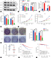
Downregulation of MPI expression enhances the anticancer effect of mannose. The expression of MPI in human prostate cancer tissues and its prognostic value. (a and b) The expression of MPI was silenced by siRNA-MPI and verified by western blotting. siRNA-MPI with different base sequences including si-1, si-2 and si-3 were used for downregulating the MPI expression in PCa cells. According to the degree of down-regulation of MPI protein, si-3 and si-2 with the best interference effect were applied to DU145 and PC3 cells, respectively. (c) Intracellular mannose concentration and (d) ATP content in cells. (e) Growth curves and (f) colony formation assays of cells. (g) The IHC scores for MPI expression in PCa tissues. (h) Kaplan–Meier curves of BCR-free survival and (i) overall survival for the low and high MPI expression groups of patients in the TCGA-PRAD dataset. *P < 0.05, **P < 0.01. NC: PCa cells cultured in normal medium. MAN: PCa cells cultured in normal medium with 25 mmol l−1 mannose for DU145 or with 50 mmol l−1 mannose for PC3. MPI: mannose phosphate isomerase; si: siRNA, small interfering RNA; IHC: immunohistochemistry; PCa: prostate cancer; BCR: biochemical recurrence; TCGA-PRAD: The Cancer Genome Atlas-Prostate Adenocarcinoma; GAPDH: glyceraldehyde-3-phosphate dehydrogenase; TMA: tissue microarray; ATP: adenosine triphosphate.

References

    1. Siegel RL, Miller KD, Fuchs HE, Jemal A. Cancer statistics, 2021. CA Cancer J Clin. 2021;71:7–33. - PubMed
    1. Cattaruzza L, Fregona D, Mongiat M, Ronconi L, Fassina A, et al. Antitumor activity of gold(III)-dithiocarbamato derivatives on prostate cancer cells and xenografts. Int J Cancer. 2011;128:206–15. - PubMed
    1. Gonzalez PS, O'Prey J, Cardaci S, Barthet VJ, Sakamaki JI, et al. Mannose impairs tumour growth and enhances chemotherapy. Nature. 2018;563:719–23. - PubMed
    1. Luo Q, Li B, Li G. Mannose suppresses the proliferation and metastasis of lung cancer by targeting the ERK/GSK-3β/β-catenin/SNAIL axis. Onco Targets Ther. 2020;13:2771–81. - PMC - PubMed
    1. Lu J, Dong W, He H, Han Z, Zhuo Y, et al. Autophagy induced by overexpression of DCTPP1 promotes tumor progression and predicts poor clinical outcome in prostate cancer. Int J Biol Macromol. 2018;118:599–609. - PubMed