

OCTA Derived Vessel Skeleton Density Versus Flux and Their Associations With Systemic Determinants of Health
- PMID: 35142788
- PMCID: PMC8842473
- DOI: 10.1167/iovs.63.2.19
OCTA Derived Vessel Skeleton Density Versus Flux and Their Associations With Systemic Determinants of Health
Abstract
Purpose: To examine the associations of optical coherence tomography angiography (OCTA)-derived retinal capillary flux with systemic determinants of health.
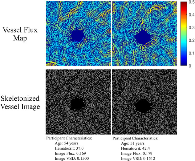
Methods: This is a cross-sectional study of subjects recruited from the African American Eye Disease Study. A commercially available swept-source (SS)-OCTA device was used to image the central 3 × 3 mm macular region. Retinal capillary perfusion was assessed using vessel skeleton density (VSD) and flux. Flux approximates the number of red blood cells moving through vessel segments and is a novel metric, whereas VSD is a previously validated measure commonly used to quantify capillary density. The associations of OCTA derived measures with systemic determinants of health were evaluated using multivariate generalized linear mixed-effects models.
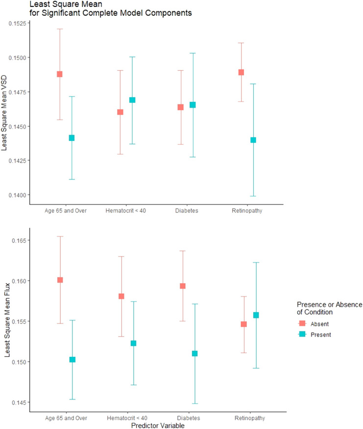
Results: A total of 154 eyes from 83 participants were enrolled. Mean VSD and flux were 0.148 ± 0.009 and 0.156 ± 0.016, respectively. In a model containing age, systolic blood pressure, diabetes status, hematocrit, and presence of retinopathy as covariates, there was a negative correlation between VSD and age (P < 0.001) and retinopathy (P = 0.02), but not with hematocrit (P = 0.85) or other factors. There was a positive correlation between flux and hematocrit (P = 0.02), as well as a negative correlation for flux with age (P < 0.001), systolic blood pressure (P = 0.04), and diabetes status (P = 0.02). A 1% decrease in hematocrit was associated with the same magnitude change in flux as ∼1.24 years of aging. Signal strength was associated with flux (P < 0.001), but not VSD (P = 0.51).
Conclusions: SS-OCTA derived flux provides additional information about retinal perfusion distinct from that obtained with skeleton density-based measures. Flux is appropriate for detecting subclinical changes in perfusion in the absence of clinical retinopathy.
Conflict of interest statement
Disclosure:
Figures
Similar articles
-
Optical Coherence Tomography Angiography-Derived Flux As a Measure of Physiological Changes in Retinal Capillary Blood Flow.Transl Vis Sci Technol. 2021 Aug 2;10(9):5. doi: 10.1167/tvst.10.9.5. Transl Vis Sci Technol. 2021. PMID: 34342607 Free PMC article.
-
Geometric Perfusion Deficits: A Novel OCT Angiography Biomarker for Diabetic Retinopathy Based on Oxygen Diffusion.Am J Ophthalmol. 2021 Feb;222:256-270. doi: 10.1016/j.ajo.2020.09.007. Epub 2020 Sep 9. Am J Ophthalmol. 2021. PMID: 32918905 Free PMC article.
-
Capillary density and caliber as assessed by optical coherence tomography angiography may be significant predictors of diabetic retinopathy severity.PLoS One. 2022 Jan 26;17(1):e0262996. doi: 10.1371/journal.pone.0262996. eCollection 2022. PLoS One. 2022. PMID: 35081154 Free PMC article. Clinical Trial.
-
Impaired Retinal Vascular Reactivity in Diabetic Retinopathy as Assessed by Optical Coherence Tomography Angiography.Invest Ophthalmol Vis Sci. 2019 Jun 3;60(7):2468-2473. doi: 10.1167/iovs.18-26417. Invest Ophthalmol Vis Sci. 2019. PMID: 31173077 Free PMC article.
-
Combined Multimodal Analysis of Peripheral Retinal and Macular Circulation in Diabetic Retinopathy (COPRA Study).Ophthalmol Retina. 2019 Jul;3(7):580-588. doi: 10.1016/j.oret.2019.03.001. Epub 2019 Mar 20. Ophthalmol Retina. 2019. PMID: 31078525
Cited by
-
OCT Angiography-Derived Retinal Capillary Perfusion Measures in the Framingham Heart Study.Ophthalmol Sci. 2024 Dec 27;5(3):100696. doi: 10.1016/j.xops.2024.100696. eCollection 2025 May-Jun. Ophthalmol Sci. 2024. PMID: 40124307 Free PMC article.
-
An open-source deep learning network AVA-Net for arterial-venous area segmentation in optical coherence tomography angiography.Commun Med (Lond). 2023 Apr 17;3(1):54. doi: 10.1038/s43856-023-00287-9. Commun Med (Lond). 2023. PMID: 37069396 Free PMC article.
-
Dimension-based quantification of aging-associated cerebral microvasculature determined by optical coherence tomography and two-photon microscopy.J Biophotonics. 2024 Mar;17(3):e202300409. doi: 10.1002/jbio.202300409. Epub 2024 Jan 4. J Biophotonics. 2024. PMID: 38176434 Free PMC article.
-
OCTA-ReVA: an open-source toolbox for comprehensive retinal vessel feature analysis in optical coherence tomography angiography.Biomed Opt Express. 2024 Sep 25;15(10):6010-6023. doi: 10.1364/BOE.537727. eCollection 2024 Oct 1. Biomed Opt Express. 2024. PMID: 39421789 Free PMC article.
-
Cross-modality Labeling Enables Noninvasive Capillary Quantification as a Sensitive Biomarker for Assessing Cardiovascular Risk.Ophthalmol Sci. 2023 Dec 5;4(3):100441. doi: 10.1016/j.xops.2023.100441. eCollection 2024 May-Jun. Ophthalmol Sci. 2023. PMID: 38420613 Free PMC article.
References
-
- Wong TY, Klein R, Couper DJ, et al. .. Retinal microvascular abnormalities and incident stroke: the Atherosclerosis Risk in Communities Study. Lancet. 2001; 358(9288): 1134–1140. - PubMed
-
- Friedenwald H. The Doyne Memorial Lecture: pathological changes in the retinal blood-vessels in arterio-sclerosis and hypertension. Trans Ophthalmol Soc UK. 1930;50452–50531.
-
- Büttner M, Schuster AKG, Vossmerbäumer U, Fischer JE.. Associations of cardiovascular risk factors and retinal vessel dimensions at present and their evolution over time in a healthy working population. Acta Ophthalmologica. 2020; 98(4): e457–e463. - PubMed
-
- Wang Lu, Wong Tien Y, Richey SA, Ronald K, Folsom Aaron R, Michael J-H. Relationship between retinal arteriolar narrowing and myocardial perfusion. Hypertension. 2008; 51(1): 119–126. - PubMed
Publication types
MeSH terms
Substances
Grants and funding
LinkOut - more resources
Full Text Sources
Medical
