. 2022 Mar 29;145(13):987-1001.
doi: 10.1161/CIRCULATIONAHA.121.058173. Epub 2022 Feb 10.

Second Heart Field-Derived Cells Contribute to Angiotensin II-Mediated Ascending Aortopathies

Affiliations

Second Heart Field-Derived Cells Contribute to Angiotensin II-Mediated Ascending Aortopathies

Hisashi Sawada et al. Circulation. .

Abstract

Background: The ascending aorta is a common location for aneurysm and dissection. This aortic region is populated by a mosaic of medial and adventitial cells that are embryonically derived from either the second heart field (SHF) or the cardiac neural crest. SHF-derived cells populate areas that coincide with the spatial specificity of thoracic aortopathies. The purpose of this study was to determine whether and how SHF-derived cells contribute to ascending aortopathies.

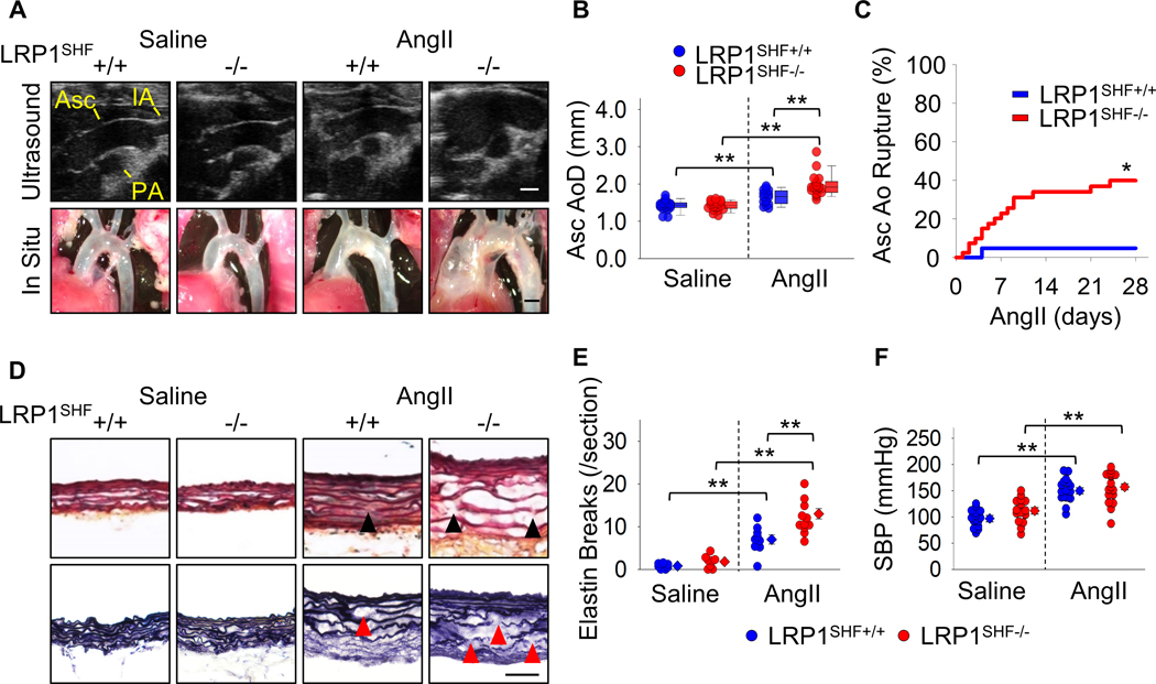
Methods: Ascending aortic pathologies were examined in patients with sporadic thoracic aortopathies and angiotensin II (AngII)-infused mice. Ascending aortas without overt pathology from AngII-infused mice were subjected to mass spectrometry-assisted proteomics and molecular features of SHF-derived cells were determined by single-cell transcriptomic analyses. Genetic deletion of either Lrp1 (low-density lipoprotein receptor-related protein 1) or Tgfbr2 (transforming growth factor-β receptor type 2) in SHF-derived cells was conducted to examine the effect of SHF-derived cells on vascular integrity.

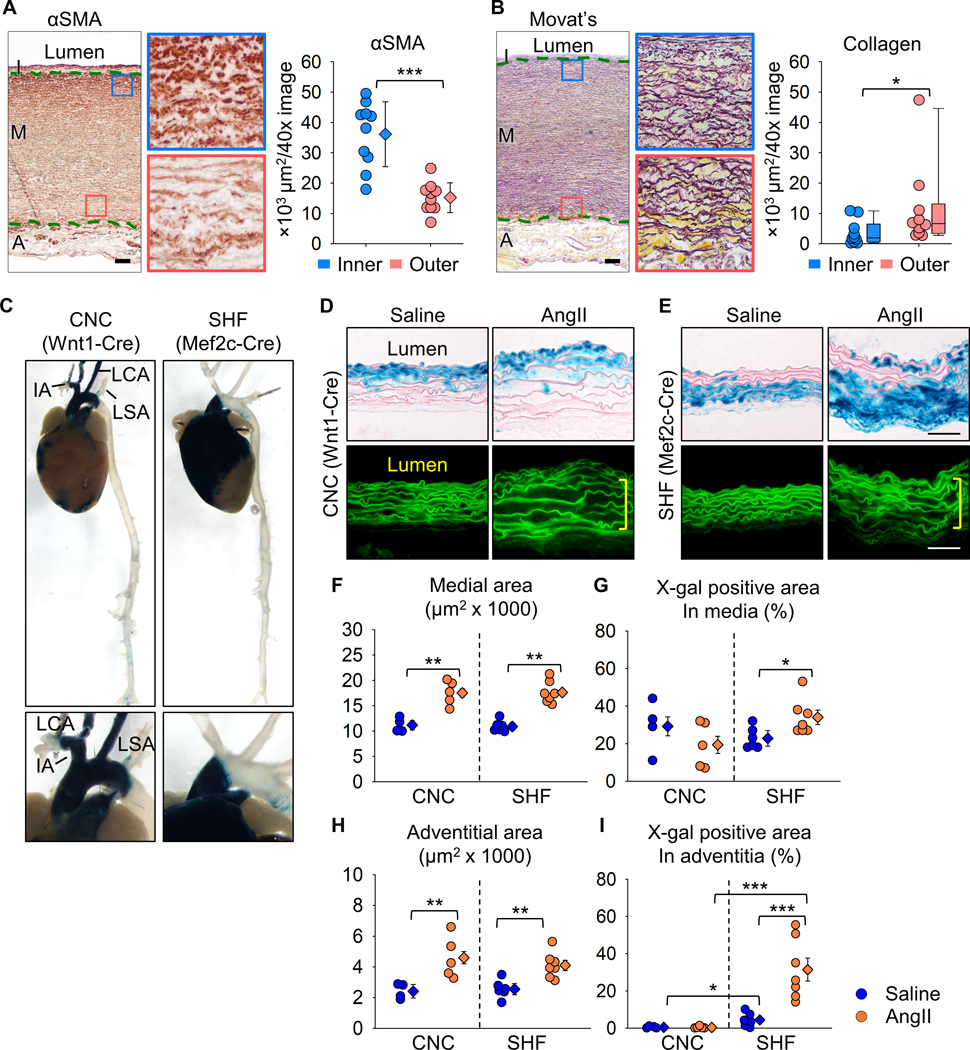
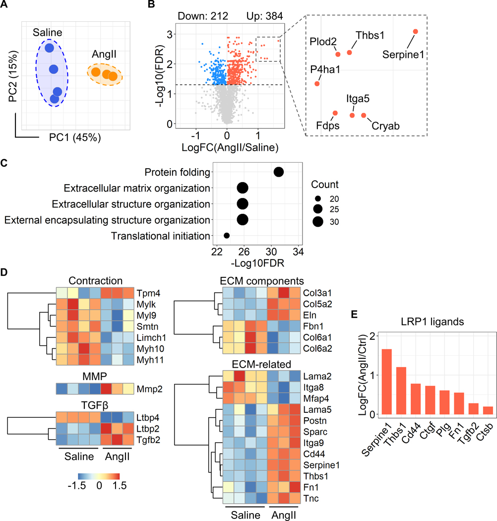
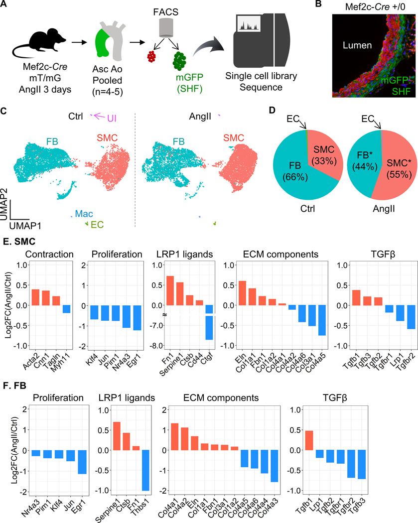
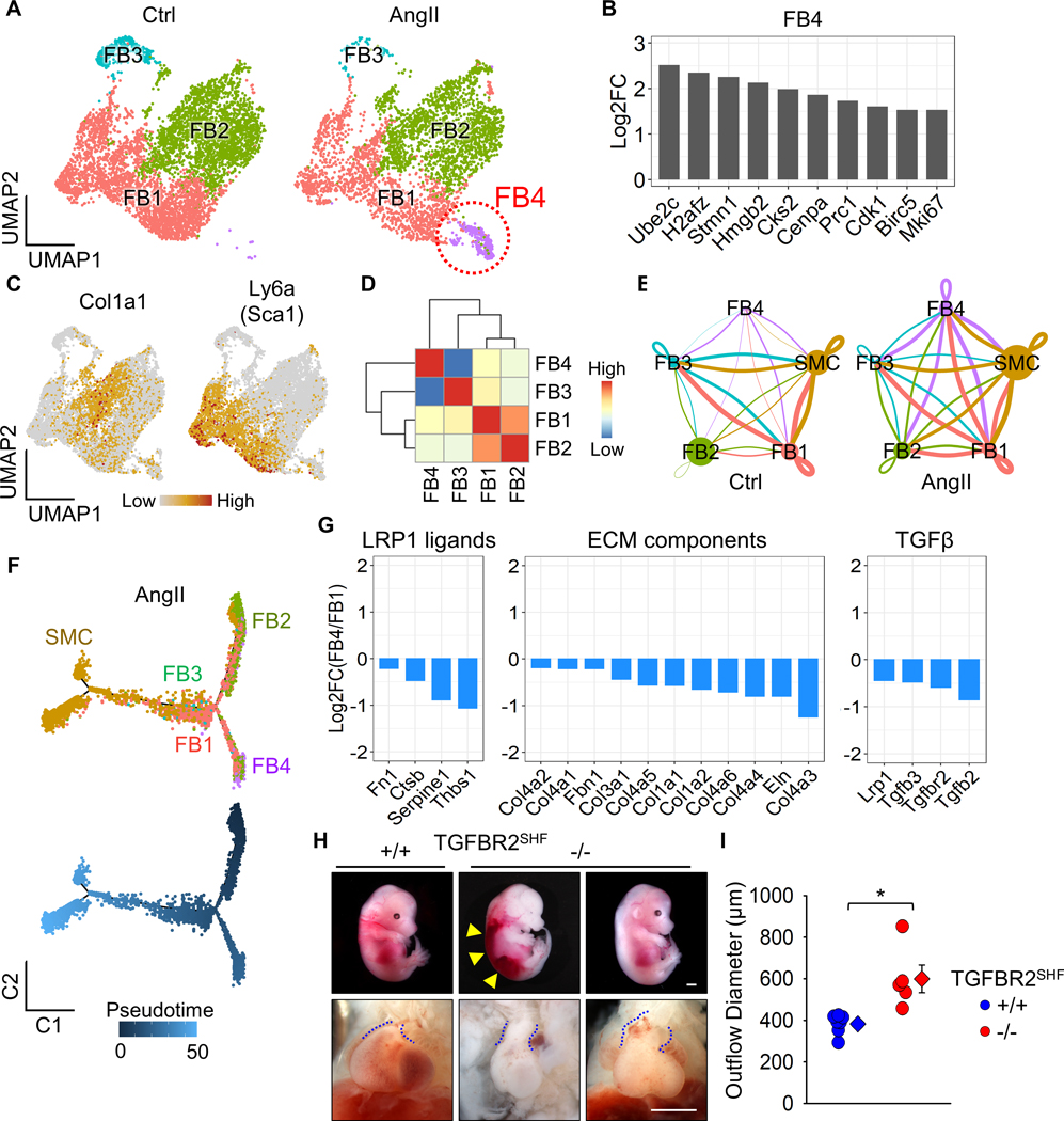
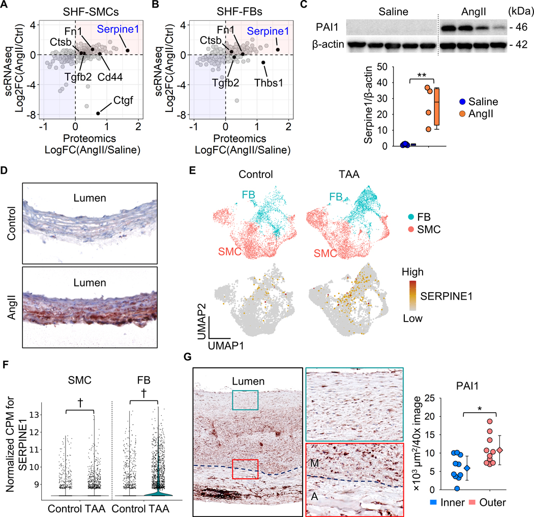
Results: Pathologies in human ascending aortic aneurysmal tissues were predominant in outer medial layers and adventitia. This gradient was mimicked in mouse aortas after AngII infusion that was coincident with the distribution of SHF-derived cells. Proteomics indicated that brief AngII infusion before overt pathology occurred evoked downregulation of smooth muscle cell proteins and differential expression of extracellular matrix proteins, including several LRP1 ligands. LRP1 deletion in SHF-derived cells augmented AngII-induced ascending aortic aneurysm and rupture. Single-cell transcriptomic analysis revealed that brief AngII infusion decreased Lrp1 and Tgfbr2 mRNA abundance in SHF-derived cells and induced a unique fibroblast population with low abundance of Tgfbr2 mRNA. SHF-specific Tgfbr2 deletion led to embryonic lethality at E12.5 with dilatation of the outflow tract and retroperitoneal hemorrhage. Integration of proteomic and single-cell transcriptomics results identified PAI1 (plasminogen activator inhibitor 1) as the most increased protein in SHF-derived smooth muscle cells and fibroblasts during AngII infusion. Immunostaining revealed a transmural gradient of PAI1 in both ascending aortas of AngII-infused mice and human ascending aneurysmal aortas that mimicked the gradient of medial and adventitial pathologies.

Conclusions: SHF-derived cells exert a critical role in maintaining vascular integrity through LRP1 and transforming growth factor-β signaling associated with increases of aortic PAI1.

Keywords: angiotensin; aortic aneurysm, thoracic; fibroblast; mice; smooth muscle.


Figures

Figure 1.
Figure 1.. Transmural gradients of aortic pathologies in patients with sporadic TAAs and AngII-infused mice.
Representative images of (A) immunostaining for α-smooth muscle actin (red) and (B) Movat’s pentachrome staining (elastin: dark purple, collagen: yellow). High magnification images are captured from the inner (blue box) and outer (red box) media (n=10). I indicates intima; M, media; A, adventitia. Green dotted lines indicate internal and external elastic layers. αSMA positive and collagen deposited areas were quantified in inner and outer media. Log transformation was applied for the data of collagen deposition. (C) Representative images of X-gal stained (blue) aortas from Wnt1- and Mef2c-Cre ROSA26RLacZ mice. IA indicates innominate artery; LCA, left common carotid artery; LSA, left subclavian artery. High magnification images of the proximal thoracic aortas are captured. Representative X-gal staining and FITC images of aortas from either saline- or AngII-infused (D) Wnt1- and (E) Mef2c-Cre ROSA26RLacZ mice (n=5 to 7 per group). Yellow square brackets indicate medial thickening. Scale bar, 50 μm. Dot plots for (F) medial and (H) adventitial areas and X-gal positive area in the (G) media and (I) adventitia. Diamonds and error bars indicate the mean and SEM, respectively. *P<0.05, **P<0.01, ***P<0.001 by Student’s t-test or two-way ANOVA followed by Holm-Sidak test.
Figure 2.
Figure 2.. AngII altered proteins related to ECM in the ascending aorta of mice.
(A) Principal component analysis of the unfiltered proteome. PC1/PC2 indicate first/second principal components (n=3 or 4 per group). (B) Volcano plots for differentiated proteins between in ascending aortas of saline and AngII-infused mice. (C) Top 5 annotations of enrichment analyses in gene ontology - biological process. (D) Heatmap with Z score coloring for proteins related to SMC contraction, MMP, TGFβ signaling, ECM-related molecules compared between saline and AngII infusion. (E) Log fold change of LRP1 ligands.
Figure 3.
Figure 3.. LRP1 deletion in SHF-derived cells augmented AngII-induced TAA formation and aortic rupture in mice.
(A) Representative ultrasound images and in situ gross appearances of proximal thoracic aortas (n=21 to 40 per group). Asc indicates ascending aorta; IA, innominate artery; PA, pulmonary artery. Scale bar, 1 mm. (B) Ascending aortic diameter (Asc AoD) measured by ultrasonography. (C) Cumulative incidence of ascending aortic (Asc Ao) rupture. (D) Representative Movat’s and Verhoeff iron hematoxylin staining. Scale bar, 50 μm. Arrow heads indicate elastin fragmentation. (E) Elastin break counts in Movat’s stained sections. (F) Systolic blood pressure. Diamonds and error bars indicate the mean and SEM, respectively. *P<0.01 by log-rank test, **P<0.001 by two-way ANOVA followed by Holm-Sidak test. Log transformation was applied for the data of ascending aortic diameters.
Figure 4.
Figure 4.. Significant reduction of Tgfbr2 mRNA in SHF-derived SMCs and FBs of AngII-infused mice.
(A) Experimental design of scRNAseq using Mef2c-Cre +/0 ROSA26RmT/mG mice. (B) Representative fluorescent image of the ascending aorta from Mef2c-Cre +/0 ROSA26RmT/mG mice. (C) UMAP plots of aortic cells at baseline (Ctrl) and after 3 days of AngII infusion. SMC indicates smooth muscle cells; FB, fibroblasts; EC, endothelial cells; Mac, macrophages; UI, unidentified cells. (D) Pie charts for the composition of each cell type. *P<0.0001 by Chi-square test. Log2 fold change of mRNA abundance related to SMC contraction, cell proliferation, LRP1 ligands, ECM components, and TGFβ ligands and receptors between Ctrl vs AngII in (E) SMCs and (F) FB clusters.
Figure 5.
Figure 5.. A distinct sub-cluster with decreased Tgfbr2 mRNA in SHF-derived fibroblasts of AngII-infused mice.
(A) UMAP plots at baseline (Ctrl) and after 3 days of AngII infusion in SHF-derived FBs. (B) Log2 fold change of mRNA abundance for top 10 unique genes in FB4 sub-cluster. (C) Featured plots for Col1a1 and Ly6a in FB sub-clusters. (D) Heatmap for cluster similarity among FB sub-clusters. (E) Ligand-receptor interactions among aortic cells. Lines connect to cell populations that express cognate ligands or receptors. The line thickness and node size correspond to the number of cognate ligand-receptor expression and cell number, respectively. (F) Trajectory analysis in SMC and FB sub-clusters from AngII-infused mice. (G) Log2 fold change of mRNA abundance related to LRP1 ligands, ECM components, and TGFβ molecules between FB1 and FB4 sub-clusters after AngII infusion. (H) Representative images of gross appearance and the outflow tract of wild type littermates (left), dead (middle), and survived (right) fetuses with SHF-specific TGFBR2 deletion at E12.5. Blue dotted lines indicate the edge of the outflow tract. Yellow triangles indicate retroperitoneal hemorrhage. (I) Outflow diameter was measured at 300 to 400 μm distal to the root of viable fetuses at termination (n=5 to 7 per group). Scale bar, 1 mm. Diamonds and error bars indicate the mean and SEM, respectively. *P<0.001 by two-tailed Student’s t-test.
Figure 6.
Figure 6.. Integration of proteomic and scRNAseq results identified PAI1 (Serpine1) as a key contributor to the medial gradient of aortic pathologies.
Scatter plots for Log or Log2 fold change of protein and mRNA abundances. Data were compared between proteomics and scRNAseq in SHF-derived (A) SMC and (B) FB clusters. Selected LRP1 ligands were highlighted. (C) Western blot analysis for PAI1 (Serpine1) and β-actin in the whole ascending aorta from saline or AngII-infused mice (n=4–5 per group). **P<0.001 by Student’s t-test after Log transformation. (D) Representative images of immunostaining for PAI1 (red) in ascending aortas from control or AngII-infused mice (n=7 per group). (E) UMAP and (F) violin plots for SERPINE1 in SMC and FB clusters of control subjects and patients with ascending TAAs (n=3 and 8 per group, scRNAseq data were downloaded from GSE155468). †P<0.001 analyzed using the Hurdle model adjusted for age implemented. (G) Representative images of immunostaining for PAI1 in patients with sporadic ascending TAA (n=10) and its quantification. Dotted lines indicate external elastic laminae; M, media; A, adventitia. Diamonds and error bars indicate the mean and SEM, respectively. *P<0.01 by Student’s t-test.

References

    1. Goldfinger JZ, Halperin JL, Marin ML, Stewart AS, Eagle KA and Fuster V. Thoracic aortic aneurysm and dissection. J Am Coll Cardiol. 2014;64:1725–1739. - PubMed
    1. Hiratzka LF, Bakris GL, Beckman JA, Bersin RM, Carr VF, Casey DE Jr., Eagle KA, Hermann LK, Isselbacher EM, Kazerooni EA, et al. 2010 ACCF/AHA/AATS/ACR/ASA/SCA/SCAI/SIR/STS/SVM Guidelines for the diagnosis and management of patients with thoracic aortic disease. A Report of the American College of Cardiology Foundation/American Heart Association Task Force on Practice Guidelines, American Association for Thoracic Surgery, American College of Radiology,American Stroke Association, Society of Cardiovascular Anesthesiologists, Society for Cardiovascular Angiography and Interventions, Society of Interventional Radiology, Society of Thoracic Surgeons,and Society for Vascular Medicine. J Am Coll Cardiol. 2010;55:e27–e129. - PubMed
    1. Isselbacher EM. Thoracic and abdominal aortic aneurysms. Circulation. 2005;111:816–828. - PubMed
    1. Albornoz G, Coady MA, Roberts M, Davies RR, Tranquilli M, Rizzo JA and Elefteriades JA. Familial thoracic aortic aneurysms and dissections--incidence, modes of inheritance, and phenotypic patterns. Ann Thorac Surg. 2006;82:1400–1405. - PubMed
    1. Trachet B, Piersigilli A, Fraga-Silva RA, Aslanidou L, Sordet-Dessimoz J, Astolfo A, Stampanoni MF, Segers P and Stergiopulos N. Ascending aortic aneurysm in angiotensin II-infused mice: Formation, progression, and the role of focal dissections. Arterioscler Thromb Vasc Biol. 2016;36:673–681. - PubMed

Publication types

Substances