. 2022 Mar 15;132(6):e149828.
doi: 10.1172/JCI149828.

Intermittent glucocorticoid treatment enhances skeletal muscle performance through sexually dimorphic mechanisms

Affiliations

Intermittent glucocorticoid treatment enhances skeletal muscle performance through sexually dimorphic mechanisms

Isabella M Salamone et al. J Clin Invest. .

Abstract

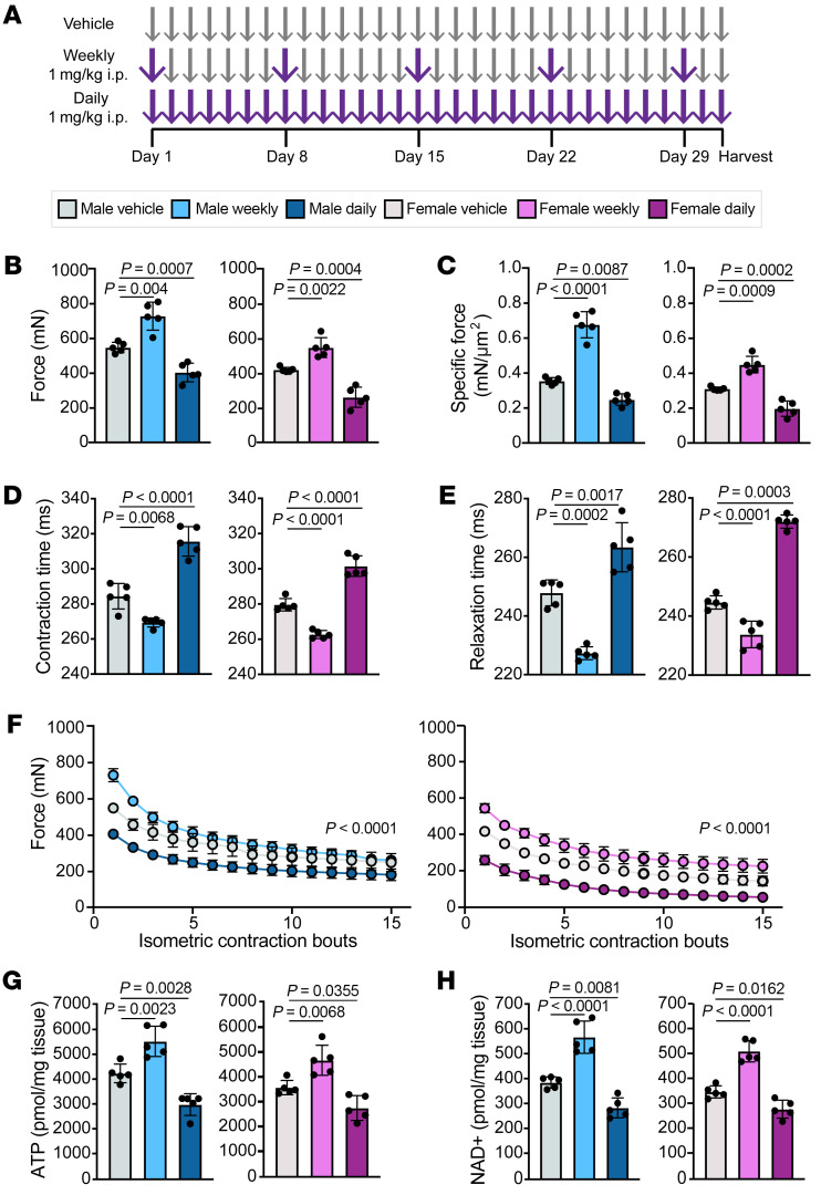
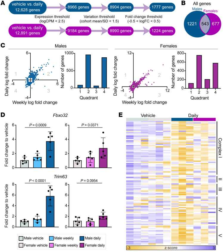
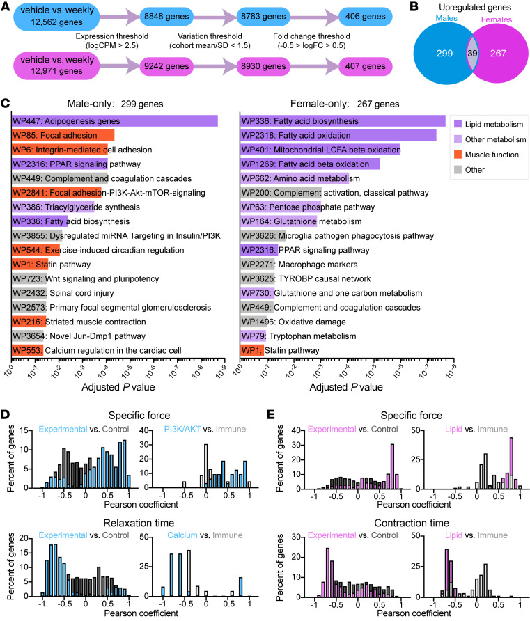
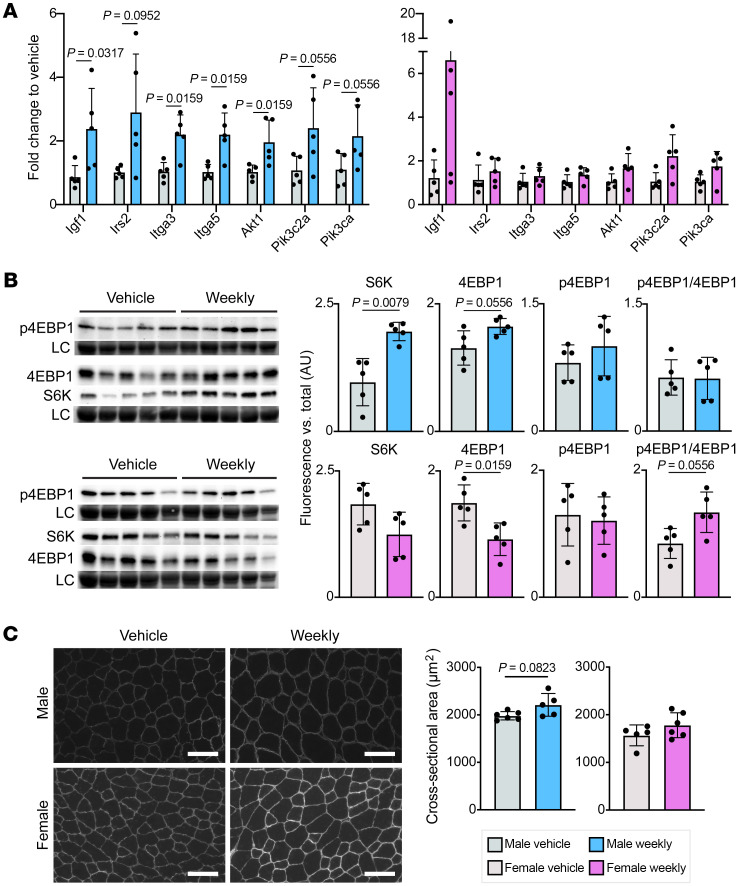
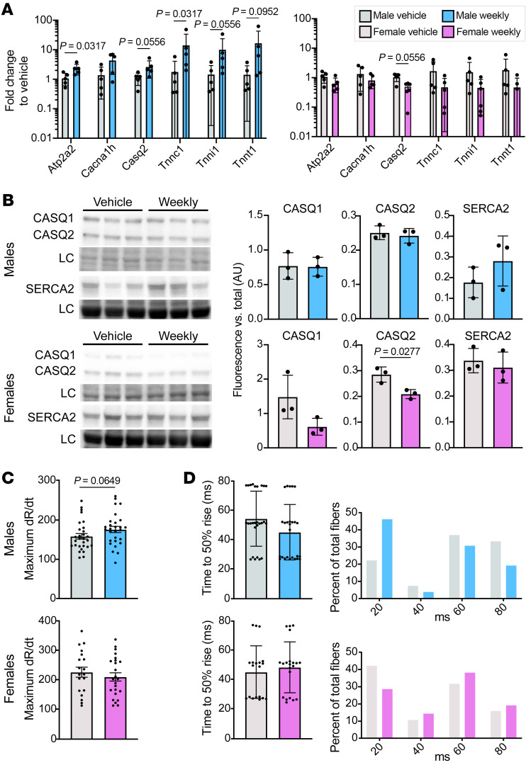
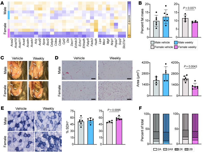
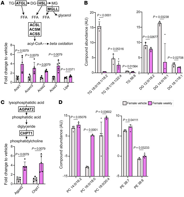
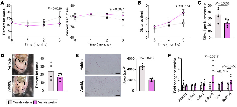
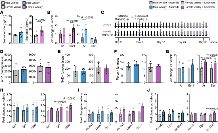
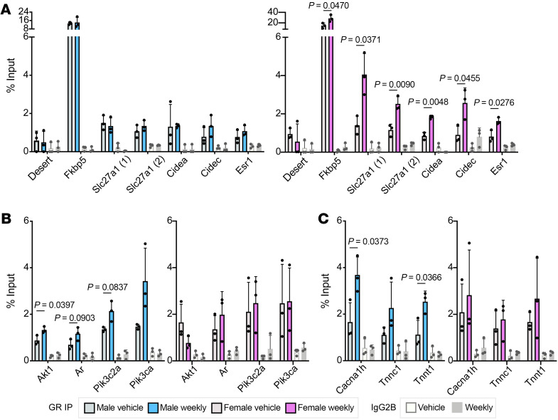
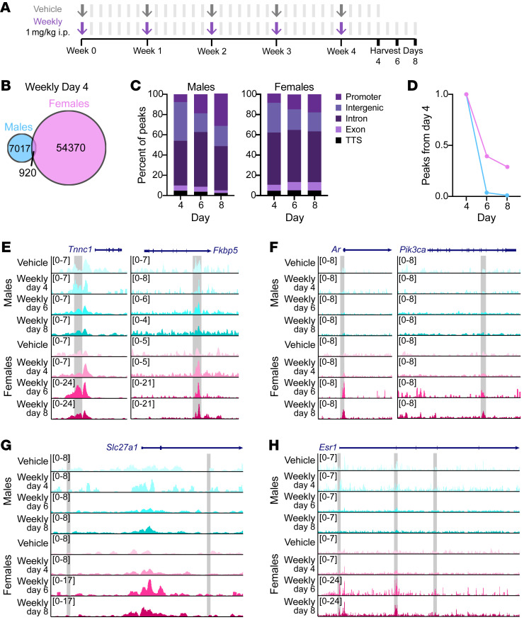
Glucocorticoid steroids are commonly prescribed for many inflammatory conditions, but chronic daily use produces adverse effects, including muscle wasting and weakness. In contrast, shorter glucocorticoid pulses may improve athletic performance, although the mechanisms remain unclear. Muscle is sexually dimorphic and comparatively little is known about how male and female muscles respond to glucocorticoids. We investigated the impact of once-weekly glucocorticoid exposure on skeletal muscle performance comparing male and female mice. One month of once-weekly glucocorticoid dosing improved muscle specific force in both males and females. Transcriptomic profiling of isolated myofibers identified a striking sexually dimorphic response to weekly glucocorticoids. Male myofibers had increased expression of genes in the IGF1/PI3K pathway and calcium handling, while female myofibers had profound upregulation of lipid metabolism genes. Muscles from weekly prednisone-treated males had improved calcium handling, while comparably treated female muscles had reduced intramuscular triglycerides. Consistent with altered lipid metabolism, weekly prednisone-treated female mice had greater endurance relative to controls. Using chromatin immunoprecipitation, we defined a sexually dimorphic chromatin landscape after weekly prednisone. These results demonstrate that weekly glucocorticoid exposure elicits distinct pathways in males versus females, resulting in enhanced performance.

Keywords: Calcium; Endocrinology; Insulin signaling; Muscle Biology; Skeletal muscle.


Conflict of interest statement

Conflict of interest: Northwestern University has filed a US patent on intermittent glucocorticoid use (provisional number 62/876,238).

Figures

Figure 1
Figure 1. Weekly prednisone improved muscle performance in both male and female mice.
(A) C57BL/6J mice were treated for 4 weeks with vehicle (DMSO) or weekly or daily prednisone, and then analyzed. (BF) Weekly treated mice exhibited significantly increased maximal tetanic force (B) and specific force (C) and reduced contraction (D) and relaxation (E) time compared with vehicle-treated mice. Once-weekly prednisone–treated mice also exhibited increased response to repetitive force (F) compared with vehicle-treated mice. (G and H) Weekly treated mice had increased concentrations of ATP (G) and NAD+ (H) compared with vehicle-treated animals, while daily treated mice had significantly reduced concentrations. Data were analyzed with 1-way ANOVA (BE, G, and H) or 2-way ANOVA (F).
Figure 2
Figure 2. Daily and weekly prednisone treatment elicited similar transcriptional profiles with differential atrogene activation.
(A) RNA sequencing analysis of daily prednisone–treated muscles (quadriceps) compared with vehicle-treated for both sexes; prednisone-responsive genes were identified as being above expression and fold-change thresholds and below a variation threshold; n = 3 animals per group. (B) Less than half of all prednisone-responsive genes were shared among daily prednisone–treated males and females. (C) The majority of prednisone-responsive genes above a log2(fold change) threshold had the same response to both daily and weekly treatment, i.e., increased (quadrant 2) or decreased (quadrant 4) expression. (D) Expression of atrogenes Fbxo32 and Trim63 was increased in daily treated muscle fibers compared with vehicle, as evaluated by qPCR and 1-way ANOVA, while weekly treated muscle fibers had no change in expression of these atrogenes. (E) Expression of genes encoding the mitochondrial respiratory chain was decreased in response to daily treatment in both sexes compared with vehicle treatment.
Figure 3
Figure 3. Weekly prednisone elicits distinct transcriptional response in male versus female muscle.
(A) Transcriptome comparison of once-weekly prednisone–treated muscle to vehicle-treated for both sexes; prednisone-responsive genes were identified as being above expression and fold-change (FC) thresholds and below a variation threshold. n = 3 animals per group. (B) The majority of prednisone-responsive upregulated genes were unique to one sex. (C) Gene ontology analysis of male- and female-only upregulated genes shows differential pathway enrichment. (D and E) Pheno-RNA analysis of genes shows high correlation between gene expression and physiological response in males (D) and females (E). Genes responsive to weekly prednisone are more correlated with physiological response than a shuffled control group and genes in pathways of interest are more correlated with physiological response than prednisone-responsive genes involved in other cellular processes.
Figure 4
Figure 4. Weekly prednisone treatment activated the IGF1/PI3K pathway in males but not females.
(A) Genes encoding IGF1/PI3K pathway members had increased expression in the muscle (quadriceps) of weekly treated male muscle but not female muscle by qPCR. (B) Total protein levels of mTOR targets S6K and 4EBP1 were increased in the gastrocnemius of weekly treated males but decreased in weekly treated females. LC, loading control. (C) Myofiber cross-sectional area in the tibialis anterior trended larger in weekly treated compared with vehicle-treated males but was unchanged in females. Scale bars: 100 μm. Data in AC were analyzed with Mann-Whitney test.
Figure 5
Figure 5. Weekly prednisone treatment improved calcium handling in males but not females.
(A) Genes encoding calcium-handling proteins were upregulated in the quadriceps of weekly treated males but downregulated in weekly treated females by qPCR. (B) Protein levels of CASQ1, CASQ2, and SERCA2 were unchanged or increased in the gastrocnemius of weekly treated males, but decreased in weekly treated females compared with vehicle-treated animals. LC, loading control. (C and D) Calcium transient measurements in isolated flexor digitorum brevis fibers trended toward a faster maximum rate of calcium release (change in ratio/time, dR/dt) (C) and time to 50% rise (D) in weekly treated males and no change in weekly treated females compared to vehicle-treated animals. Data were analyzed with Mann-Whitney test (A and C) or Welch’s t test (B).
Figure 6
Figure 6. Weekly prednisone treatment improved lipid metabolism in females but not males.
(A) Results from RNA sequencing analysis revealed increased expression of lipid metabolism–related genes in weekly prednisone–treated females compared with vehicle-treated females and weekly prednisone–treated males. (B) Body composition analysis after 4 weeks of treatment demonstrated reduced whole-body percentage fat mass in once-weekly prednisone–treated females, while comparably treated males had no change. (C and D) The visceral fat pad of weekly treated females was smaller than vehicle-treated females and weekly treated males (C) and the adipocytes in this fat pad had significantly reduced cross-sectional area (D). (E) Weekly treated females had an increased proportion of succinate dehydrogenase–positive (SDH+) fibers in the tibialis anterior compared with vehicle-treated females, while the males exhibited no change. Scale bars: 100 μm (D) and 50 μm (E). (F) Neither sex had changes in fiber type proportion following weekly treatment. Data in B, D, and E were analyzed with Mann-Whitney test.
Figure 7
Figure 7. Weekly prednisone–treated females had increased glyceride catabolism and mitochondrial phospholipid abundance.
(A) Muscles (quadriceps) from weekly prednisone–treated females had significantly increased expression of enzymes involved in triglyceride catabolism and acyl-CoA synthesis. (B) Females also had significantly reduced abundance of triglyceride and diglyceride species in the quadriceps. (C) Enzymes involved in phosphatidylcholine synthesis were significantly upregulated by weekly prednisone treatment. (D) Phosphatidylcholine (PC) and phosphatidylethanolamine (PE) abundance was increased in weekly prednisone–treated females compared with vehicle-treated females. Top 3 most abundant lipid compounds are presented as (number of carbon atoms):(number of double bonds); less abundant compounds are listed in Supplemental Table 4. Data were analyzed with Mann-Whitney test (A and C) or Welch’s t test (B and D).
Figure 8
Figure 8. Long-term weekly prednisone treatment increased endurance in female mice.
(A) Weekly prednisone–treated females had reduced whole-body percentage fat mass and increased percentage lean mass after 3 months of treatment. (B) Weekly prednisone–treated females showed increased distance to exhaustion in a treadmill run-to-exhaustion test compared with vehicle-treated females. (C) Weekly prednisone–treated females required fewer stimuli per kilometer run than vehicle-treated females. (D) After 9 months of treatment, weekly treated females had a slightly smaller visceral fat pad and moderately reduced percentage fat mass than vehicle-treated females. (E) Adipocytes in the visceral fat pad of weekly treated females had significantly reduced cross-sectional area compared with vehicle-treated females. Scale bars: 100 μm. (F) Some lipid metabolism genes continued to be upregulated in response to weekly prednisone. Data were analyzed with 2-way ANOVA (A and B) or Mann-Whitney test (C, E, and F).
Figure 9
Figure 9. Sex steroid receptor antagonism attenuated the effects of weekly prednisone.
(A) Circulating levels of testosterone and estrogen were increased in weekly prednisone–treated animals. (B) Expression of the gene encoding the androgen receptor (Ar) was increased in weekly prednisone–treated males, while expression of the gene encoding estrogen receptor α (Esr1) was increased in weekly treated females. (C) C57BL/6J mice were cotreated for 4 weeks with sex steroid inhibitors (males, flutamide; females, fulvestrant). Concomitantly, half of the cohort received weekly prednisone or vehicle. Arrows indicate i.p. injections. (D and E) Weekly prednisone–treated mice had no change in concentrations of ATP (D) and NAD+ (E) compared to vehicle-treated animals. (F) Weekly treated animals had no change in whole-body percentage fat mass when cotreated with sex steroid inhibitors. (GJ) After 4 weeks of weekly prednisone, males cotreated with flutamide had no change in gene expression profiles for sex steroid receptors (G), IGF1 pathway (H), calcium handling (I), or lipid metabolism (J). Females cotreated with fulvestrant had some increased sex steroid receptor (G), IGF1 pathway (H), and lipid metabolism (J) gene expression. Data were analyzed with Mann-Whitney test (A, B, and DJ).
Figure 10
Figure 10. Sex-specific GR binding patterns in skeletal muscle treated with weekly prednisone.
(A) ChIP-qPCR showed low occupancy of GR at a negative control locus on chromosome 5 (desert) and high occupancy at the canonical GRE near Fkbp5 in both males and females. Putative GREs near lipid metabolism genes had increased occupancy of GR in weekly treated females compared with vehicle-treated females; males had no change in GR occupancy. (B and C) ChIP-qPCR showed increased occupancy at putative GREs near IGF1/PI3K pathway (B) and calcium-handling (C) genes in weekly treated males, while females had no change in binding with treatment. Data were analyzed with Welch’s t test (AC).
Figure 11
Figure 11. Skeletal muscle sex-specific enhancer landscape after weekly prednisone.
(A) C57BL/6J mice were treated for 4 weeks with vehicle or weekly prednisone and then analyzed 4, 6, or 8 days after the final injection. Arrows indicate i.p. injections; bars indicate no injection. (B and C) Enhancers present 4 days after the final prednisone injection but not present in vehicle-treated myofibers were mostly unique to one sex (B) and predominantly intergenic or intronic (C). TTS, transcription termination site. (D) Percentage of enhancers present 4 days after final prednisone injection that were still present after 6 and 8 days. (EG) ChIP-Seq tracks showing presence (E) or absence (F and G) of H3K27ac enrichment at putative GREs (gray boxes). (H) ChIP-Seq tracks showing female-specific enhancer peaks (gray boxes) near Esr1.

Similar articles

Cited by

References

    1. Strehl C, et al. Defining conditions where long-term glucocorticoid treatment has an acceptably low level of harm to facilitate implementation of existing recommendations: viewpoints from an EULAR task force. Ann Rheum Dis. 2016;75(6):952–957. doi: 10.1136/annrheumdis-2015-208916. - DOI - PubMed
    1. Bodine SC, et al. Identification of ubiquitin ligases required for skeletal muscle atrophy. Science. 2001;294(5547):1704–1708. doi: 10.1126/science.1065874. - DOI - PubMed
    1. Braun TP, et al. Cancer- and endotoxin-induced cachexia require intact glucocorticoid signaling in skeletal muscle. FASEB J. 2013;27(9):3572–3582. doi: 10.1096/fj.13-230375. - DOI - PMC - PubMed
    1. Lonard DM, O’Malley BW. Nuclear receptor coregulators: judges, juries, and executioners of cellular regulation. Mol Cell. 2007;27(5):691–700. doi: 10.1016/j.molcel.2007.08.012. - DOI - PubMed
    1. Lonard DM, O’Malley BW. Expanding functional diversity of the coactivators. Trends Biochem Sci. 2005;30(3):126–132. doi: 10.1016/j.tibs.2005.01.001. - DOI - PubMed

Publication types