. 2022 Feb 10;17(2):e0263588.
doi: 10.1371/journal.pone.0263588. eCollection 2022.

Three-dimensional heads-up surgery in ab-interno trabeculotomy: Image processing-assisted trabeculotomy

Affiliations

Three-dimensional heads-up surgery in ab-interno trabeculotomy: Image processing-assisted trabeculotomy

Takafumi Suzuki et al. PLoS One. .

Abstract

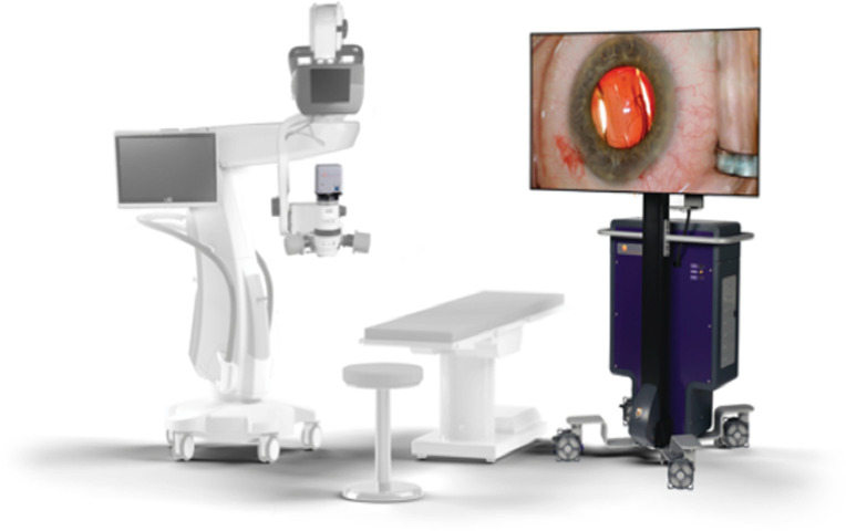
Purpose: We compared the visibility and surgeon posture between image-processing-assisted trabeculotomy (IP-LOT) using the NGENUITY® 3D visual system and conventional microsurgery (microscope-assisted trabeculotomy; MS-LOT).

Methods: IP-LOT was performed for five pig eyes. The visibility of the trabecular mesh work was evaluated on images of the trabecular mesh work and the posterior surface of the cornea (Cor) obtained under three different conditions. Images were then analyzed using ImageJ® to measure differences in luminance between the trabecular mesh work and Cor. IP-LOT was also performed for eleven human eyes, and the data were analyzed using the same approach as that used for the pig eyes. The length from the surgeon's abdomen to the operative eye (working distance) during MS-LOT and IP-LOT was measured for 12 different surgeons and compared to evaluate surgeon posture.

Results: Image processing significantly increased the difference in luminance between the trabecular mesh work and Cor in both pig and human eyes (p < 0.05). Moreover, the working distance in IP-LOT was significantly shorter than that in MS-LOT (p < 0.05).

Conclusion: Our findings suggest that the NGENUITY® 3D visual system provides better trabecular mesh work visibility than a normal microscope in conventional surgical methods, and it allows surgeons to operate without moving far from the operative eye.


Conflict of interest statement

no competing interests exist.

Figures

Fig 1
Fig 1. Pictures of the microscope and display monitor.
The operating microscope (left) with the image capture module (ICM) sends up to 3 GB image information per second to the embedded processing unit (EPU), and the sent image was projected onto the 3D display (right).
Fig 2
Fig 2. Images of the angle in pig eyes under various conditions and examples of measurements of luminance with ImageJ®.
(A) Trabecular meshwork (TM) area (without image processing). (B) Posterior corneal (Cor) area (without image processing). (C) TM area (Condition 1). (D) Cor area (Condition 1). (E) TM area (Condition 2). (F) Cor area (Condition 2). The imaging conditions for NGENUITY® are described as follows. Both brightness and contrast were changed for each condition. Without image processing: The brightness was 47.8 cd/m2, and the contrast was 54.9 arb. units. Condition 1: The brightness was reduced from 47.8 cd/m2 to 40.5 cd/m2, and the contrast was increased from 54.9 arb. units to 65.2 arb. units. Condition 2: The brightness was reduced from 47.8 cd/m2 to 40.3 cd/m2, and the contrast was increased from 54.9 arb. units to 73.8 arb. units. The 0.5 mm × 3.5 mm area of the angle was used as the analysis area in all images.
Fig 3
Fig 3. Images of the angle in human eyes under various conditions and examples of measurements of luminance with ImageJ®.
(A) Trabecular meshwork (TM) area (without image processing). (B) Posterior corneal (Cor) area (without image processing). (C) TM area (Condition 1’). (D) Cor area (Condition 1’). (E) TM area (Condition 2’). (F) Cor area (Condition 2’). The imaging conditions of NGENUITY® are described as follows. Only the contrast was changed for each condition. Without image processing’: The brightness was 47.8 cd/m2, and the contrast was 54.9 arb. units. Condition 1’: Brightness was kept at 47.8 cd/m2, and contrast was increased from 54.9 arb. units to 65.5 arb. units. Condition 2’: Brightness was kept at 47.8 cd/m2, and contrast was increased from 54.9 arb. units to 75.5 arb. units. The 0.5 mm × 3.5 mm area of the angle was used as the analysis area in all images.
Fig 4
Fig 4. Example of surgeon posture and measurement of working distance in microscope-assisted trabeculotomy (MS-LOT) and image-processing-assisted trabeculotomy (IP-LOT).
(A) Surgeon’s posture in MS-LOT. (B) Surgeon’s posture in IP-LOT. To evaluate the working distance, the distance from the operative eye to the surgeon’s abdomen was measured, as indicated by the length of the arrow above.
Fig 5
Fig 5. The average difference in luminance in pig and human eyes under each condition and in the working distances.
(A) Image-processing-assisted trabeculotomy (IP-LOT) for 5 pig eyes. There were significant differences between no processing and condition 1 and between no processing and condition 2 (n = 5, p < 0.05). (B) IP-LOT for 11 human eyes. There were significant differences between no processing’ and condition 1’ (n = 11, p < 0.05). However, there were no significant differences between no processing’ and condition 2’. (C) Working distance in microscope-assisted trabeculotomy (MS-LOT) and IP-LOT. The working distance in IP-LOT was significantly shorter than that in MS-LOT (n = 12, p < 0.05).

Similar articles

Cited by

References

    1. Coleman AL. Glaucoma. Lancet. 1999;354: 1803–1810. doi: 10.1016/S0140-6736(99)04240-3 - DOI - PubMed
    1. De Moraes CG, Liebmann JM, Levin LA. Detection and measurement of clinically meaningful visual field progression in clinical trials for glaucoma. Prog Retin Eye Res. 2017;56: 107–147. doi: 10.1016/j.preteyeres.2016.10.001 - DOI - PMC - PubMed
    1. Chauhan BC, Mikelberg FS, Balaszi AG, LeBlanc RP, Lesk MR, Trope GE, et al.. Canadian Glaucoma Study: 2. risk factors for the progression of open-angle glaucoma. Arch Ophthalmol. 2008;126: 1030–1036. doi: 10.1001/archopht.126.8.1030 - DOI - PubMed
    1. Ismail R, Azuara-Blanco A, Ramsay CR. Variation of clinical outcomes used in glaucoma randomised controlled trials: a systematic review. Br J Ophthalmol. 2014;98: 464–468. doi: 10.1136/bjophthalmol-2013-304323 - DOI - PubMed
    1. Comparison of glaucomatous progression between untreated patients with normal-tension glaucoma and patients with therapeutically reduced intraocular pressures. Collaborative Normal-Tension Glaucoma Study Group. Am J Ophthalmol. 1998;126: 487–497. doi: 10.1016/s0002-9394(98)00223-2 - DOI - PubMed

Publication types