. 2022 Apr 4;220(4):iyac024.
doi: 10.1093/genetics/iyac024.

ccd-5, a novel cdk-5 binding partner, regulates pioneer axon guidance in the ventral nerve cord of Caenorhabditis elegans

Affiliations

ccd-5, a novel cdk-5 binding partner, regulates pioneer axon guidance in the ventral nerve cord of Caenorhabditis elegans

Abigail H Feresten et al. Genetics. .

Abstract

During nervous system development, axons navigate complex environments to reach synaptic targets. Early extending axons must interact with guidance cues in the surrounding tissue, while later extending axons can interact directly with earlier "pioneering" axons, "following" their path. In Caenorhabditis elegans, the AVG neuron pioneers the right axon tract of the ventral nerve cord. We previously found that aex-3, a rab-3 guanine nucleotide exchange factor, is essential for AVG axon navigation in a nid-1 mutant background and that aex-3 might be involved in trafficking of UNC-5, a receptor for the guidance cue UNC-6/netrin. Here, we describe a new gene in this pathway: ccd-5, a putative cdk-5 binding partner. ccd-5 mutants exhibit increased navigation defects of AVG pioneer as well as interneuron and motor neuron follower axons in a nid-1 mutant background. We show that ccd-5 acts in a pathway with cdk-5, aex-3, and unc-5. Navigation defects of follower interneuron and motoneuron axons correlate with AVG pioneer axon defects. This suggests that ccd-5 mostly affects pioneer axon navigation and that follower axon defects are largely a secondary consequence of pioneer navigation defects. To determine the consequences for nervous system function, we assessed various behavioral and movement parameters. ccd-5 single mutants have no significant movement defects, and nid-1 ccd-5 double mutants are less responsive to mechanosensory stimuli compared with nid-1 single mutants. These surprisingly minor defects indicate either a high tolerance for axon guidance defects within the motor circuit and/or an ability to maintain synaptic connections among commonly misguided axons.

Keywords: axon guidance; axon navigation; cdk5; growth cone; nervous system development; pioneer; trafficking.


Figures

Fig. 1.
Fig. 1.
Schematic representation of relevant neurons and axons in the VNC. RIF (burgundy) axons pioneer the path from the retrovesicular ganglion (rvg) toward the nerve ring in the head. The AVG (green) axon pioneers the right VNC from the rvg, and the PVPR (orange) axon pioneers the left VNC from the posterior. Interneuron axons (red) from the nerve ring follow RIFR and RIFL into the rvg, then follow the AVG to continue posteriorly along the right VNC. Motoneurons (DA and DB in blue; DD and VD in purple) extend neurites anteriorly and posteriorly within the right VNC, and laterally toward the dorsal nerve cord (data not shown). A, anterior; P, posterior.
Fig. 2.
Fig. 2.
Exon–intron structure of the ccd-5 gene showing the location of the mutations. Exons are represented as boxes, introns are represented as connecting lines. Blue exons code for the highly conserved C2 calcium binding domain needed for membrane interaction. The 4 ccd-5 alleles discussed are shown: hd152 is a T to G point mutation at position 2570136 X, gk700962 is a G to A point mutation at 2570127 X. gk5256 is a deletion that removes most of exons 1–12, gk5976 is a deletion that removes most of exons 1–8 (see Materials and Methods section for details).
Fig. 3.
Fig. 3.
AVG axon guidance defects in nid-1 ccd-5 double mutants. a) Penetrance for each AVG phenotype in nid-1 ccd-5 (green) or nid-1 (gray). N > 100, **P < 0.005 (chi square test). b) Representative images of nid-1 ccd-5 mutants exhibiting various phenotypes. Arrows point to crossovers. Marker used: Pinx-18::GFP. Scale bar: 10 μm.
Fig. 4.
Fig. 4.
CI axon guidance defects are elevated in nid-1 ccd-5 double mutants compared with nid-1 single mutants. a) Penetrance for each phenotype in nid-1 ccd-5 (red) or nid-1 (gray). N > 100, **P < 0.005 (chi square test). Defects in ccd-5 single mutant and parental control line are <5% (data not shown). b) Images of representative nid-1 ccd-5 mutants. Arrows point to crossovers. “V” indicates location of vulva where present. Marker used: Pglr-1::mCherry. Scale bar: 20 μm.
Fig. 5.
Fig. 5.
DD/VD motoneuron axon guidance defects in ccd-5 mutants. a) Penetrance for each phenotype in ccd-5 (light purple), nid-1 ccd-5 (dark purple), nid-1 (gray), and parental marker strain (MS: white). N > 100, *P < 0.05, **P < 0.0001 (chi square test). B) Images of representative nid-1; ccd-5 mutants exhibiting quantified phenotypes (bottom). Arrows point to crossovers. Marker used: Punc-47::DsRed2. Scale bar 10 μm.
Fig. 6.
Fig. 6.
DA/DB motoneuron axon guidance defects in ccd-5 mutants. Penetrance for each phenotype in ccd-5 (light blue), nid-1 ccd-5 (dark blue), nid-1 (gray), and parental marker strain (MS: white). N > 100, *P<0.05, **P < 0.0001 (chi square test).
Fig. 7.
Fig. 7.
CI crossover defects correlated with AVG crossovers. AVG (green) and CI (red) in nid-1 ccd-5 mutants. a) Normally, AVG and CI axons fasciculate in the right VNC. b) Fasciculated CI cross with AVG. c) Defasciculated CI cross with AVG. d) Independent AVG crossover. e) Independent defasciculated CI crossover. Arrows mark crossover. Arrows point to crossovers. Location of vulva indicated with “V.” Markers used: Pglr-1::mCherry and Pinx-18::GFP. Scale bar: 20 μm.
Fig. 8.
Fig. 8.
DD/VD crossover defects correlated with AVG crossovers. AVG (green) and DD/VD (purple) in nid-1 ccd-5 mutants. a) Normally, AVG and DD/VD axons fasciculate in the right VNC. b–e) Examples of 4 different DD/VD crossover types assessed. Open arrows: DD/VD defasciculation. Arrows point to crossovers. Location of vulva indicated with “V.” Markers used: Punc-47::DsRed2 and Pinx-18::GFP. Scale bar: 20 μm.
Fig. 9.
Fig. 9.
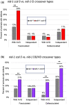
CI and DD/VD follow AVG across the midline. Graphs showing correlation between AVG and follower axon crossovers. Follower axon crossovers in nid-1 ccd-5 (color) and nid-1 (gray) are shown, differentiating between: crossovers coinciding with AVG crossovers (“with AVG”) vs separate crossovers (“independent”), and crossovers that result is a split of the follower axon fascicle (defasciculated) vs a single fascicle containing all follower axons (fasciculated). a) CI crossover penetrance in each category. b) DD/VD crossover penetrance in each category. **P < 0.005 (chi square test).
Fig. 10.
Fig. 10.
ccd-5 and cdk-5 genetically interact with unc-18, rab-3, and unc-5. AVG navigation defects in double and triple mutants. All double and triple mutants shown are significantly (P < 0.005, N > 100) more affected than nid-1 single mutants or parental marker strain (MS), and do not significantly differ from each other. None of the single mutants show significantly elevated AVG defects.
Fig. 11.
Fig. 11.
Naive motor response of ccd-5 mutants. Behavior of: marker strain (MS: light gray), nid-1 (dark gray), ccd-5 (light blue), nid-1 ccd-5 (dark blue), and lin-11 (red). a) Average forward movement speed. b) Proportion of animals engaged in forward locomotion at each timepoint, averaged across timepoints. Error bars mark upper limit of 95% confidence interval. *P < 0.05 when compared via ANOVA with the marker strain Pinx-18::GFP.
Fig. 12.
Fig. 12.
Response to mechanosensory stimuli in ccd-5 mutants. Behavior of: marker strain (MS: light gray), nid-1 (dark gray), ccd-5 (light blue), nid-1 ccd-5 (dark blue), and lin-11 (red). Plates received 5 consecutive taps in 10 s intervals. Data represent averages of tap response probability (a), average speed of reversal (b), and reversal duration (c). Error bars mark upper limit of 95% confidence interval. *P  < 0.05 when compared via ANOVA with the parental marker strain Pinx-18::GFP.
Fig. 13.
Fig. 13.
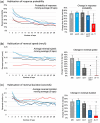
Habituation in ccd-5 mutants. Habituation of: marker strain (MS: light gray), nid-1 (dark gray), ccd-5 (light blue), nid-1; ccd-5 (dark blue), and lin-11 (red). Thirty taps were administered in 10 s intervals to induce habituation to mechanosensory stimuli. Shown here are probability of response (a), speed of reversal (b), reversal duration (c). Left: moving 3-tap average of responses from each tap. Right: bar graphs show the percent change from the first response to the averaged last 3 responses. Error bars mark upper limit of 95% confidence interval. Significance *P < 0.05 compared via ANOVA with parental marker strain Pinx-18::GFP.
Fig. 14.
Fig. 14.
Schematic of the genetic pathway regulating AVG navigation. The figure describes the observed genetic interactions from the current study and our previous study (Bhat and Hutter 2016) in the context of the known molecular functions of the proteins. Proteins from the current publication contributing to this larger picture are bolded.

References

    1. Au V, Li-Leger E, Raymant G, Flibotte S, Chen G, Martin K, Fernando L, Doell C, Rosell FI, Wang S, et al.CRISPR/Cas9 methodology for the generation of knockout deletions in Caenorhabditis elegans. G3 (Bethesda). 2019;9(1):135–144. doi:10.1534/g3.118.200778. - DOI - PMC - PubMed
    1. Bhaskar K, Shareef MM, Sharma VM, Shetty APK, Ramamohan Y, Pant HC, Raju TR, Shetty KT.. Co-purification and localization of Munc18-1 (p67) and Cdk5 with neuronal cytoskeletal proteins. Neurochem Int. 2004;44(1):35–44. doi:10.1016/S0197-0186(03)00099-8. - DOI - PubMed
    1. Bhat JM, Hutter H.. Pioneer axon navigation is controlled by AEX-3, a guanine nucleotide exchange factor for RAB-3 in Caenorhabditis elegans. Genetics. 2016;203(3):1235–1247. doi:10.1534/genetics.115.186064. - DOI - PMC - PubMed
    1. Brenner S. The Genetics of Caenorhabditis Elegans. Genetics. 1974;77(1):71–94. - PMC - PubMed
    1. Cao J, Packer JS, Ramani V, Cusanovich DA, Huynh C, Daza R, Qiu X, Lee C, Furlan SN, Steemers FJ, et al.Comprehensive single-cell transcriptional profiling of a multicellular organism. Science. 2017;357(6352):661–667. doi:10.1126/science.aam8940. - DOI - PMC - PubMed

Publication types

MeSH terms