. 2022 Mar 2;14(3):evac026.
doi: 10.1093/gbe/evac026.

A New Gene Family Diagnostic for Intracellular Biomineralization of Amorphous Ca Carbonates by Cyanobacteria

Affiliations

A New Gene Family Diagnostic for Intracellular Biomineralization of Amorphous Ca Carbonates by Cyanobacteria

Karim Benzerara et al. Genome Biol Evol. .

Abstract

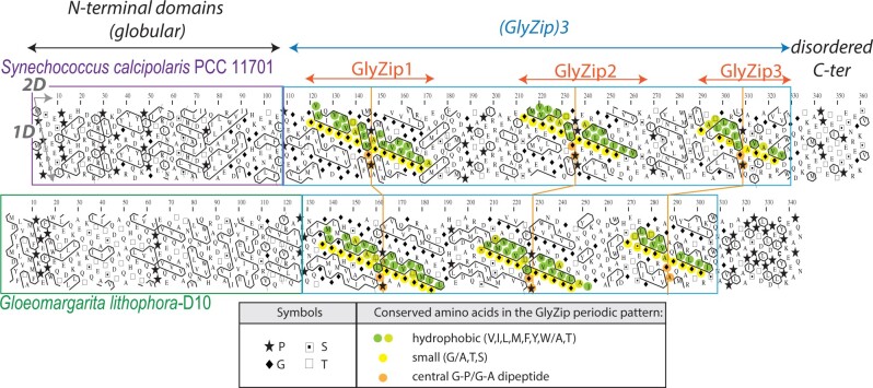
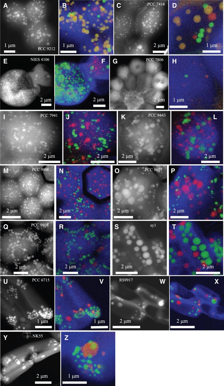
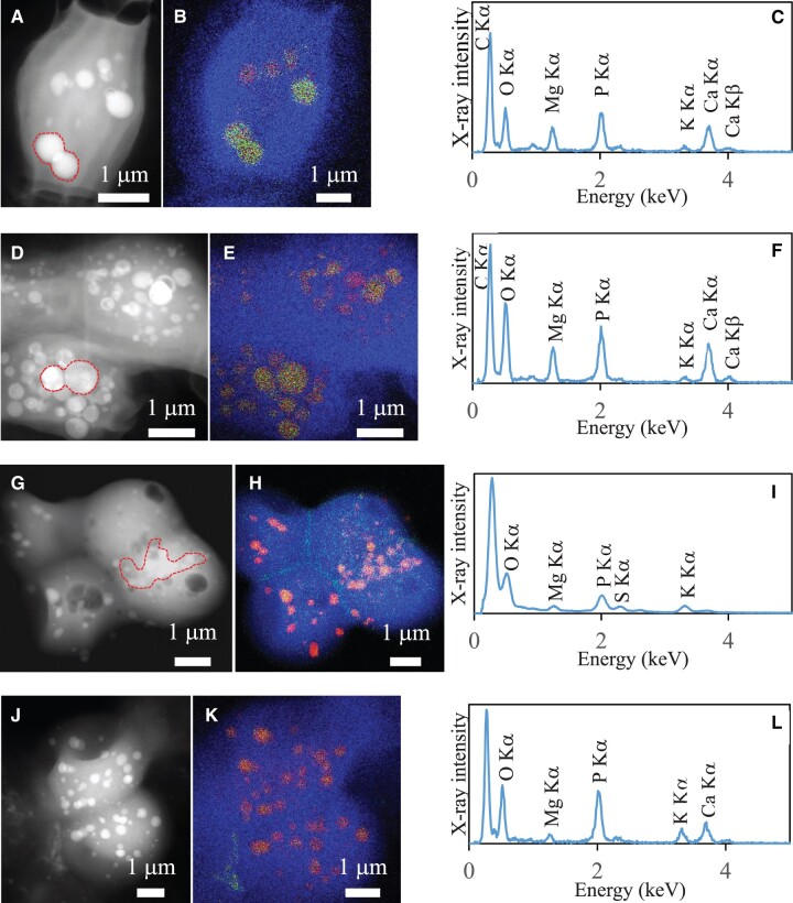
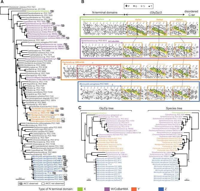
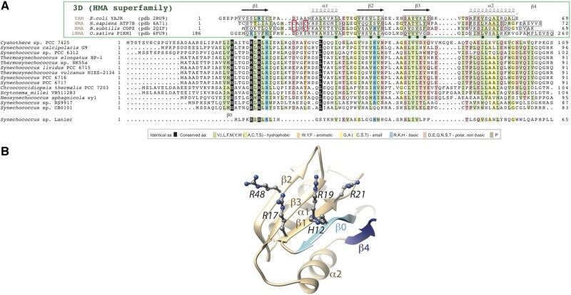
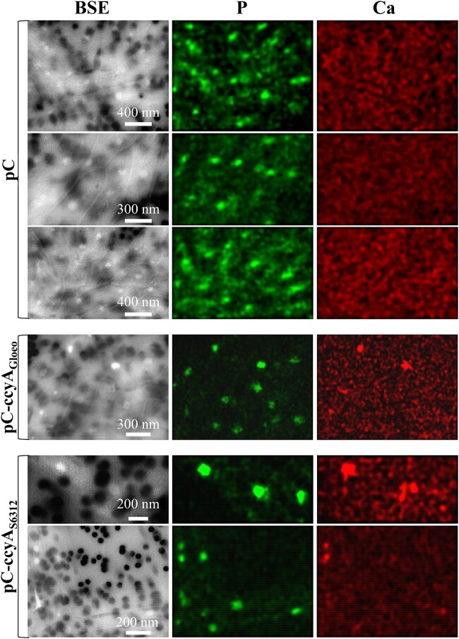
Cyanobacteria have massively contributed to carbonate deposition over the geological history. They are traditionally thought to biomineralize CaCO3 extracellularly as an indirect byproduct of photosynthesis. However, the recent discovery of freshwater cyanobacteria-forming intracellular amorphous calcium carbonates (iACC) challenges this view. Despite the geochemical interest of such a biomineralization process, its molecular mechanisms and evolutionary history remain elusive. Here, using comparative genomics, we identify a new gene (ccyA) and protein family (calcyanin) possibly associated with cyanobacterial iACC biomineralization. Proteins of the calcyanin family are composed of a conserved C-terminal domain, which likely adopts an original fold, and a variable N-terminal domain whose structure allows differentiating four major types among the 35 known calcyanin homologs. Calcyanin lacks detectable full-length homologs with known function. The overexpression of ccyA in iACC-lacking cyanobacteria resulted in an increased intracellular Ca content. Moreover, ccyA presence was correlated and/or colocalized with genes involved in Ca or HCO3- transport and homeostasis, supporting the hypothesis of a functional role of calcyanin in iACC biomineralization. Whatever its function, ccyA appears as diagnostic of intracellular calcification in cyanobacteria. By searching for ccyA in publicly available genomes, we identified 13 additional cyanobacterial strains forming iACC, as confirmed by microscopy. This extends our knowledge about the phylogenetic and environmental distribution of cyanobacterial iACC biomineralization, especially with the detection of multicellular genera as well as a marine species. Moreover, ccyA was probably present in ancient cyanobacteria, with independent losses in various lineages that resulted in a broad but patchy distribution across modern cyanobacteria.

Keywords: amorphous calcium carbonates; biomineralization; cyanobacteria; glycine zipper motifs, comparative genomics; phylogeny; protein structure prediction.


Figures

Fig. 1.
Fig. 1.
Domain architecture of calcyanins, as viewed by HCA. HCA plots of the calcyanin sequences of Synechococcus calcipolaris PCC 11701 and Gloeomargarita lithophora D10. The protein amino acid sequences (one-letter code) are displayed on a duplicated alpha-helical net, on which the strong hydrophobic amino acids (V, I, L, F, M, Y, and W) are contoured. The latter form clusters, which mainly correspond to the internal faces of regular secondary structures (α-helices and β-strands). The way to read the primary (1D) and secondary (2D) structures is shown with arrows (one amino acid or one hydrophobic cluster after another, respectively), whereas special symbols used for four amino acids with specific structural properties (P, G, S, and T) are described in the inset, together with the color code used to highlight conserved amino acids within the periodic patterns of the two calcyanin sequences. The two distinct CcyA folded domains (∼1/3 strong hydrophobic amino acids) are boxed.
Fig. 2.
Fig. 2.
EM detection of iACC in 13 calcyanin-bearing cyanobacterial strains not previously known to biomineralize carbonates. STEM-HAADF images of the 13 newly identified iACC-forming strains and overlays of C (blue), Ca (green), and P (red) chemical maps as obtained by EDXS. The name of the strains is provided on the STEM-HAADF image. Numbers in parenthesis correspond to replicate numbers of SEM-EDXS, STEM-EDXS, or both analyses. (A and B) Chlorogloeopsis fritschii PCC 9212 (13); (C and D) Fischerella muscicola PCC 7414 (4); (E and F) Fischerella sp. NIES-4106 (5); (G and H) Microcystis aeruginosa PCC 7806 (9); (I and J) M. aeruginosa PCC 7941 (7); (K and L) M. aeruginosa PCC 9443 (3); (M and N) M. aeruginosa PCC 9806 (4); (O and P) M. aeruginosa PCC 9807 (3); (Q and R) M. aeruginosa PCC 9808 (4); (S and T) Neosynechococcus sphagnicola sy1 (4); (U and V) Synechococcus lividus PCC 6715 (3); (W and X) Synechococcus sp. RS9917 (4); (Y and Z) Thermosynechococcus sp. NK55 (6).
Fig. 3.
Fig. 3.
TEM analyses of the four ccyA-harboring strains not forming iACC. Each row corresponds to one strain. The first column shows STEM-HAADF images. The second column shows overlays of C, Ca, and P EDXS maps. The third column shows EDXS spectra of inclusions detected in the cells. (A, B, and C) Fischerella sp. NIES-3754. EDXS spectrum is extracted from the area indicated in (A) by a dashed line; (D, E, and F) Chlorogloeopsis fritschii PCC 6912. (G, H, and I) Microcystis aeruginosa PCC 9432; (J, K, and L) M. aeruginosa PCC 9717.
Fig. 4.
Fig. 4.
Phylogenetic analysis and domain architecture of the calcyanin protein family. (A) Maximum-likelihood phylogenetic tree of Cyanobacteria based on 58 conserved proteins; the strains containing the ccyA gene are highlighted in bold and color. (B) HCA plots of representative calcyanin sequences (see fig. 1 for details of the HCA representation). The positions of the domains are indicated, with red boxes corresponding to the duplicated subdomain composing domain Y (labels a–e refer to equivalent hydrophobic clusters). The periodic patterns, made of glycine (or small amino acids—yellow) and hydrophobic amino acids (green) are highlighted for each GlyZip, with conserved signatures specific of each GlyZip shown with other colors. GlyZip2, which is present in only one species in the Y family, is indicated with a dotted box. (C) Unrooted maximum-likelihood phylogenetic tree of the GlyZip domain of calcyanin (left) compared with the species tree based on 58 conserved proteins (right). Numbers on branches indicate bootstrap support (BS, only values >50% are shown), BS of 100% are indicated by black circles. The species names and HCA profiles are color-coded according to the type of N-terminal domain of calcyanins (the code is shown at the bottom of the figure).
Fig. 5.
Fig. 5.
The CoBaHMA domain. (A) Multiple sequence alignment of calcyanins and members of the HMA superfamily with known 3D structures. Identical amino acids are shown in white on a black background, similarities are colored according to amino acid properties (inset). Sequences of proteins of the HMA superfamily, whose 3D structures are known and with which the CoBaHMA sequences can be aligned, are shown on top. PDB identifiers are provided. Observed 2D structures are boxed. The two cysteines of the CXXC motif specific of the HMA family are boxed in red. Green dots highlight the positions in which the hydrophobic character is strongly conserved, corresponding to amino acids participating in the hydrophobic core of the ferredoxin fold. An additional β-strand, named β0, is predicted in the CoBaHMA sequences, including a strictly conserved histidine. (B) Model of the CoBaHMA 3D structure, illustrated here with the Synechococcus sp. RS9917 sequence. The HMA common core is colored in beige, whereas specific secondary structures of the CoBaHMA family are in blue. The four highly conserved basic amino acids are shown with atomic details.
Fig. 6.
Fig. 6.
SEM analyses of mutants overexpressing ccyA. SEM-EDXS images (in BSE mode), P (green), and Ca (red) maps of Synechochoccus elongatus PCC 7942 mutants. The scale bar provided on the BSE images is the same for the corresponding P and Ca maps on each row. The 0.2 µm pores of the filters appear as dark disks in the BSE images. At the accelerating voltage used for these analyses, S. elongatus cells appear as relatively transparent, packed rods. Polyphosphate inclusions appear as brighter dots. The first three rows show cells of a S. elongatus PCC 7942 mutant harboring the empty pC plasmid. No Ca-rich inclusions are observed in these cells as shown by the homogeneous background in the Ca maps. In contrast, Ca-rich inclusions (polyphosphates) are observed in cells of S. elongatus PCC 7942 mutants harboring the plasmids pC-ccyAGloeo (fourth row) or pC-ccyAS6312 (fifth and sixth rows), appearing as hotspots in Ca maps. See supplementary data 3, Supplementary Material online for details concerning the plasmid and strains.

References

    1. Aizenberg J, Lambert G, Weiner S, Addadi L.. 2002. Factors involved in the formation of amorphous and crystalline calcium carbonate: a study of an ascidian skeleton. J Am Chem Soc. 124(1):32–39. - PubMed
    1. Altermann W, Kazmierczak J, Oren A, Wright DT.. 2006. Cyanobacterial calcification and its rock-building potential during 3.5 billion years of Earth history. Geobiology 4(3):147–166.
    1. Altschul SF, et al. 1997. Gapped BLAST and PSI-BLAST: a new generation of protein database search programs. Nucleic Acids Res. 25(17):3389–3402. - PMC - PubMed
    1. Benzerara K, et al. 2014. Intracellular Ca-carbonate biomineralization is widespread in cyanobacteria. Proc Natl Acad Sci U S A. 111(30):10933–10938. - PMC - PubMed
    1. Bitard-Feildel T, Lamiable A, Mornon J-P, Callebaut I.. 2018. Order in disorder as observed by the “hydrophobic cluster analysis” of protein sequences. Proteomics 18(21–22):e1800054. - PMC - PubMed

Publication types