. 2022 Feb 10;23(1):25.
doi: 10.1186/s12931-022-01944-8.

Pulmonary vascular inflammation with fatal coronavirus disease 2019 (COVID-19): possible role for the NLRP3 inflammasome

Affiliations

Pulmonary vascular inflammation with fatal coronavirus disease 2019 (COVID-19): possible role for the NLRP3 inflammasome

Oindrila Paul et al. Respir Res. .

Abstract

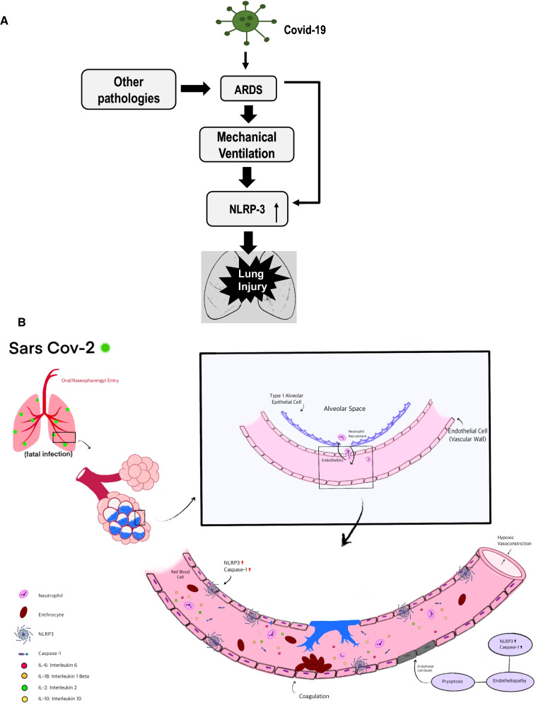
Background: Pulmonary hyperinflammation is a key event with SARS-CoV-2 infection. Acute respiratory distress syndrome (ARDS) that often accompanies COVID-19 appears to have worse outcomes than ARDS from other causes. To date, numerous lung histological studies in cases of COVID-19 have shown extensive inflammation and injury, but the extent to which these are a COVID-19 specific, or are an ARDS and/or mechanical ventilation (MV) related phenomenon is not clear. Furthermore, while lung hyperinflammation with ARDS (COVID-19 or from other causes) has been well studied, there is scarce documentation of vascular inflammation in COVID-19 lungs.

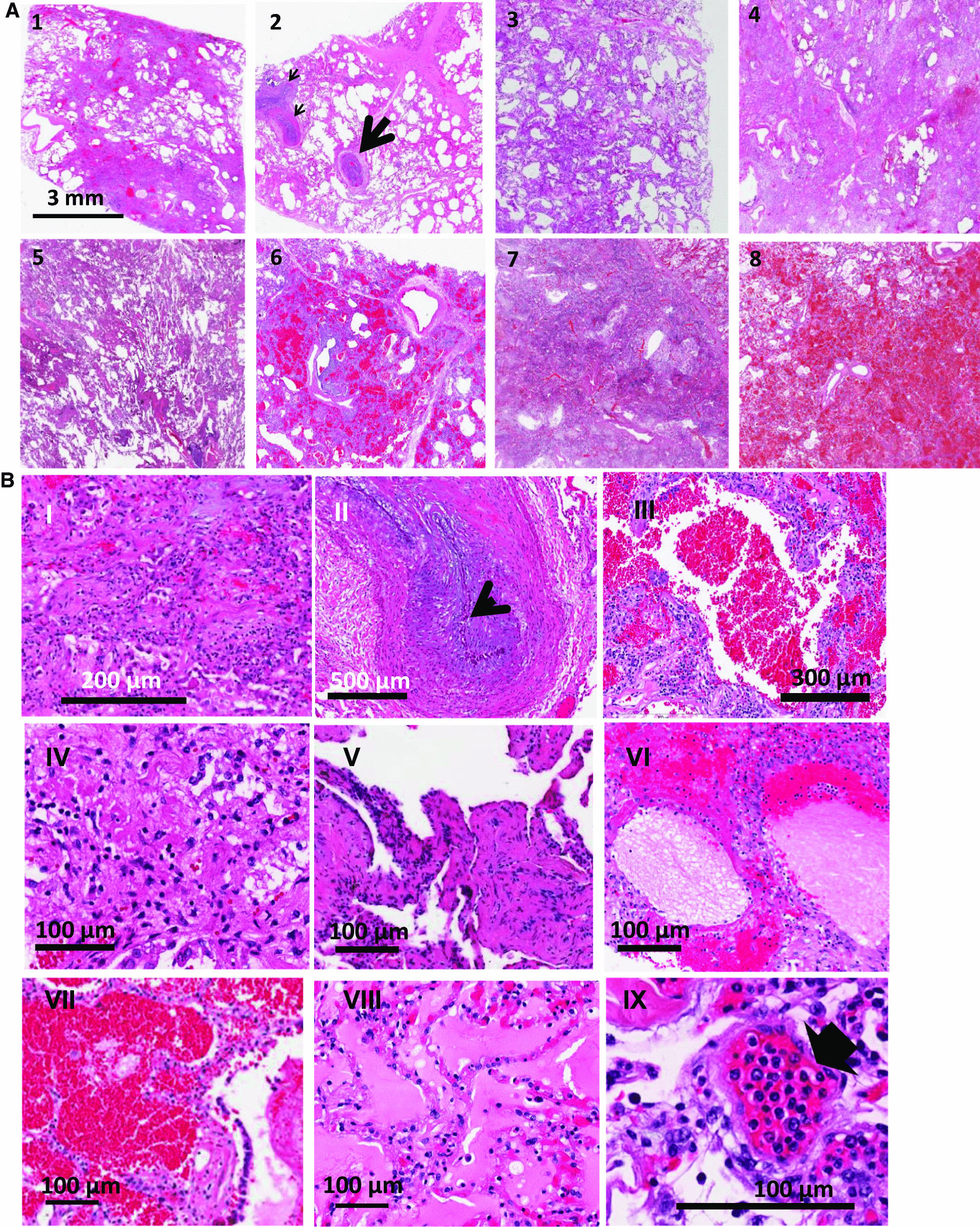
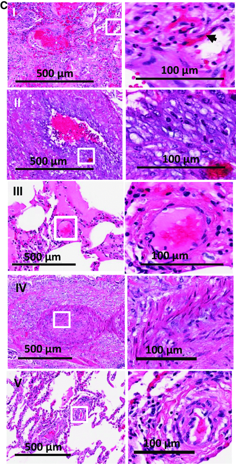
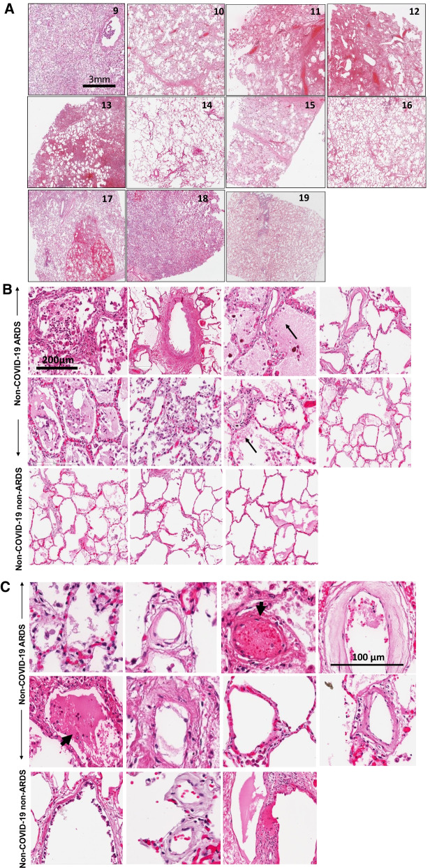
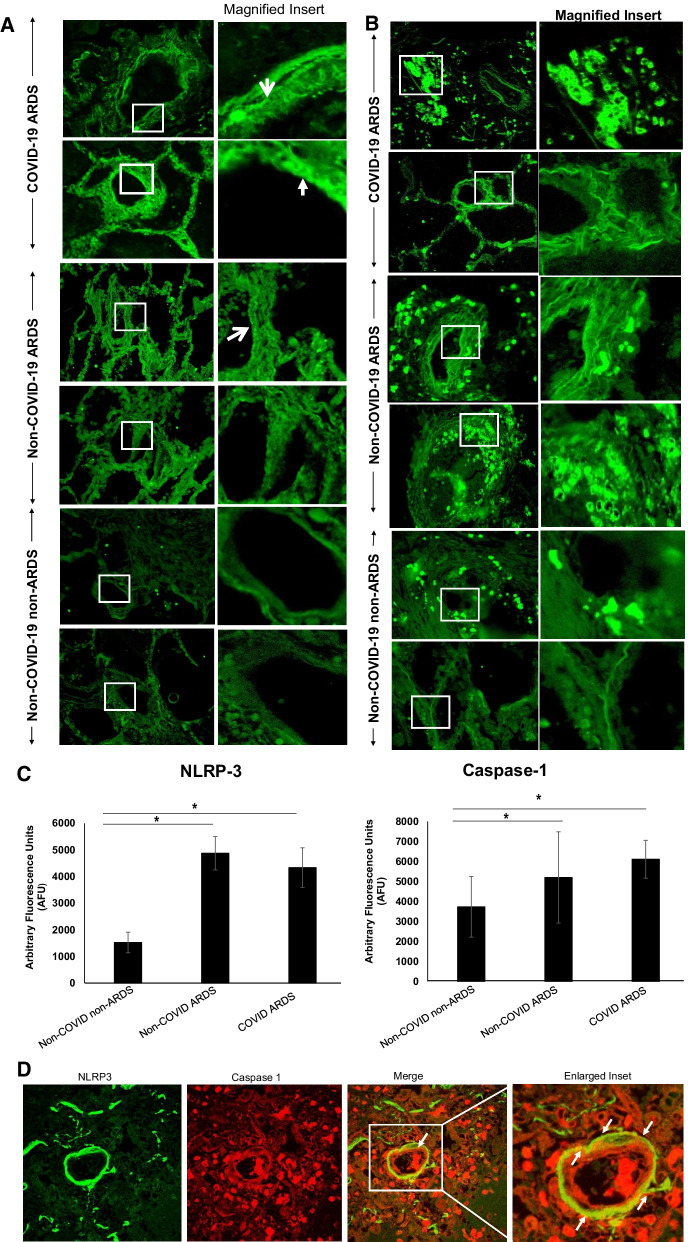
Methods: Lung sections from 8 COVID-19 affected and 11 non-COVID-19 subjects, of which 8 were acute respiratory disease syndrome (ARDS) affected (non-COVID-19 ARDS) and 3 were from subjects with non-respiratory diseases (non-COVID-19 non-ARDS) were H&E stained to ascertain histopathological features. Inflammation along the vessel wall was also monitored by expression of NLRP3 and caspase 1.

Results: In lungs from COVID-19 affected subjects, vascular changes in the form of microthrombi in small vessels, arterial thrombosis, and organization were extensive as compared to lungs from non-COVID-19 (i.e., non-COVID-19 ARDS and non-COVID-19 non-ARDS) affected subjects. The expression of NLRP3 pathway components was higher in lungs from COVID-19 ARDS subjects as compared to non-COVID-19 non-ARDS cases. No differences were observed between COVID-19 ARDS and non-COVID-19 ARDS lungs.

Conclusion: Vascular changes as well as NLRP3 inflammasome pathway activation were not different between COVID-19 and non-COVID-19 ARDS suggesting that these responses are not a COVID-19 specific phenomenon and are possibly more related to respiratory distress and associated strategies (such as MV) for treatment.

Keywords: COVID-19; Lung inflammation; Mechanical ventilation; Microthrombosis; NLRP3 inflammasome; Vascular endothelium.


Conflict of interest statement

The authors declare that they have no competing interests.

Figures

Fig. 1
Fig. 1
Hematoxylin and eosin-stained sections staining from representative regions of the lung parenchyma of post-mortem lung tissue of 8 COVID-19 patients (Patient 1 to 8). A All patients show extensive alteration of lung microstructure in the form of alveolar damage, fibrin exudation into alveolar space, thrombi and fibroblastic proliferation. The septa are thickened and there is presence of hyaline membranes and dense infiltrates. Scale bar is 3 mm. 1: patient 1- Alveolar damage with collagen deposition and exudative pattern of damage, 2. patient 2- Large thrombi and smaller caliber arteries showing fibrin thrombi (arrows), 3. patient 3-Alveolar damage pattern arising from fibroblastic proliferations, 4 and 5. patient 4 and 5- Exudate in the entire lung, 6. patient 6-Necrosis with blood and exudate in the lung parenchyma, 7. patient 7-Hemorrhagic infarction of lung tissue adjacent to a pulmonary artery with thrombotic material, 8. patient 8-Pulmonary hemorrhage with blood and fibrin exudation into the parenchyma. B H and E staining at higher magnification to show representative areas of extensive diffuse alveolar damage, microthrombi and edema in regions of the lungs from various COVID-19 ARDS samples. I. Fibroblastic proliferation, II. Plugged airway due to remodeling, III. Coagulation necrosis with blood in the lung tissue, IV. Proliferative phase of diffuse alveolar damage, V. Patchy distribution of damage, VI. Proteinaceous exudates in alveolar spaces, VII. Blood and fibrin exudation into parenchyma, VIII. Proteinaceous exudates in alveolar spaces, IX. Endotheliitis of small vessel < 100 μm with infiltration of the vessel wall by lymphocytes (arrow shows infiltrated cells). C Thrombi and microthrombi were identified in lung sections of 7 of the 8 patients. Images of vessels were chosen to emphasize the microthrombi. Box is magnified in the right panel. Arrow shows microthrombi on alveolar septa
Fig. 1
Fig. 1
Hematoxylin and eosin-stained sections staining from representative regions of the lung parenchyma of post-mortem lung tissue of 8 COVID-19 patients (Patient 1 to 8). A All patients show extensive alteration of lung microstructure in the form of alveolar damage, fibrin exudation into alveolar space, thrombi and fibroblastic proliferation. The septa are thickened and there is presence of hyaline membranes and dense infiltrates. Scale bar is 3 mm. 1: patient 1- Alveolar damage with collagen deposition and exudative pattern of damage, 2. patient 2- Large thrombi and smaller caliber arteries showing fibrin thrombi (arrows), 3. patient 3-Alveolar damage pattern arising from fibroblastic proliferations, 4 and 5. patient 4 and 5- Exudate in the entire lung, 6. patient 6-Necrosis with blood and exudate in the lung parenchyma, 7. patient 7-Hemorrhagic infarction of lung tissue adjacent to a pulmonary artery with thrombotic material, 8. patient 8-Pulmonary hemorrhage with blood and fibrin exudation into the parenchyma. B H and E staining at higher magnification to show representative areas of extensive diffuse alveolar damage, microthrombi and edema in regions of the lungs from various COVID-19 ARDS samples. I. Fibroblastic proliferation, II. Plugged airway due to remodeling, III. Coagulation necrosis with blood in the lung tissue, IV. Proliferative phase of diffuse alveolar damage, V. Patchy distribution of damage, VI. Proteinaceous exudates in alveolar spaces, VII. Blood and fibrin exudation into parenchyma, VIII. Proteinaceous exudates in alveolar spaces, IX. Endotheliitis of small vessel < 100 μm with infiltration of the vessel wall by lymphocytes (arrow shows infiltrated cells). C Thrombi and microthrombi were identified in lung sections of 7 of the 8 patients. Images of vessels were chosen to emphasize the microthrombi. Box is magnified in the right panel. Arrow shows microthrombi on alveolar septa
Fig. 2
Fig. 2
A. Hematoxylin and Eosin-stained sections staining from representative regions of the lung parenchyma of post-mortem lung tissue of 8 non-COVID-19 ARDS (patient numbers 10–15, 17,18) and 3 non-COVID-19 non-ARDS (patient numbers 9,16,19) patients. Scale bar is 3 mm. B H and E staining at higher magnification: Diffuse alveolar damage, microthrombi and edema were observed. Arrows show proteinaceous exudate in the airspaces. Scale bar is 200 microns. C Vascular structures in lungs from non-COVID-19 sources. Arrows show thrombi in vessels. About 40% of the fields from non-COVID ARDS showed thrombi. Very few microthrombi were observed in non-COVID non-ARDS lungs. Scale bar is 100 microns
Fig. 3
Fig. 3
Inflammasome in the lungs of patients with COVID-19 ARDS, non-COVID-19 ARDS and non-COVID-19 non-ARDS. Representative images of the immunofluorescence in lung sections stained with anti-NLRP3 and Caspase 1. A The NLRP3 subunit (green) along the walls of arterioles (arrow). Upper panels: COVID-19 ARDS lungs. Middle Panels: Lungs from non-COVID-19 ARDS subjects. Lower Panels: Lungs from non-COVID-19 non-ARDS subjects, without respiratory disease. B Caspase staining (green). Upper panels: Upper panels: COVID-19 ARDS lungs. Middle Panels: Lungs from non-COVID-19 ARDS subjects. Lower Panels: Lungs from non-COVID 19 non-ARDS subjects, without respiratory disease. C Quantitation of the fluorescence intensity of the images using MetaMorph Imaging Program. *p < 0.01 as compared to non-COVID non-ARDS lungs. Results are presented as mean ± standard deviation (SD). Group differences were evaluated by ANOVA followed by Tukey post hoc comparisons. Statistical significance was accepted as p < 0.05. D NLRP3 (green) and caspase 1 (red) in lung sections of patients with COVID-19. Arrows show colocalization (yellow) in regions along the vascular wall
Fig. 4
Fig. 4
A ARDS (and mechanical ventilation) associated with COVID-19 seems to be largely responsible for activation of the NLRP3 inflammasome. This pathway seems to be ARDS related and not COVID-19 (SARS CoV2 virus infection) specific. B Overview of SARS-CoV-2 entry, infection and endothelial inflammation and cell death. As is well established, oral nasopharyngeal entry of SARS-CoV-2 is followed by its binding to the alveolar epithelium. The infected pneumocytes secrete cytokine and chemokines, which attract neutrophils to the alveolar space, leading to a possible breach of the alveolar wall. Meanwhile, endothelial cells overexpress NLRP3 as we observed in the autopsies (either by infection, or via increased amounts of chemokines and cytokines). The NLRP3 pathway drives endothelial pyroptosis. This leads to breakdown of the endothelial-alveolar barrier and causes interstitial and alveolar space flooding. Endothelial cell death and debris activate coagulation cascades that promotes thrombi formation

Update of

References

    1. Xie J, Tong Z, Guan X, Du B, Qiu H. Clinical characteristics of patients who died of coronavirus disease 2019 in China. JAMA Netw Open. 2020;3:e205619. - PMC - PubMed
    1. Guan WJ, Ni ZY, Hu Y, Liang WH, Ou CQ, He JX, Liu L, Shan H, Lei CL, Hui DSC, et al. Clinical characteristics of coronavirus disease 2019 in China. N Engl J Med. 2020;382:1708–1720. - PMC - PubMed
    1. Fox SE, Akmatbekov A, Harbert JL, Li G, Quincy Brown J, Vander Heide RS. Pulmonary and cardiac pathology in African American patients with COVID-19: an autopsy series from New Orleans. Lancet Respir Med. 2020;8:681–686. - PMC - PubMed
    1. Jin Y, Yang H, Ji W, Wu W, Chen S, Zhang W, Duan G. Virology, epidemiology, pathogenesis, and control of COVID-19. Viruses. 2020;12:372. - PMC - PubMed
    1. Gustine JN, Jones D. Immunopathology of hyperinflammation in COVID-19. Am J Pathol. 2021;191:4–17. - PMC - PubMed

Substances