. 2022 Feb 10;13(1):817.
doi: 10.1038/s41467-022-28512-4.

Superior Colliculus to VTA pathway controls orienting response and influences social interaction in mice

Affiliations

Superior Colliculus to VTA pathway controls orienting response and influences social interaction in mice

Clément Solié et al. Nat Commun. .

Abstract

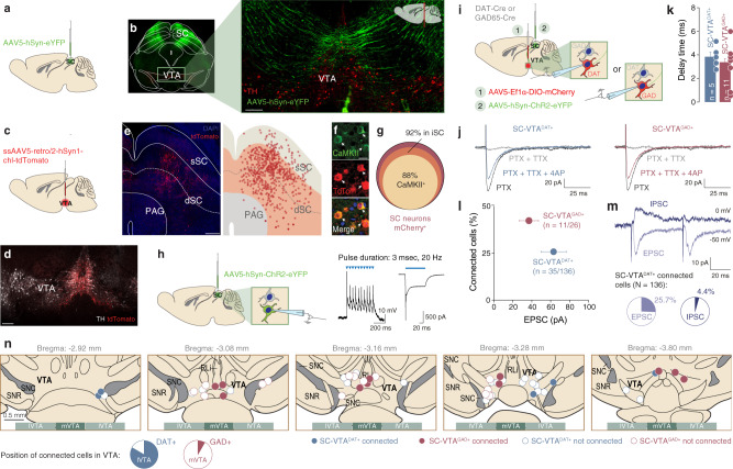
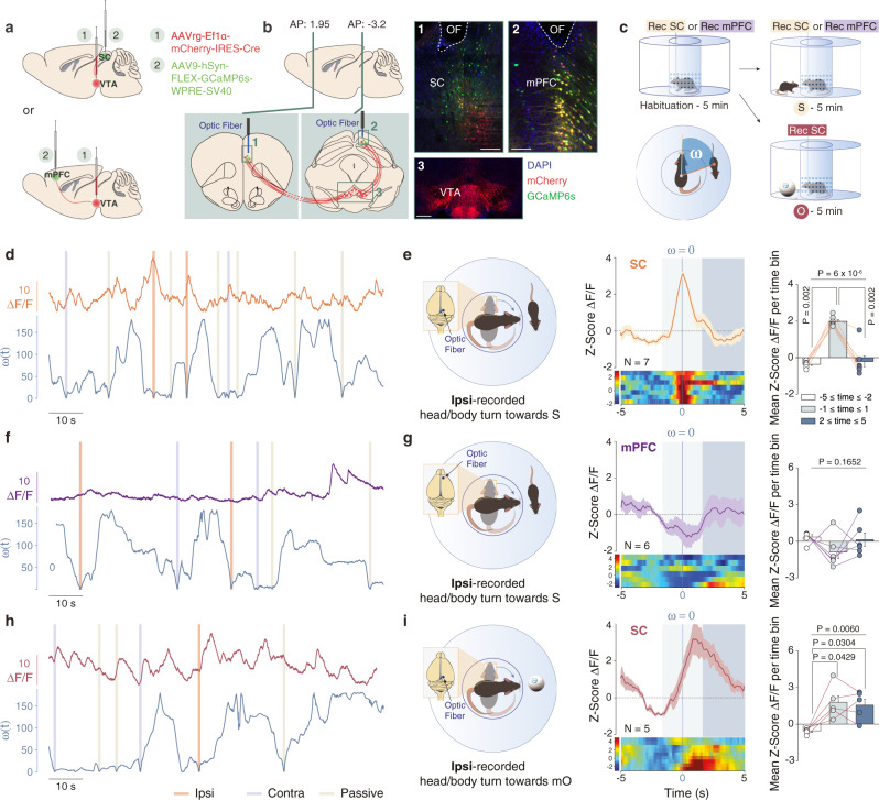
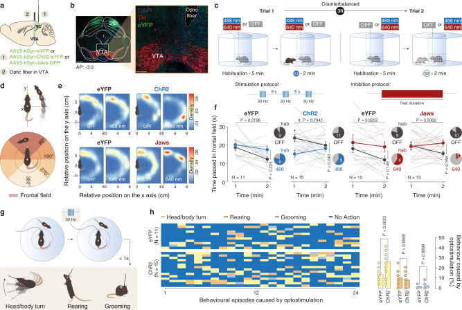
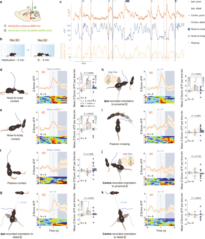
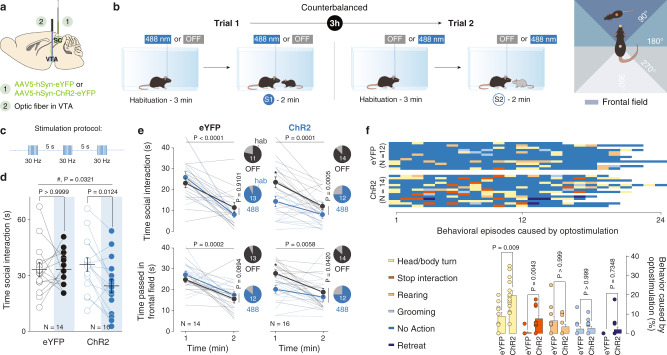
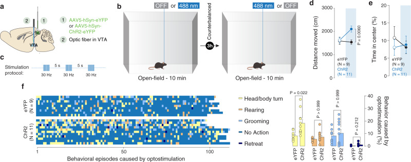
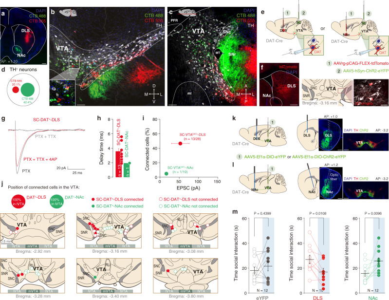
Social behaviours characterize cooperative, mutualistic, aggressive or parental interactions that occur among conspecifics. Although the Ventral Tegmental Area (VTA) has been identified as a key substrate for social behaviours, the input and output pathways dedicated to specific aspects of conspecific interaction remain understudied. Here, in male mice, we investigated the activity and function of two distinct VTA inputs from superior colliculus (SC-VTA) and medial prefrontal cortex (mPFC-VTA). We observed that SC-VTA neurons display social interaction anticipatory calcium activity, which correlates with orienting responses towards an unfamiliar conspecific. In contrast, mPFC-VTA neuron population activity increases after initiation of the social contact. While protracted phasic stimulation of SC-VTA pathway promotes head/body movements and decreases social interaction, inhibition of this pathway increases social interaction. Here, we found that SC afferents mainly target a subpopulation of dorsolateral striatum (DLS)-projecting VTA dopamine (DA) neurons (VTADA-DLS). While, VTADA-DLS pathway stimulation decreases social interaction, VTADA-Nucleus Accumbens stimulation promotes it. Altogether, these data support a model by which at least two largely anatomically distinct VTA sub-circuits oppositely control distinct aspects of social behaviour.


Conflict of interest statement

The authors declare no competing interests.

Figures

Fig. 1
Fig. 1. Anatomo-functional connectivity from SC to VTA dopaminergic and GABAergic neurons.
a Schema of injection in the Superior Colliculus (SC) with AAV5-hSyn-eYFP. b Right: Representative coronal image of immunostaining experiments against Tyrosine Hydroxylase (TH) enzyme (in red) performed on midbrain slices of adult mice infected with AAV5-hSyn-eYFP (green) in the SC. The projecting fibers from SC to VTA are visible. Left: Image at higher magnification of the coronal slice (scale bar: 100 µm). The fibers project from SC to VTA (this experiment was reproduced at least three times). c Schema of injection in the VTA with ssAAV5-retro/2-hSyn1-chl-tdTomato. d Representative coronal image of ssAAV5-retro/2-hSyn1-chl-tdTomato injection site in the VTA. Immunostaining labelled TH-positive neurons (scale bar: 200 µm. This experiment was reproduced at least three times). e Left: Representative image of the infected cells with ssAAV5-retro/2-hSyn1-chl-tdTomato in the SC (scale bar: 200 µm. This experiment was reproduced at least three times). Right: schema reporting the position of tdTomato positive cells in the superficial (sSC) and intermediate/deeper layers (dSC) of the SC and in the periaqueductal grey (PAG) for six infected brains. f Representative image of immunostaining against Ca2+/calmodulin-dependent protein kinase II (CAMKII) in the SC and infected cells with ssAAV5-retro/2-hSyn1-chl-tdTomato (scale bar: 10 µm. This experiment was reproduced at least three times). g Quantification of tdTomato infected cells in the SC for six infected brains. The cells are preferentially located in the intermediate layer of the SC and are predominantly CAMKII+. h Whole-cell patch-clamp from SC ChR2-expressing neurons. Protocol of stimulation indicates that SC neurons follow 20 Hz light stimulation protocol. i Schema of injection in the SC with AAV5-hSyn-ChR2-eYFP and patch of the VTA DAT+ and GAD+ neurons (depending on the mice line). j Example traces of optogenetically elicited excitatory postsynaptic currents (EPSCs) in VTA DAT+ and GAD+ neurons in presence of PTX, TTX, and 4AP. k Delay time of the EPSCs for VTA DAT+ and GAD+ neurons. l Quantification of connected cells from SC onto VTA DAT+ and GAD+ neurons in relation of the amplitude of EPSCs. m Upper panel: Example traces of optogenetically elicited EPSCs or inhibitory postsynaptic currents (IPSCs) in VTA DAT+ neurons. Lower panel: Percentage of evoked EPSCs or IPSCs in the VTA DAT+ neurons connected with the SC. n Position of some patched VTA DAT+ and GAD+ neurons. SC-VTADAT+ connected neurons are mainly in the lateral part of the VTA (lVTA) while SC-VTAGAD+ connected neurons are more medially located (mVTA). n indicates number of cells. All the data are shown as the mean ± s.e.m. as error bars. Source data are provided as a Source Data file.
Fig. 2
Fig. 2. Calcium activity of SC to VTA-projecting neurons during social and non-social orientation tests.
a Schema of injections of AAVrg-Ef1α-mCherry-IRES-Cre in the VTA and AAV9-hSyn-FLEX-GCaMP6s-WPRE-SV40 in the SC or mPFC. Representative coronal images of midbrain slices of adult mice infected with AAVrg-Ef1α-mCherry-IRES-Cre in the VTA (b panel 3, scale bar: 100 µm) and AAV9-hSyn-FLEX-GCaMP6s-WPRE-SV40 in the SC (b panel 1, scale bar: 50 µm) and in the mPFC (b panel 2, scale bar: 50 µm). The location of the optic fiber (OF) is indicated. Similar viral expression and OF location were observed in all the mice that performed the experiment described in Figs. 2c, 4b. c Schema of the social and non-social orientation test. Bottom left panel: Schema representing the points and vectors used for the calculation of the oriented angle towards the stimulus (ω). d, f, h Example traces. ΔF/F signals recorded in SC- or mPFC-VTA-projecting neurons aligned with instantaneous head orientation ω(t) during orientation test. Ipsi-recorded and contra-recorded head/body turn episodes are reported as well as the passive crossing events. e Left panel: Schema of the ipsi-recorded head/body turns towards the social stimulus (S). Middle panel: Peri-event time histogram (PETH) of normalized ΔF/F for SC-VTA-projecting neurons, centered on ipsi-recorded orientation towards social stimulus. Right panel: Mean ΔF/F (Z-score) before, during and after ipsi-recorded orientation. Repeated measures (RM) one-way ANOVA (Events main effect: F(2,6) = 44.34, P < 0.0001) followed by Bonferroni-Holm post-hoc test correction. g Left panel: Schema of the ipsi-recorded head/body turns towards the social stimulus (S). Middle panel: PETH of normalized ΔF/F for mPFC-VTA-projecting neurons, centered on ipsi-recorded orientation towards social stimulus. Right panel: Mean ΔF/F (Z-score) before, during and after ipsi-recorded orientation. RM one-way ANOVA (Events main effect: F(2,5) = 2.17, P = 0.1652). i Left panel: Schema of the ipsi-recorded head/body turns towards the moving object (mO). Middle panel: PETH of normalized ΔF/F for SC-VTA-projecting neurons, centered on ipsi-recorded orientation towards moving object. Right panel: Mean ΔF/F (Z-score) before, during and after ipsi-recorded orientation. RM one-way ANOVA (Events main effect: F(2,4) = 10.34, P = 0.0060) followed by Bonferroni-Holm post-hoc test correction. N indicates the number of mice. All the data are shown as the mean ± s.e.m. as error bars or error bands. Source data are provided as a Source Data file.
Fig. 3
Fig. 3. Optogenetic manipulation of SC-VTA pathway alters orienting response.
a Schema of injections sites in SC with AAV5-hSyn-eYFP, AAV5-hSyn-ChR2-eYFP, or AAV5-hSyn-Jaws-GFP, and optic fiber implantation above the VTA. b Representative image of coronal midbrain slices of adult mice infected with AAV5-hSyn-eYFP (green) in the SC. In red is visible the immunostaining anti-Tyrosine Hydroxylase (TH) (Left panel: scale bar = 500 µm. Right panel: image at higher magnification, scale bar = 100 µm). Similar viral expression and OF location were observed in all the mice that performed the experiment described in Figs. 3c, 5b. c Top panel: Schema of the social orientation test. The eYFP-, ChR2- and Jaws-expressing mice oriented towards two different unfamiliar mice under both stimulation conditions. Bottom left panel: Stimulation and inhibition protocols. 8 pulses of 488 nm light (30 Hz) were separated by 5 s in the light ON condition. Bottom right panel: Continuous inhibition was instead provoked with 640 nm light. d Upper panel: schema representing the relative position of the social stimulus when the center body point of the experimental animals is fixed at (0, 0) and the nose point is fixed along the y-axis. Lower panel: schema representing the position of the frontal field. e Heatmaps reporting the relative position of the social stimulus during orientation test for the 1st and 2nd minute in the different conditions. f Time passed with the social stimulus in the frontal field for the 1st and 2nd minute of the social orienting test in light and no-light conditions. RM two-way ANOVA (eYFP488 nm: Light main effect: F(1,10) = 1.683, P = 0.2236; Time main effect: F(1,10) = 7.711, P = 0.0196; Light × Time Interaction: F(1,10) = 1.254, P = 0.2890. ChR2: Light main effect: F(1,14) = 5.138, P = 0.0398; Time main effect: F(1,14) = 0.070, P = 0.7947; Light × Time Interaction: F(1,14) = 4.868, P = 0.0446. eYFP640 nm: Light main effect: F(1,9) = 2.537, P = 0.1456; Time main effect: F(1,9) = 7.185, P = 0.0252; Light × Time Interaction: F(1,9) = 0.6144, P = 0.4533. Jaws: Light main effect: F(1,12) = 2.266, P = 0.1581; Time main effect: F(1,12) = 0.4696, P = 0.5062; Light × Time Interaction: F(1,12) = 4.572, P = 0.0538) followed by Bonferroni’s multiple comparisons post-hoc test. Pie charts represent the percentage of mice that decrease the orientation between 1st and 2nd minute. g Possible behaviors caused by the optostimulation and observed less than 1 sec after a burst of light. h Left panel: identification and report of the behavioral episodes caused by optostimulation. Right panel: quantification of the behavioral episodes caused by optostimulation. Two-way ANOVA (Behavior main effect: F(1.874,43.09) = 60.95, P < 0.0001; Group main effect: F(1, 23) = 3.684, P = 0.0674; Behavior × Group Interaction: F(2, 46) = 11.29, P = 0.0001) followed by Bonferroni’s multiple comparisons post-hoc test. N indicates the number of mice. # indicates significantly different interaction. All the data are shown as the mean ± s.e.m. as error bars. Source data are provided as a Source Data file.
Fig. 4
Fig. 4. Calcium activity of SC and mPFC to VTA-projecting neurons during free social interaction.
a Schema of injections of AAVrg-Ef1α-mCherry-IRES-Cre in the VTA and AAV9-hSyn-FLEX-GCaMP6s-WPRE-SV40 in the SC. b Schema of free social interaction test. c Example traces. ΔF/F signals recorded in SC-VTA-projecting neurons aligned with instantaneous head orientation ω(t) and the distance between the nose of the experimental mouse and the gravity center of the stimulus mouse during the free social interaction test. Ipsi-recorded (proximal and distal) and contra-recorded (proximal and distal) head/body turn episodes are reported as well as the manual scoring of the nose-to-nose, nose-to-body and passive contacts. d Left: Schema of nose-to-nose contact. Middle: Peri-event time histogram (PETH) of normalized ΔF/F for SC-VTA-projecting neurons, centered on nose-to-nose contacts. Right: Mean ΔF/F (Z-score) before, during and after nose-to-nose. RM one-way ANOVA (Events main effect: F(2,7) = 14.15, P = 0.0004) followed by Bonferroni-Holm post-hoc test correction. e Left: Schema of nose-to-body contact. Middle: PETH of normalized ΔF/F for SC-VTA-projecting neurons, centered on nose-to-body contacts. Right: Mean ΔF/F (Z-score) before, during and after nose-to-body. RM one-way ANOVA (Events main effect: F(2,7) = 10.3, P = 0.0018) followed by Bonferroni-Holm post-hoc test correction. f Left: Schema of passive contact. Middle: PETH of normalized ΔF/F for SC-VTA-projecting neurons, centered on passive contacts. Right: Mean ΔF/F (Z-score) before, during and after passive contacts. RM one-way ANOVA (Events main effect: F(2,7) = 2.15, P = 0.1528). g Left: Schema of ipsilateral recorded orientation to distal social stimulus. Middle: Peri-event time histogram (PETH) of normalized ΔF/F for SC-VTA-projecting neurons, centered on distal ipsilateral orientation. Right: Mean ΔF/F (Z-score) before, during and after distal ipsilateral orientation. RM one-way ANOVA (Events main effect: F(2,7) = 3.8947, P = 0.0452) followed by Bonferroni-Holm post-hoc test correction. h Left: Schema of ipsilateral recorded orientation to proximal social stimulus. Middle: Peri-event time histogram (PETH) of normalized ΔF/F for SC-VTA-projecting neurons, centered on proximal ipsilateral orientation. Right: Mean ΔF/F (Z-score) before, during and after proximal ipsilateral orientation. RM one-way ANOVA (Events main effect: F(2,7) = 14.45, P = 0.0004) followed by Bonferroni-Holm post-hoc test correction. i Left: Schema of passive crossing of social stimulus in the frontal field. Middle: Peri-event time histogram (PETH) of normalized ΔF/F for SC-VTA-projecting neurons, centered on passive crossing. Right: Mean ΔF/F (Z-score) before, during and after passive crossing. RM one-way ANOVA (Events main effect: F(2,7) = 1.3413, P = 0.2931) followed by Bonferroni-Holm post-hoc test correction. j Left: Schema of contralateral recorded orientation to proximal social stimulus. Middle: Peri-event time histogram (PETH) of normalized ΔF/F for SC-VTA-projecting neurons, centered on proximal contralateral orientation. Right: Mean ΔF/F (Z-score) before, during and after proximal contralateral orientation. RM one-way ANOVA (Events main effect: F(2,7) = 0.3474, P = 0.7125) followed by Bonferroni-Holm post-hoc test correction. k Left: Schema of contralateral recorded orientation to distal social stimulus. Middle: Peri-event time histogram (PETH) of normalized ΔF/F for SC-VTA-projecting neurons, centered on distal contralateral orientation. Right: Mean ΔF/F (Z-score) before, during and after distal contralateral orientation. RM one-way ANOVA (Events main effect: F(2,7) = 0.1090, P = 0.8975) followed by Bonferroni-Holm post-hoc test correction. N indicates the number of mice. All the data are shown as the mean ± s.e.m. as error bars or error bands. Source data are provided as a Source Data file.
Fig. 5
Fig. 5. Optogenetic manipulation of SC-VTA pathway alters social interaction and perturbs head orientation towards conspecific.
a Schema of injections sites in SC with AAV5-hSyn-eYFP or AAV5-hSyn-ChR2-eYFP, and optic fiber implantation above the VTA. b Left panel: Schema of free social interaction. The eYFP- and ChR2-expressing mice freely interacted with two different unfamiliar mice under both stimulation conditions. Right panel: schema representing the position of the frontal field. c Stimulation protocols: 8 pulses of 488 nm light (30 Hz) were separated by 5 s in the light ON condition. d Time social interaction during the free social interaction test for eYFP- and ChR2-expressing mice in the SC. RM two-way ANOVA (Light main effect: F(1,28) = 5.0855, P = 0.0321; Virus main effect: F(1,28) = 0.8528, P = 0.3637; Light × Virus Interaction: F(1,28) = 4.1962, P = 0.0500) followed by Bonferroni-Holm post-hoc test correction. e Upper panels: time passed interacting with the social stimulus for the 1st and 2nd minute of the free social interaction test in light and no-light conditions. RM two-way ANOVA (eYFP: Light main effect: F(1,13) = 0.013, P = 0.9101; Time main effect: F(1,13) = 34.64, P < 0.0001; Light × Time Interaction: F(1,13) = 3.487, P = 0.085. ChR2: Light main effect: F(1,15) = 19.43, P = 0.0005; Time main effect: F(1,15) = 26.38, P = 0.0001; Light × Time Interaction: F(1,15) = 1.878, P = 0.1907) followed by Bonferroni’s multiple comparisons post-hoc test. Lower panels: time passed with the social stimulus in the frontal field for the 1st and 2nd minute of the free social interaction test in light and no-light conditions. RM two-way ANOVA (eYFP: Light main effect: F(1,13) = 3.368, P = 0.0894; Time main effect: F(1,13) = 25.31, P = 0.0002; Light × Time Interaction: F(1,13) = 0.01355, P = 0.9091. ChR2: Light main effect: F(1,15) = 5.006, P = 0.042; Time main effect: F(1,15) = 10.59, P = 0.0058; Light × Time Interaction: F(1,15) = 2.749, P = 0.1196) followed by Bonferroni’s multiple comparisons post-hoc test. Pie charts represent the percentage of mouse that decreases the interaction/orientation between 1st and 2nd minute. f Upper panel: identification and report of the behavioral episodes caused by a burst of optostimulation. Lower panel: quantification of the behavioral episodes caused by a burst of optostimulation. Two-way ANOVA (Behavior main effect: F(2.791, 66.99) = 23.43, P < 0.0001; Group main effect: F(1, 24) = 12.73, P = 0.0016; Behavior × Group Interaction: F(4, 46) = 6.539, P = 0.0001) followed by Bonferroni’s multiple comparisons post-hoc test. N indicates the number of mice. # indicates significantly different interaction. All the data are shown as the mean ± s.e.m. as error bars. Source data are provided as a Source Data file.
Fig. 6
Fig. 6. SC-VTA optostimulation increases exploratory behavior.
a Schema of injections sites in SC with AAV5-hSyn-eYFP or AAV5-hSyn-ChR2-eYFP, and optic fiber implantation above the VTA. b Schema of the open-field arena. The mice are free to explore the apparatus for 10 min in both stimulations’ conditions. c Stimulation protocols: 8 pulses of 488 nm light (30 Hz) were separated by 5 s in the light ON condition. d Distance moved in the open-field arena for eYFP and ChR2 mice under light and no-light stimulation. RM two-way ANOVA (Light main effect: F(1,18) = 4.0046, P = 0.0607; Virus main effect: F(1,18) = 4.8210, P = 0.0415; Light × Virus interaction: F(1,18) = 4.8217, P = 0.0415) followed by Bonferroni-Holm post-hoc test correction. e Time passed in the center of the open-field arena for eYFP and ChR2 mice under light and no-light stimulation. RM two-way ANOVA (Light main effect: F(1,18) = 0.1628, P = 0.6913; Virus main effect: F(1,18) = 0.145, P = 0.7078; Light × Virus interaction: F(1,18) = 0.546, P = 0.4695). f Right panel: identification and report of the behavioral episodes caused by a burst of optostimulation. Left panel: quantification of the behavioral episodes caused by a burst of optostimulation. Two-way ANOVA (Behavior main effect: F(2.004, 36.08) = 20.47, P < 0.0001; Group main effect: F(1, 18) = 5.370, P = 0.0325; Behavior × Group Interaction: F(3, 54) = 3.364, P = 0.0251) followed by Bonferroni’s multiple comparisons post-hoc test. N indicates the number of mice. # indicates significantly different interaction. All the data are shown as the mean ± s.e.m. as error bars. Source data are provided as a Source Data file.
Fig. 7
Fig. 7. SC is mainly connected to VTA DAT+ neurons which project to DLS.
a Representative image of the NAc and DLS in coronal slice infected with the CTB-488 and CTB-555 (scale bar  500 µm. This experiment was reproduced at least three times). b Coronal plane image of the VTA with TH staining (white) and infected cells projecting to the NAc or DLS (scale bar = 100 µm. This experiment was reproduced at least three times). c Horizontal plane image of the VTA with TH staining (white) and infected cells projecting to the NAc or DLS (scale bar = 100 µm. This experiment was reproduced at least three times). d Proportion of TH+ and either TH+/CTB-488+ (NAc-projecting VTA DAT+ neurons) or TH+/CTB-555+ (DLS-projecting VTA DAT+ neurons). e Schema of injections in DAT-Cre mice. The SC was infected with AAV5-hSyn-ChR2-eYFP and the NAc or the DLS with the AAVrg-pCAG-FLEX-tdTomato. Whole-cell patch was then performed in DAT+ neurons projecting to two different regions. f Left panel: Representative image of the AAVrg-pCAG-FLEX-tdTomato site of injection in the DLS (scale bar = 500 µm). Right panel: example of DAT+ neurons projecting to DLS (scale bar = 20 µm). This experiment was reproduced at least three times. g Example traces of optogenetically elicited excitatory postsynaptic currents (EPSCs) in VTA DAT+ neurons projecting to DLS in presence of PTX, TTX, and 4AP. h Delay time of the EPSCs for VTA DAT+-NAc or DAT+-DLS neurons. i Quantification of connected cells from SC onto VTA DAT+-NAc or DAT+-DLS neurons in relation of the amplitude of EPSCs. The VTA DAT neurons receiving projections from the SC are mainly projecting to the DLS with the highest current amplitude. j Position of some patched VTA DAT+-NAc or DAT+-DLS neurons. SC-VTADAT+-DLS connected neurons are mainly in the lateral part of the VTA (lVTA). k Left panel: Schema of injections sites in the VTA of DAT-Cre mice with AAV5-Ef1α-DIO-eYFP or AAV5-Ef1α-DIO-ChR2-eYFP, and optic fiber implantation above the DLS. Middle panel: representative image showing the fiber optic’s track in the DLS (scale bar = 500 µm). Right panel: representative image of the site of injection in the VTA (scale bar = 500 µm). Similar viral expression and OF location were observed in all the mice that performed the experiment which results are shown in Fig. 7l. l Left panel: Schema of injections sites in the VTA of DAT-Cre mice with AAV5-Ef1α-DIO-ChR2-eYFP or AAV5-Ef1α-DIO-eYFP, and optic fiber implantation above the NAc. Middle panel: representative image showing the fiber optic’s track in the NAc (scale bar = 500 µm). Right panel: representative image of the site of injection in the VTA (scale bar = 500 µm). Similar viral expression and OF location were observed in all the mice that performed the experiment which results are shown in Fig. 7l. m Left panel: VTA DAT+-NAceYFP and VTA DAT+-DLSeYFP mice do not change time of social interaction between the two stimulation conditions. Paired t-test two-sided (t(11) = −0.8013). Middle panel: VTA DAT+-DLSChR2 mice decrease the time of social interaction during light ON condition. Paired t-test two-sided (t(11) = 3.2015). Right panel: VTA DAT+-NAcChR2 mice increase the time of social interaction during light ON condition. Paired t-test two-sided (t(11) = −2.8177). N, n indicate number of mice and cells respectively. All the data are shown as the mean ± s.e.m. as error bars. Source data are provided as a Source Data file.

Similar articles

Cited by

References

    1. Chen P, Hong W. Neural circuit mechanisms of social behavior. Neuron. 2018;98:16–30. doi: 10.1016/j.neuron.2018.02.026. - DOI - PMC - PubMed
    1. Robinson DL, Heien MLAV, Wightman RM. Frequency of dopamine concentration transients increases in dorsal and ventral striatum of male rats during introduction of conspecifics. J. Neurosci. 2002;22:10477–10486. doi: 10.1523/JNEUROSCI.22-23-10477.2002. - DOI - PMC - PubMed
    1. Robinson DL, Zitzman DL, Smith KJ, Spear LP. Fast dopamine release events in the nucleus accumbens of early adolescent rats. Neuroscience. 2011;176:296–307. doi: 10.1016/j.neuroscience.2010.12.016. - DOI - PMC - PubMed
    1. Gunaydin LA, et al. Natural neural projection dynamics underlying social behavior. Cell. 2014;157:1535–1551. doi: 10.1016/j.cell.2014.05.017. - DOI - PMC - PubMed
    1. Bariselli S, et al. Role of VTA dopamine neurons and neuroligin 3 in sociability traits related to nonfamiliar conspecific interaction. Nat. Commun. 2018;9:3173. doi: 10.1038/s41467-018-05382-3. - DOI - PMC - PubMed

Publication types