

Central obesity is selectively associated with cerebral gray matter atrophy in 15,634 subjects in the UK Biobank
- PMID: 35145215
- PMCID: PMC9050590
- DOI: 10.1038/s41366-021-00992-2
Central obesity is selectively associated with cerebral gray matter atrophy in 15,634 subjects in the UK Biobank
Abstract
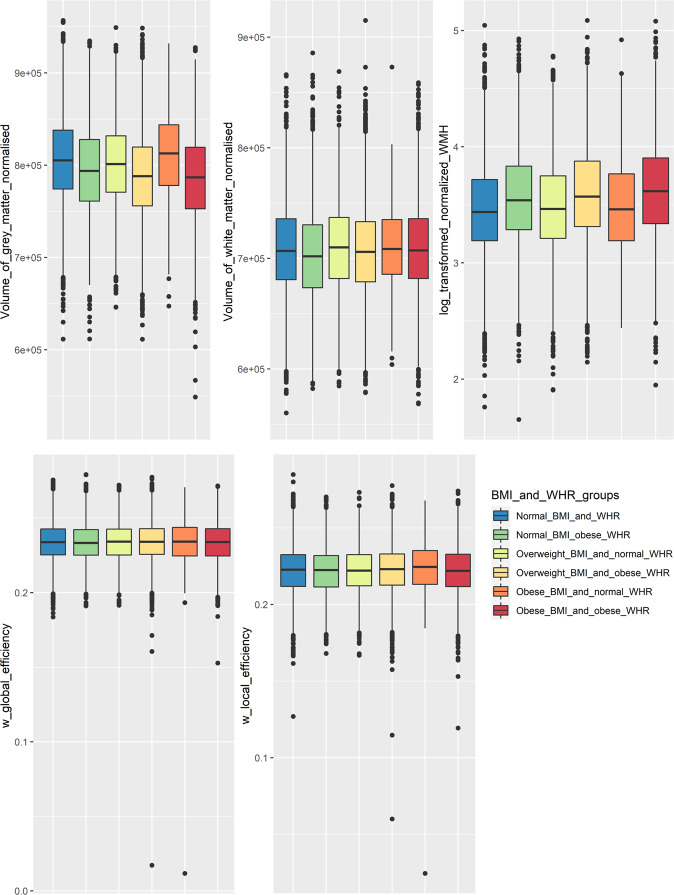
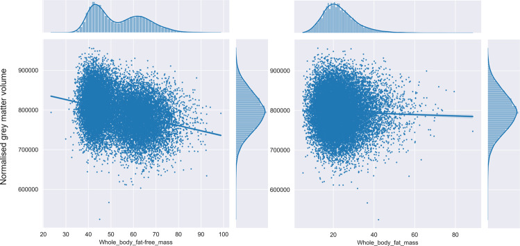
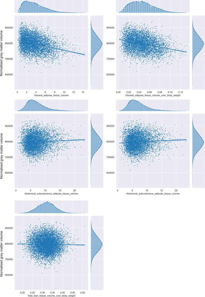
Background: Obesity is a risk factor for both cardiovascular disease and dementia, but the mechanisms underlying this association are not fully understood. We examined associations between obesity, including estimates of central obesity using different modalities, with brain gray matter (GM) volume in the UK Biobank, a large population-based cohort study.
Methods: To determine relationships between obesity and the brain we used brain MRI, abdominal MRI, dual-energy X-ray absorptiometry (DXA), and bioelectric whole-body impedance. We determined whether obesity was associated with any change in brain gray matter (GM) and white matter (WM) volumes, and brain network efficiency derived from the structural connectome (wiring of the brain) as determined from diffusion-tensor MRI tractography. Using Waist-Hip Ratio (WHR), abdominal MRI and DXA we determined whether any associations were primarily with central rather than peripheral obesity, and whether associations were mediated by known cardiovascular risk factors. We analyzed brain MRI data from 15,634.
Results: We found that central obesity, was associated with decreased GM volume (anthropometric data: p = 6.7 × 10⁻¹⁶, DXA: p = 8.3 × 10-81, abdominal MRI: p = 0.0006). Regional associations were found between central obesity and with specific GM subcortical nuclei (thalamus, caudate, pallidum, nucleus accumbens). In contrast, no associations were found with WM volume or structure, or brain network efficiency. The effects of central obesity on GM volume were not mediated by C-reactive protein or blood pressure, glucose, lipids.
Conclusions: Central body-fat distribution rather than the overall body-fat percentage is associated with gray matter changes in people with obesity. Further work is required to identify the factors that mediate the association between central obesity and GM atrophy.
© 2022. The Author(s).
Conflict of interest statement
The authors declare no competing interests.
Figures
References
Publication types
MeSH terms
Grants and funding
LinkOut - more resources
Full Text Sources
Research Materials
