. 2022 Feb;24(2):268-278.
doi: 10.1038/s41556-021-00836-1. Epub 2022 Feb 10.

dCas9-based gene editing for cleavage-free genomic knock-in of long sequences

Affiliations

dCas9-based gene editing for cleavage-free genomic knock-in of long sequences

Chengkun Wang et al. Nat Cell Biol. 2022 Feb.

Abstract

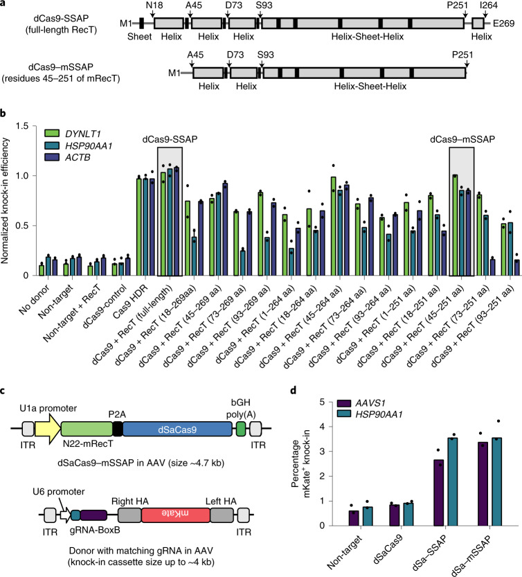
Gene editing is a powerful tool for genome and cell engineering. Exemplified by CRISPR-Cas, gene editing could cause DNA damage and trigger DNA repair processes that are often error-prone. Such unwanted mutations and safety concerns can be exacerbated when altering long sequences. Here we couple microbial single-strand annealing proteins (SSAPs) with catalytically inactive dCas9 for gene editing. This cleavage-free gene editor, dCas9-SSAP, promotes the knock-in of long sequences in mammalian cells. The dCas9-SSAP editor has low on-target errors and minimal off-target effects, showing higher accuracy than canonical Cas9 methods. It is effective for inserting kilobase-scale sequences, with an efficiency of up to approximately 20% and robust performance across donor designs and cell types, including human stem cells. We show that dCas9-SSAP is less sensitive to inhibition of DNA repair enzymes than Cas9 references. We further performed truncation and aptamer engineering to minimize its size to fit into a single adeno-associated-virus vector for future application. Together, this tool opens opportunities towards safer long-sequence genome engineering.


Conflict of interest statement

Stanford University has filed patent applications with L.C., C.W. and Y.Q. as inventors based on this work.

Figures

Fig. 1
Fig. 1. Development of a cleavage-free dCas9-based gene editor using microbial SSAPs.
a, Schematic model of the dCas9–SSAP editor. b, Design of the genomic knock-in assay to measure the level of gene-editing efficiency. FL, fluorescent; PAM, protospacer adjacent motif. c, Construct designs for screening the gene-editing efficiency of SSAPs using a genomic knock-in assay with an 800-bp 2A–mKate transgene. NLS, nuclear localization sequence. d, Knock-in efficiency of the initial screen of three SSAPs: Bet protein from lambda phage (LBet), RecT protein from Rac prophage (RecT) and gp2.5 from T7 phage (gp2.5). NTC, non-target control. Donor templates with HA lengths of approximately 200 bp (DYNLT1) and 300 bp (HSP90AA1 and ACTB) were added in all groups, except the no-donor controls. The error bars represent the s.e.m. of n = 3 biologically independent experiments. e, Imaging to verify mKate knock-in at endogenous genome loci using the dCas9–SSAP editor. Data represent n = 3 biologically independent experiments. dsDNA, double-stranded DNA. Source data
Fig. 2
Fig. 2. Measurement of the on- and off-target editing errors of dCas9–SSAP.
a, On-target indel errors (800-kp knock-in). Deep sequencing was used to measure the levels of indel formation when using dCas9–SSAP and Cas9 references at the endogenous targets DYNLT1 (left) and HSP90AA1 (right). HDR templates with a 200-bp HA were used as the donor template. Details of the assay are provided in Methods; n = 3 biologically independent experiments. b, On-target knock-in errors (800 kp knock-in). Clonal Sanger sequencing analysis of the accuracy of knock-in editing using dCas9–SSAP and Cas9 references with different MMEJ and HDR templates. The MMEJ and HDR donor templates had HAs of 25 bp and approximately 200 bp, respectively (Methods). ce, Genome-wide detection of insertion sites of the knock-in cassette using unbiased sequencing. The workflow (c), representative reads aligned at the knock-in genomic site (d) and summary of the detected on-target and off-target insertion sites (e) are presented. f,g, Workflow (f) and results (g) of the measurement of the cell-fitness effect, defined by the percentage of live cells after editing (normalized to the mock controls). Statistical analyses and comparisons were performed using a Student’s t-test; *P < 0.05; ***P < 0.001; n = 5 biologically independent experiments. MESL, maximum edit site likelihood. Asterisks next to gene names indicate that the insertion site is within the transcription unit of the gene. a,g, The error bars represent s.e.m. h, Summary of the knock-in accuracy of the dCas9–SSAP editor in comparison with the Cas9 HDR and Cas9 MMEJ methods. Accuracy is defined as the overall yield (%) of correct knock-in in all edited outcomes (correct knock-in, knock-in with indels and NHEJ indels). NGS, next-generation sequencing. Source data
Fig. 3
Fig. 3. Validation and benchmarking of dCas9–SSAP across donor designs and cell types.
a, Comparison of the knock-in efficiencies of dCas9–SSAP and other alternative Cas9, nCas9 and HDR-enhancing tools. Cas9-HE, CtIP-fusion Cas9; Cas9-Gem, Geminin-fusion Cas9; nCas9, Cas9-D10A nickase reference; and nCas9-hRAD51, an improved Cas9 nickase editor. The same donor templates as those used in Fig. 1 were used. Statistical analyses and comparisons were performed using a Student’s t-test; *P < 0.05 and **P < 0.01. b, Design of knock-in donors with different transgene lengths. c, Gene-editing efficiencies at the DYNLT1 (left) and HSP90AA1 (right) loci in HEK293T cells for three types of donor designs with different HA lengths. a,c, The error bars represent the s.e.m. of n = 3 biologically independent experiments. d, Knock-in efficiencies for different transgene lengths using the dCas9–SSAP editors. Donor-HA lengths of approximately 200 bp (DYNLT1) and 300 bp (HSP90AA1) were used; n = 2 biologically independent experiments. e,f, Knock-in gene editing in hESC (H9) cells using the dCas9–SSAP editor. The knock-in efficiencies of the Cas9, Cas9 HDR and dCas9–SSAP editors (e; n = 2 biologically independent experiments), and flow cytometry analyses of the Cas9 HDR and dCas9–SSAP editors (f) are shown. Donor-HA lengths of approximately 200 bp (HSP90AA1 and ACTB) and 212 bp + 253 bp for OCT4 were used. Data were collected in duplicate. Source data
Fig. 4
Fig. 4. Optimization of dCas9–SSAP for efficient and durable gene editing.
a, Knock-in efficiencies for SSAP-dosage optimization. Donor-HA lengths of approximately 200 bp (DYNLT1), 300 bp (HSP90AA1) and 200 bp (ACTB) were used; n = 3 biologically independent experiments. b, Performance (knock-in efficiency) of the dCas9–SSAP editor compared with Cas9 references across seven endogenous loci in HEK293T cells after SSAP-dosage optimization and donor-HA extension. Donor-HA lengths of 673 bp + 750 bp for HSP90AA1, 500 bp + 800 bp for ACTB, 608 bp + 740 bp for BCAP31, 212 bp + 413 bp for HIST1H2BK, 705 bp + 602 bp for CLTA, 464 bp + 440 bp for RAB11A and approximately 200 bp for DYNLT1 were used. All knock-in donors targeted the carboxy termini of the endogenous proteins, except for the CLTA and RAB11A donors which targeted the N termini. The error bars represent the s.e.m. of n = 3 biologically independent experiments. c, Long-term stability of transgene expression at HSP90AA1 (left) and ACTB (right) post sorting on Day 3 after dCas9–SSAP knock-in. Variable sorting efficiencies led to different starting mKate+ rates (full time course in Extended Data Fig. 9). Source data
Fig. 5
Fig. 5. Validation of the dCas9–SSAP editor using protein functional assays.
a, Design of the genomic puromycin- and blasticidin-resistance-cassette knock-in assay to validate functional on-target editing by dCas9–SSAP. b, Immunoblotting confirmation of the presence and sizes of on-target dCas9–SSAP knock-in products at the HSP90AA1 and ACTB loci, performed with antibody to V5, which recognizes in-frame fusion with endogenous protein. Data are representative of n = 3 biologically independent experiments. Schematics (not to scale) of the knock-in proteins are shown (left). c,d, Validation and quantification of on-target knock-in using dCas9–SSAP via colony formation assays. Cells were selected by the knock-in resistance cassettes, stained with crystal violet (c) and quantified (d). Scale bar, 500 µm. The error bars represent the s.e.m. of n = 4 biologically independent experiments. Source data
Fig. 6
Fig. 6. Chemical perturbation to probe the editing mechanism of dCas9–SSAP.
ad, Schematics showing experiment designs to measure the influence of endogenous DNA repair enzymes and cell cycle progression on dCas9-SSAP editing (a,c). Gene-editing efficiency (b,d) of the dCas9–SSAP editor when treated with DNA repair-pathway inhibitors (mirin, RI-1 and B02) without (a,b) or with (c,d) cell-cycle synchronization (DTB). The donors were the same as those used in Fig. 1. b,d, Statistical analyses were from the t-test results with a false-detection rate of 1% from the two-stage step-up method of Benjamini, Krieger and Yekutieli. The error bars represent the s.e.m. of n = 4 biologically independent experiments. Statistical analyses and comparisons were performed using a Student’s t-test; ***P < 0.001 and NS, not significant. Source data
Fig. 7
Fig. 7. Minimization of dCas9–SSAP as a compact editing tool for convenient delivery.
a, Predicted secondary structure and priming sites for constructing truncated EcRecT protein. b, Relative knock-in efficiencies of the truncation designs. All groups were normalized to the Cas9 references (individually for each target). aa, amino-acid residue. c,d, Schematic of the dSaCas9–mSSAP system in AAV construct using the compact SaCas9 (c; not shown to scale) and its knock-in (800 bp) efficiencies at AAVS1 and HSP90AA1 using in vitro delivery of AAV2 vectors carrying the original and minimized dSaCas9–SSAP editors in HEK293T cells (d). b,d, Data are for n = 2 biologically independent experiments. Source data
Extended Data Fig. 1
Extended Data Fig. 1. Gel electrophoresis and sequencing verification of knock-in-specific PCR products using dCas9–SSAP.
(a) Agarose gel results of knock-in-specific junction PCR at DYNLT1 locus. (be) Sanger sequencing chromatogram of genomic junctions from knock-in experiments at DYNLT1 locus. For all samples, we amplified the 5′ (b, c) and 3′ (d, e) end of genomic DNA using junction-spanning primers outside of the donor DNAs to confirm knock-in. The assay has been performed 3 times with similar results.
Extended Data Fig. 2
Extended Data Fig. 2. RecT-like SSAP candidate screening.
(a) Representative FACS plots of gating strategy for the current study. (b) Phylogenetic tree and amino acid alignment of representative RecT homologues along with the protein conserved domain annotated. (c) Screening RecT like SSAP candidates via metagenomics homologue mining and knock-in assay. The most active candidate is labelled as dCas9–SSAP. Data from 2 biologically independent experiments are shown. Source data
Extended Data Fig. 3
Extended Data Fig. 3. Confirmation of knock-in using sanger sequencing.
Schematic showing the workflows used in Sanger sequencing of knock-in products (a) and the sequencing method used in deep on-target indel assay (b). Assays described here correspond to Fig. 2. gPCR, genomic PCR. Seq-F/seq-R are primers for Sanger sequencing binding upstream/downstream of the knock-in donors. Sanger sequencing chromatogram of genomic junctions from dCas9–SSAP experiments at DYNLT1 (c and d) and HSP90AA1 (e and f) locus. For all samples, we amplified the 5′ (c and e) and 3′ (d and f) end of genomic DNA using junction-spanning primers to confirm knock-in precision. The sequences in the red boxes were not precisely repaired. The genomic-binding primers used are completely outside of the donor DNAs to avoid contamination. The assay has been performed 3 times with similar results.
Extended Data Fig. 4
Extended Data Fig. 4. Unbiased genome-wide insertion quantification.
(a) Overall workflow for unbiased genome-wide insertion site mapping process. On-target and off-target insertions sites are recovered from reads that align to the reference genome (hg38). Full protocol and data analysis pipeline are detailed in Methods. (b) Quantification of genome-wide insertion sites counting all aligned reads (with valid UMI) showed decreased insertion site abundance using Cas9-SSAP compared with Cas9 HDR, across two genomic loci (DYNLT1 and HSP90AA1). The abundance of insertion sites is measured as RPKU, or Reads Per Thousand UMIs. n = 3 biologically independent experiments. All results in this figure are from replicate experiments with error bars representing standard error of the mean (S.E.M.). The statistical analysis and comparison were performed using t-test. Source data
Extended Data Fig. 5
Extended Data Fig. 5. Comparation and optimization of dCas9–SSAP editor.
Quantified knock-in efficiency for the comparation of dCas9–SSAP with Cas9/nCas9 based SSAP editor at DYNLT1 (a) and HSP90AA1 (b) locus. The donor DNAs used are the same as shown in Fig. 3a with 800-bp knock-in design. n = 3 biologically independent experiments. All results in this figure are from replicate experiments with error bars representing standard error of the mean (S.E.M.). Testing dCas9–SSAP editor tool using single-guide (c) and dual-guide (d) designs across three genomic targets (shown on the top). The distance between guides is 19 bp for HSP90AA1, 21 bp for DYNLT1, and 31 bp for ACTB. The donor DNAs used are the same as shown in Fig. 3a with 800-bp knock-in design. Data from 2 biologically independent experiments are shown. Source data
Extended Data Fig. 6
Extended Data Fig. 6. Deep sequencing of short-sequence editing comparing dCas9–SSAP and Cas9 editors.
(a) donor design of 16-bp replacement at EMX1. (b) Analyse the precision HDR and indel editing outcomes using deep sequencing at EMX1 genomic locus. The first round of PCR used sequencing primers completely outside of the donor to ensure the sequencing results will be free from the donor template contamination, validated by the non-target control (where the donor DNAs are delivered into the cells). n = 2 biologically independent experiments.
Extended Data Fig. 7
Extended Data Fig. 7. Validation of dCas9–SSAP knock-in efficiencies in different cell lines.
(ac) Results in HepG2, HeLa, and U2OS cell lines. The knock-in experiments used similar donor DNA with ~800-bp cassettes encoding 2A-mKate transgene for all cell lines tested. n = 2 biologically independent experiments. (df) Flow cytometry analysis of knock-in gene editing at HSP90AA1, ACTB, OCT4 endogenous loci in human embryonic stem cells (hESC, H9) using dCas9–SSAP compared with non-target controls and Cas9 (Cas9 HDR) references. Source data
Extended Data Fig. 8
Extended Data Fig. 8. Optimization of donor dosages and homology arms of donor DNA.
(a) Quantify genomic mKate knock-in efficiency at DYNLT1, HSP90AA1, ACTB loci for donor dosage optimization when using dCas9–SSAP editor. non target, non-target controls. Donor-HA lengths are 200 bp+200 bp for DYNLT1, 200 bp + 400 bp for HSP90AA1, 200 bp + 400 bp for ACTB. Quantify mKate knock-in efficiency at HSP90AA1 (b) and ACTB (c) locus for donor homology arm (HA) optimization when using dCas9–SSAP editor. non target, non-target controls. Donor-HA lengths are 200 bp + 200 bp or 673 bp + 750 bp for HSP90AA1, 200 bp + 400 bp or 500 bp + 800 bp for ACTB. n = 3 biologically independent experiments. All results in this figure are from replicate experiments with error bars representing standard error of the mean (S.E.M.). Source data
Extended Data Fig. 9
Extended Data Fig. 9. Validation the stability of on-target editing.
(a) Workflow of the long-term time-course experiments to evaluate the editing outcome stability using dCas9–SSAP editor. (b) Flow cytometry analysis of knock-in gene editing at HSP90AA1, ACTB endogenous loci at different time points post delivery of dCas9–SSAP and donor DNA. (c, d) Representative crystal violet staining images for the on-target puromycin knock-in at HSP90AA1 and ACTB locus. Scale bar = 500 um. The assay has been performed 3 times with similar results. (e) Quantify HSP90AA1 and ACTB gene expression levels in HEK293T cells by bulk RNA-seq analysis, demonstrating notable higher levels of HSP90AA1 expression. This led to the better cell survival in the HSP90AA1 group compared with ACTB group. Data from 2 biologically independent experiments are presented. Source data
Extended Data Fig. 10
Extended Data Fig. 10. Schematic showing the RecT protein secondary structure predicted using online tool (CFSSP, see Methods).
The prediction results (secondary structure visualized at top, alignment at bottom) formed the basis for developing a truncated functional RecT variant.

References

    1. Carroll D. Genome engineering with targetable nucleases. Annu. Rev. Biochem. 2014;83:409–439. - PubMed
    1. Pickar-Oliver A, Gersbach CA. The next generation of CRISPR–Cas technologies and applications. Nat. Rev. Mol. Cell Biol. 2019;20:490–507. - PMC - PubMed
    1. Barrangou R, Doudna JA. Applications of CRISPR technologies in research and beyond. Nat. Biotechnol. 2016;34:933–941. - PubMed
    1. Hsu PD, Lander ES, Zhang F. Development and applications of CRISPR–Cas9 for genome engineering. Cell. 2014;157:1262–1278. - PMC - PubMed
    1. Doudna JA, Charpentier E. The new frontier of genome engineering with CRISPR–Cas9. Science. 2014;346:1258096. - PubMed

Publication types

MeSH terms

LinkOut - more resources