. 2022 Feb;24(2):205-216.
doi: 10.1038/s41556-021-00835-2. Epub 2022 Feb 10.

METTL16 exerts an m6A-independent function to facilitate translation and tumorigenesis

Affiliations

METTL16 exerts an m6A-independent function to facilitate translation and tumorigenesis

Rui Su et al. Nat Cell Biol. 2022 Feb.

Abstract

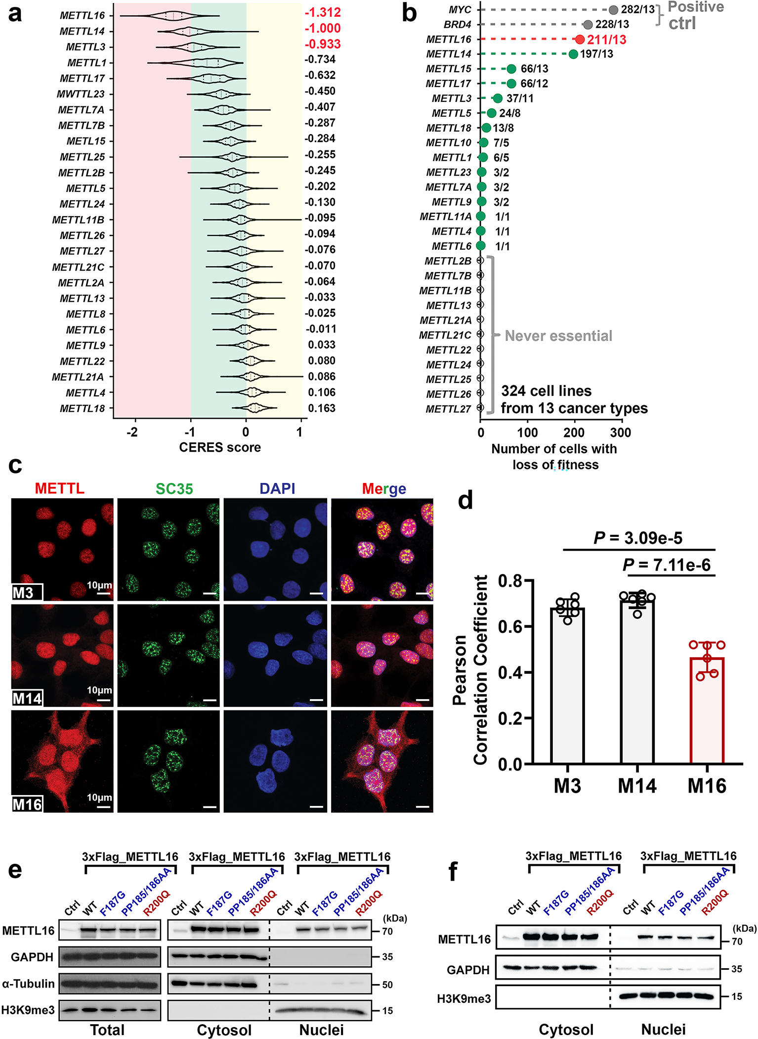
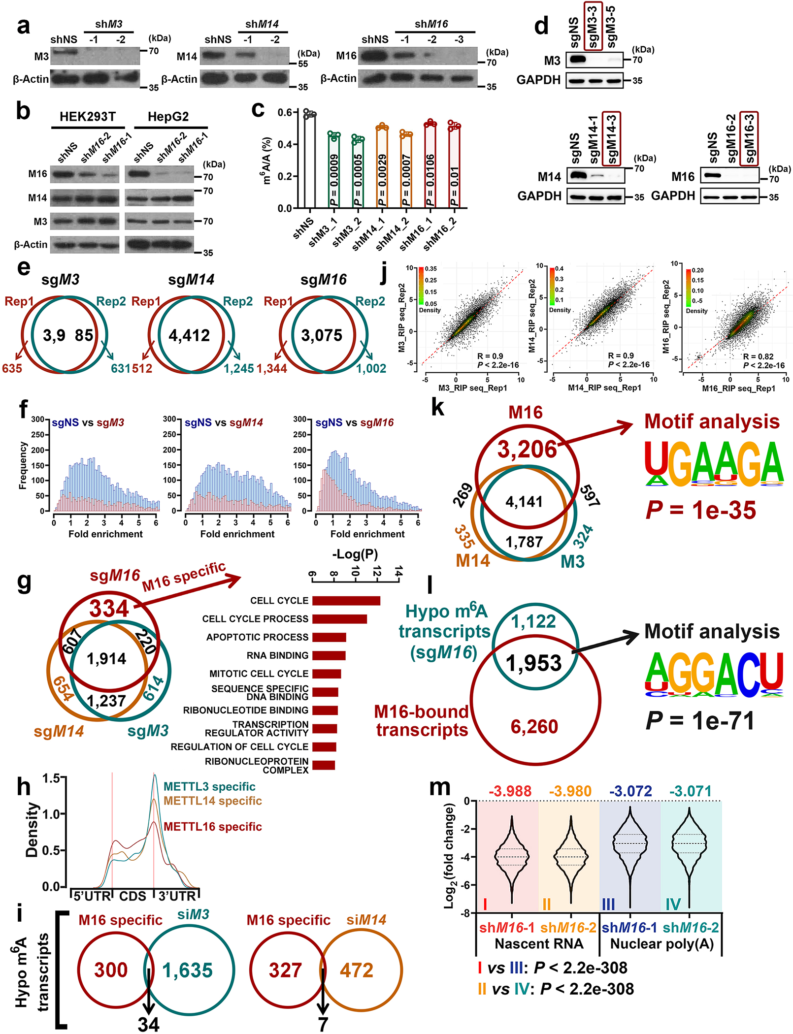
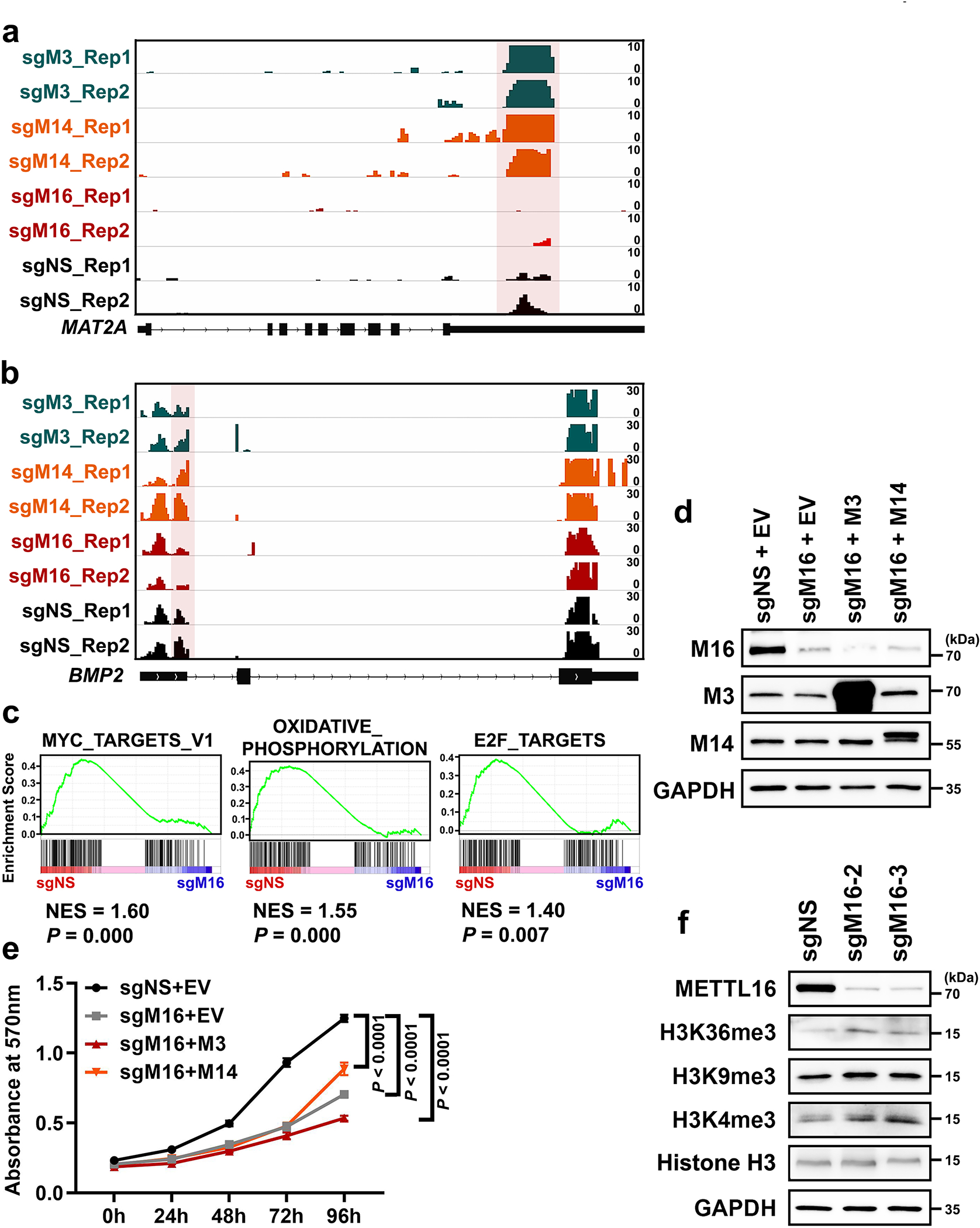
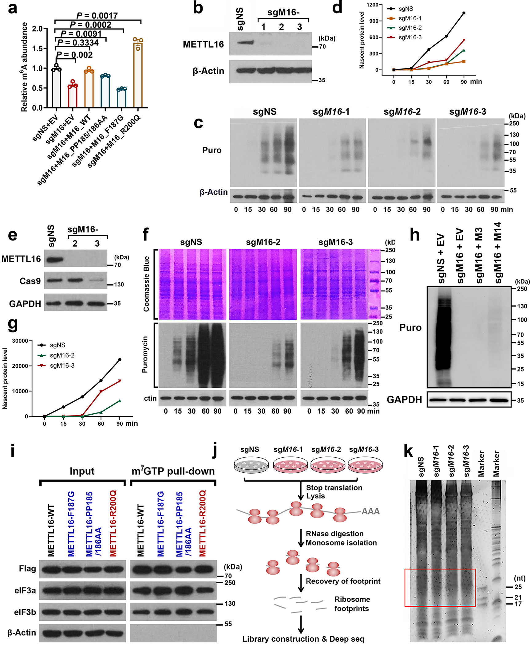
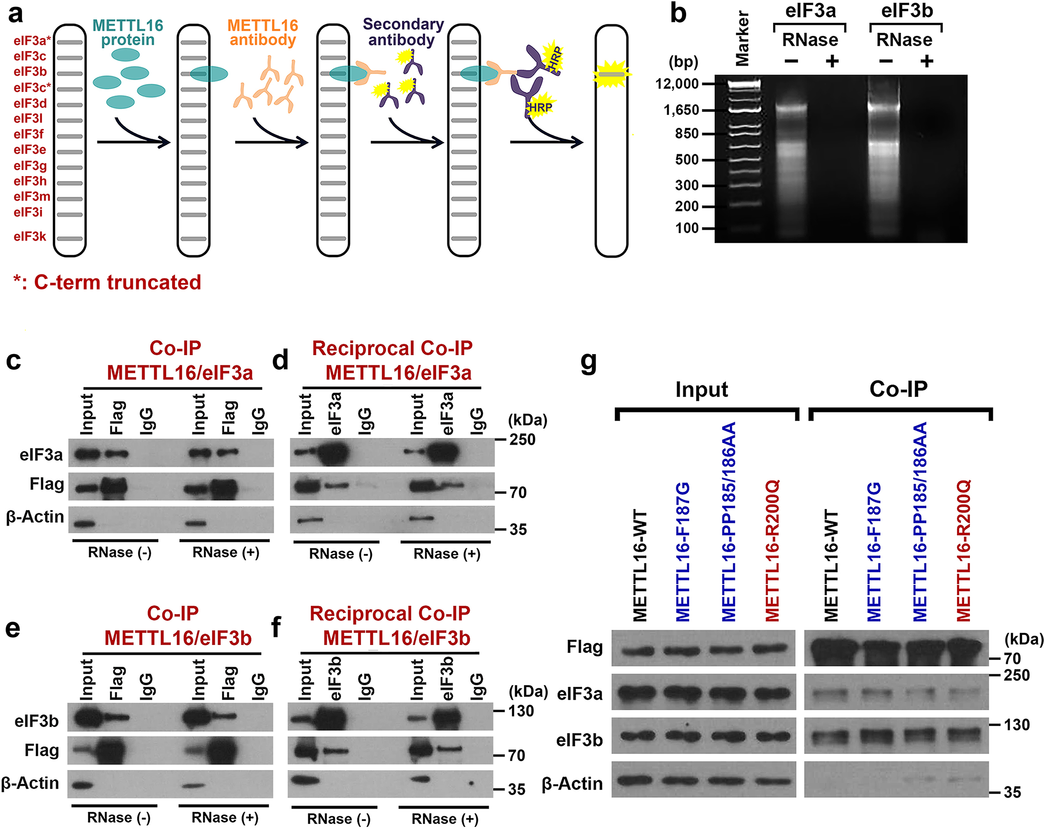
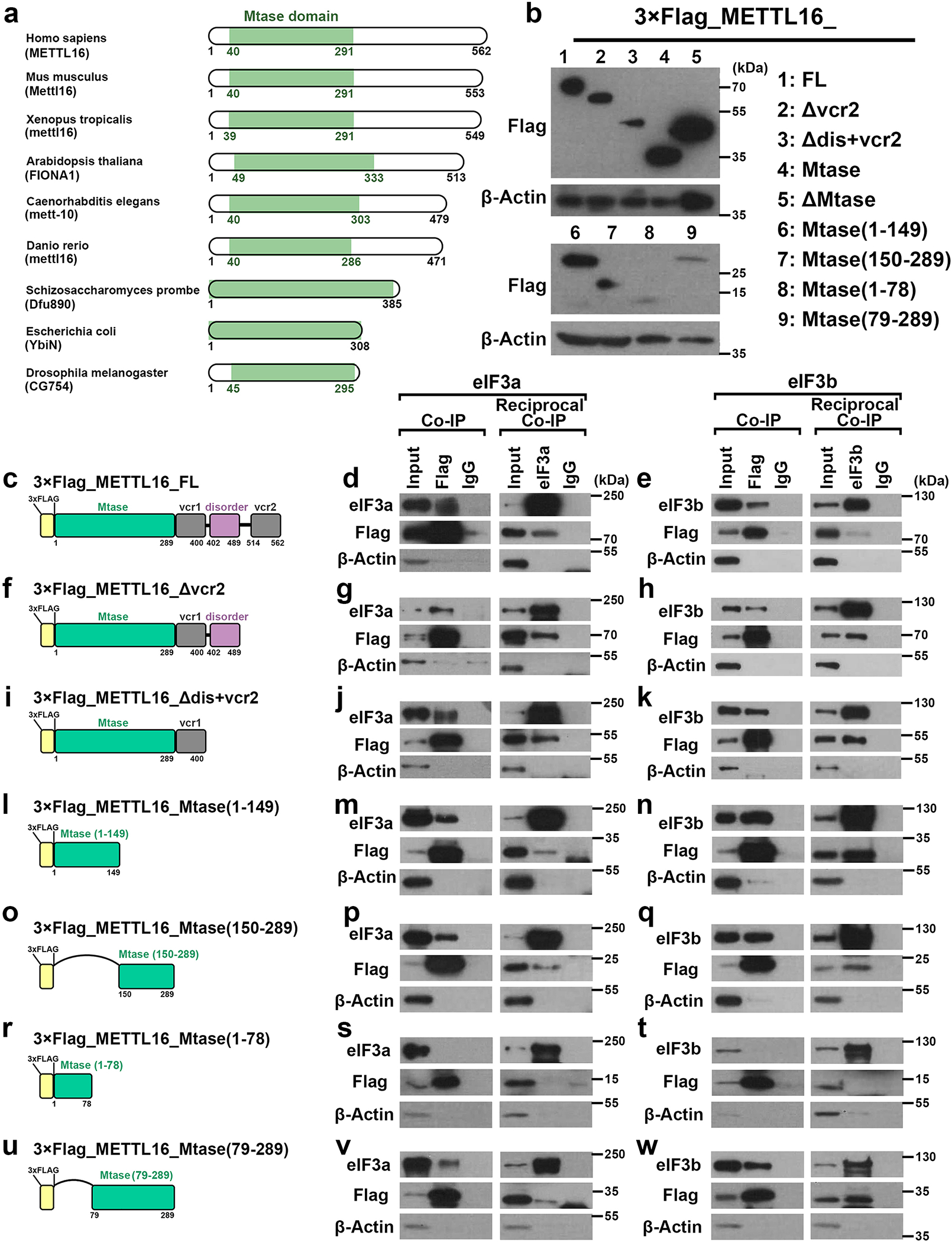
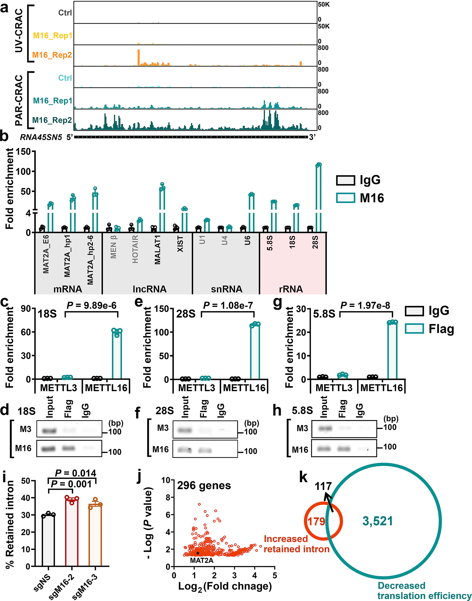
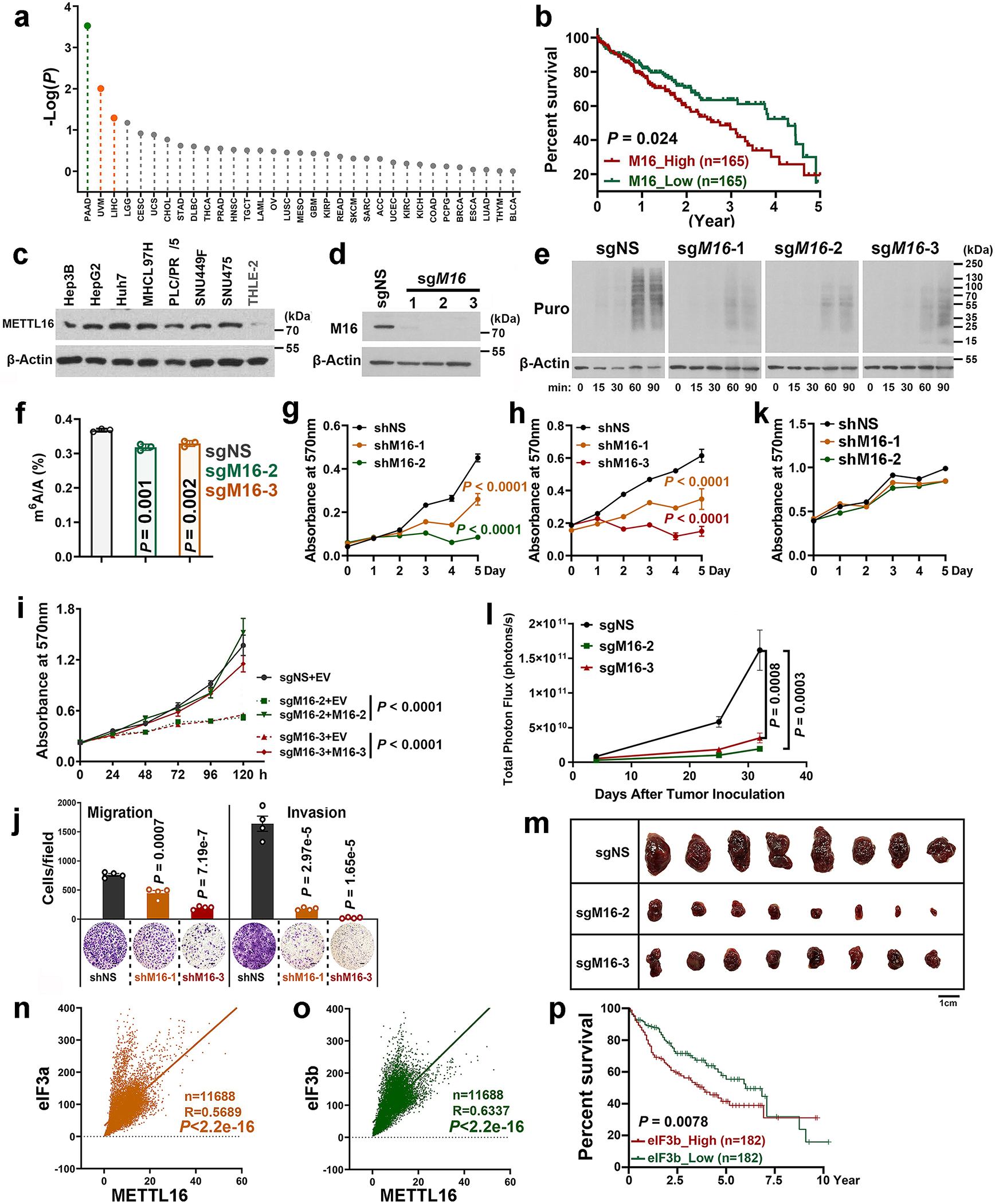
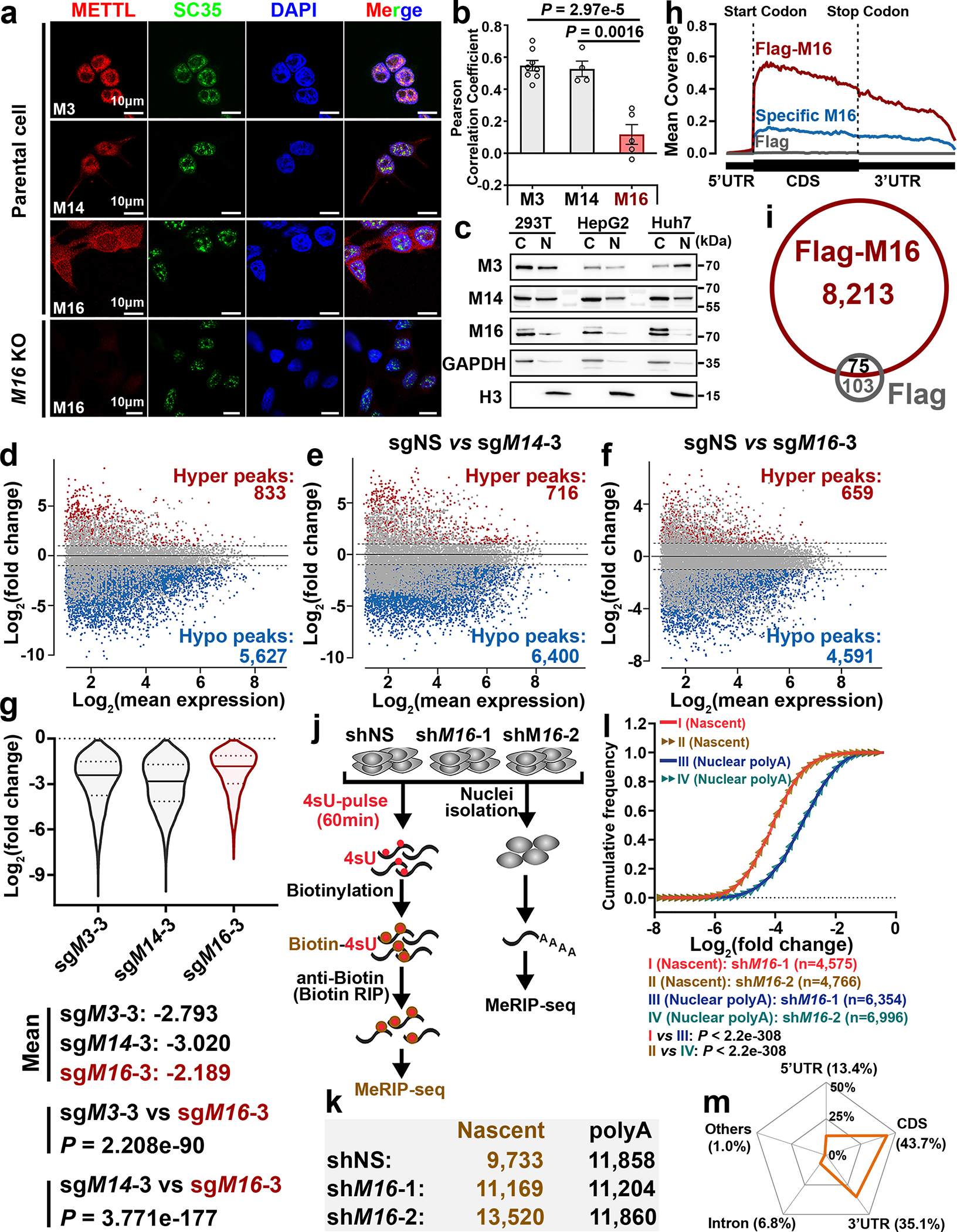
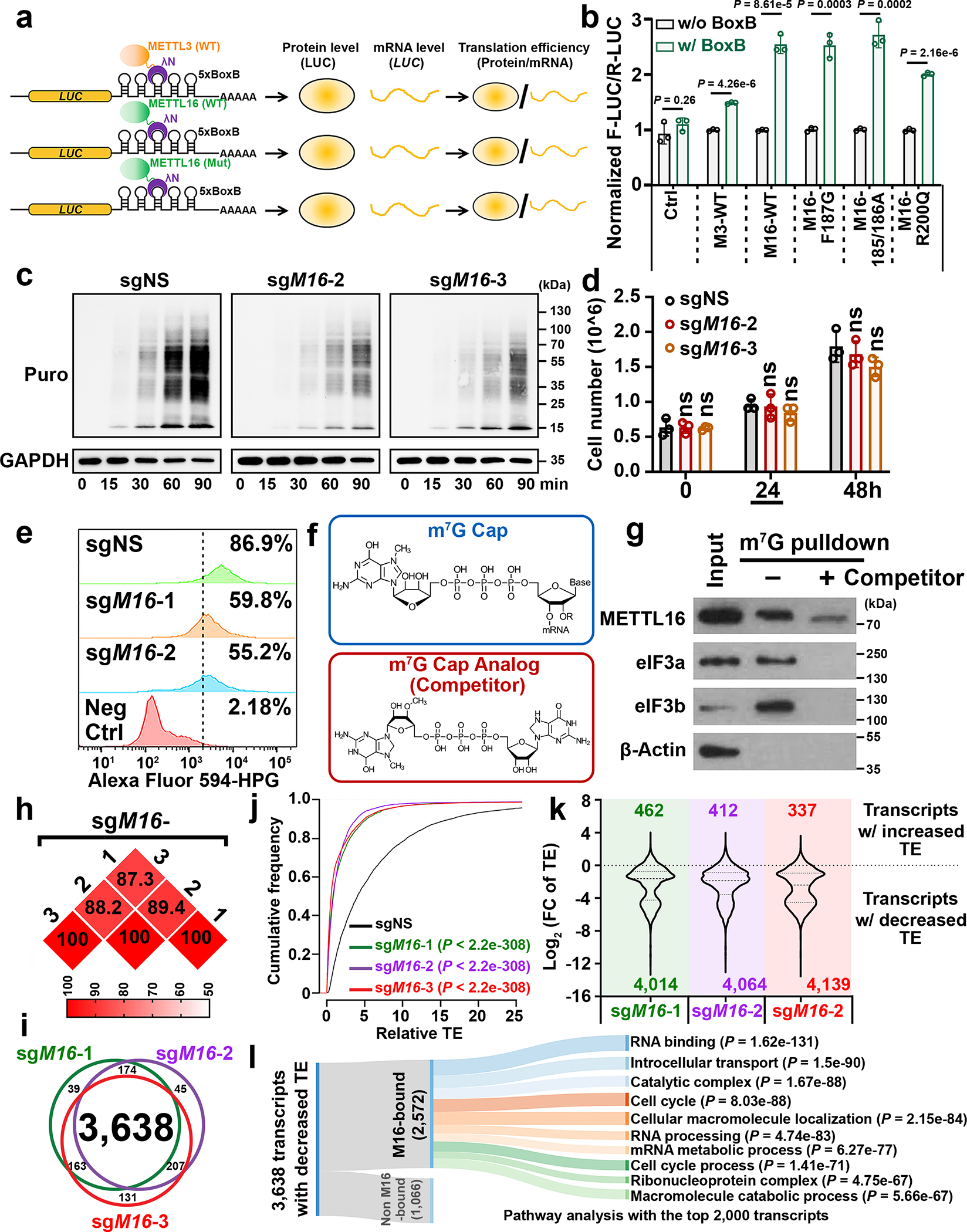
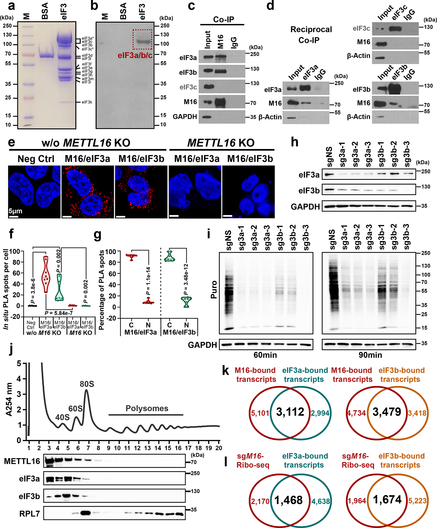
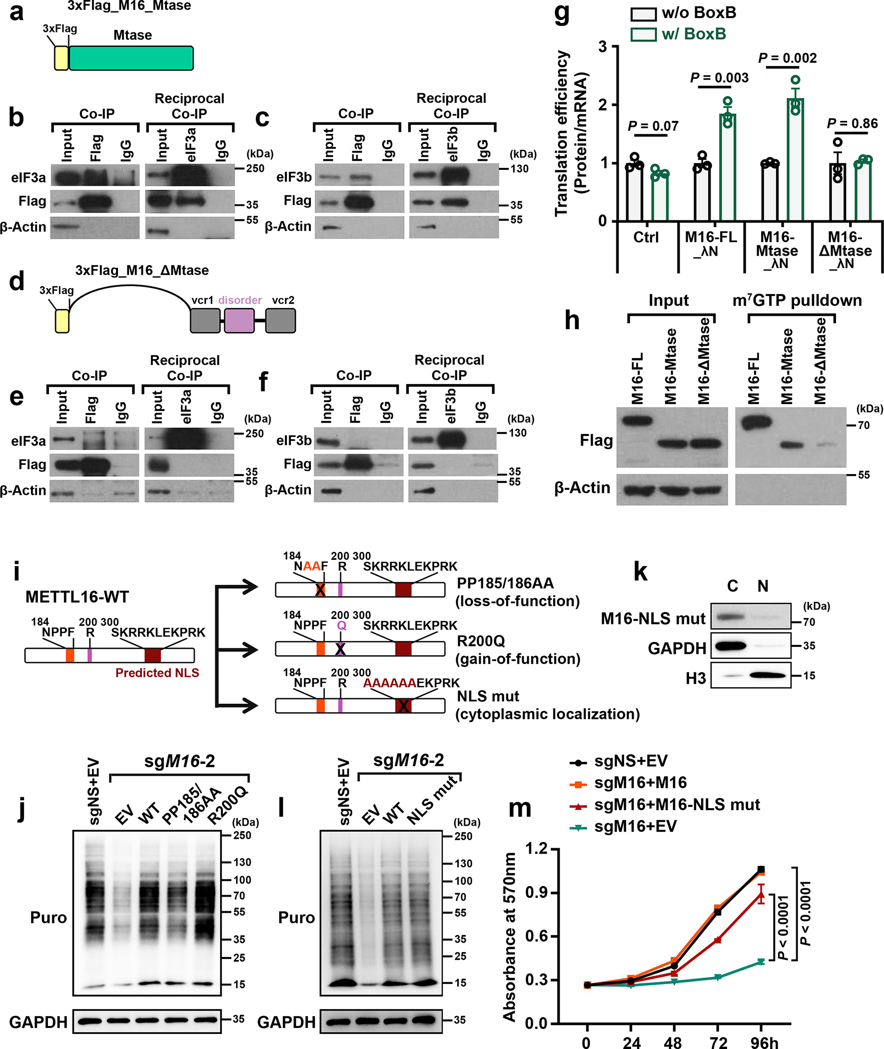
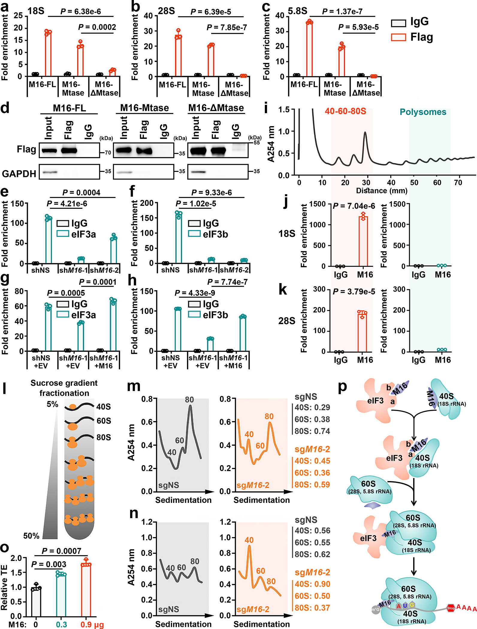
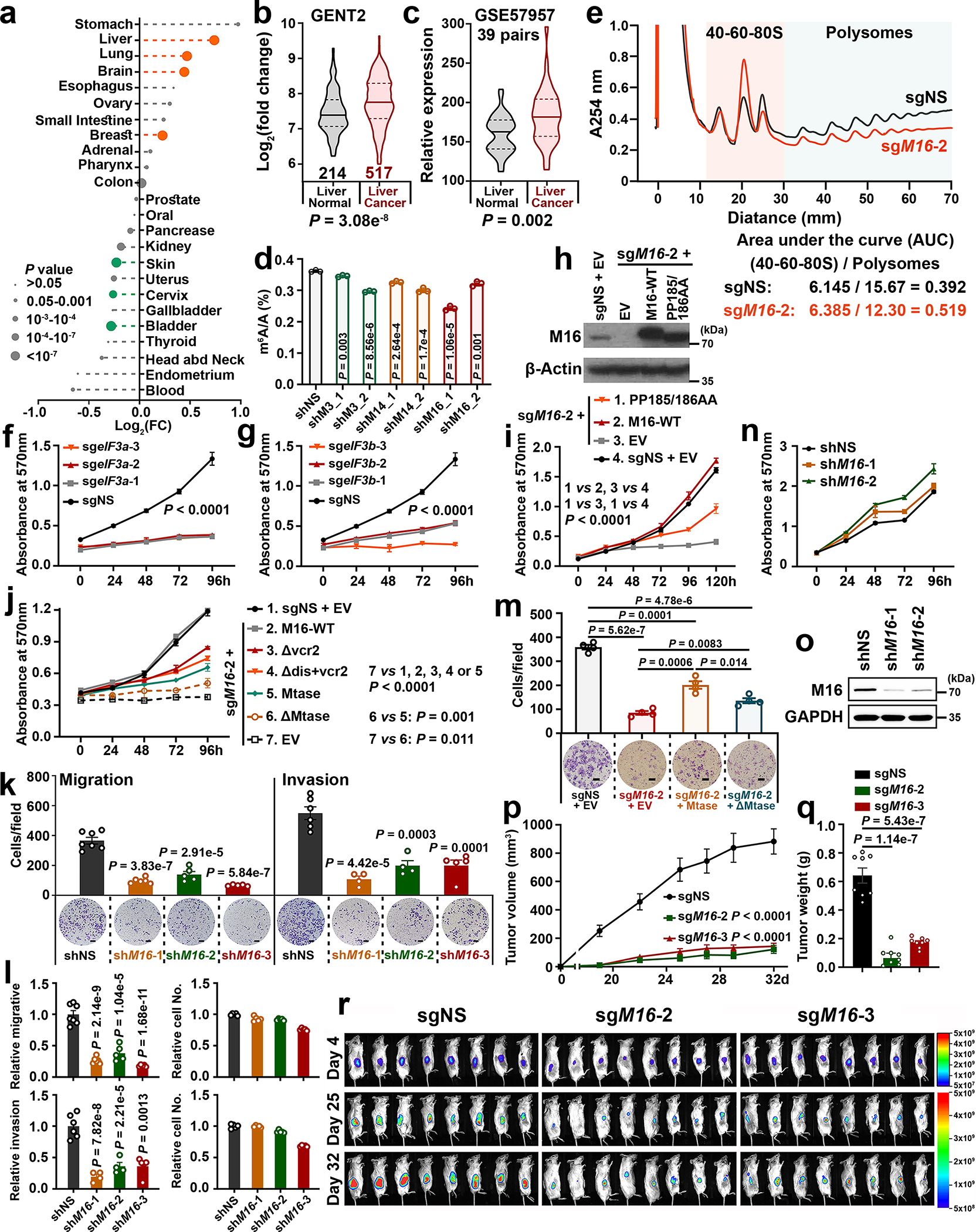
METTL16 has recently been identified as an RNA methyltransferase responsible for the deposition of N6-methyladenosine (m6A) in a few transcripts. Whether METTL16 methylates a large set of transcripts, similar to METTL3 and METTL14, remains unclear. Here we show that METTL16 exerts both methyltransferase activity-dependent and -independent functions in gene regulation. In the cell nucleus, METTL16 functions as an m6A writer to deposit m6A into hundreds of its specific messenger RNA targets. In the cytosol, METTL16 promotes translation in an m6A-independent manner. More specifically, METTL16 directly interacts with the eukaryotic initiation factors 3a and -b as well as ribosomal RNA through its Mtase domain, thereby facilitating the assembly of the translation-initiation complex and promoting the translation of over 4,000 mRNA transcripts. Moreover, we demonstrate that METTL16 is critical for the tumorigenesis of hepatocellular carcinoma. Collectively, our studies reveal previously unappreciated dual functions of METTL16 as an m6A writer and a translation-initiation facilitator, which together contribute to its essential function in tumorigenesis.


Conflict of interest statement

Competing interests

C.H. is a scientific founder and a scientific advisory board member of Accent Therapeutics, Inc., and holds equities with the company. J.C. is a scientific advisory board member of Race Oncology. The remaining authors declare no competing interests.

Figures

Extended Data Fig. 1
Extended Data Fig. 1. The dependency of METTL16 in human cancers and its preferential localization in cytoplasm.
a, CERES scores of a set of METTL family members from genome-scale CRISPR-Cas9 essentiality screens across 769 human cancer cell lines. The raw data were downloaded from DepMap (https://depmap.org/portal/). As the CERES scores, 0 and −1 represent the median effects of nonessential genes and common core essential genes, respectively. The lower CERES score indicates the higher cancer dependency of the specific gene. For each violin, the minimum, first quartile, median, third quartile, and maximum were displayed. The average of CERES scores for each METTL family member was shown. b, METTL16 was also detected by another genome-scale CRISPR-Cas9 screen with 324 cancer cell lines as a common essential gene in majority of human cancer cell lines in all 13 cancer types. The raw data were derived from https://score.depmap.sanger.ac.uk/. In this screen, 324 cancer cell lines from 13 cancer types were included. MYC and BRD4 were shown as positive controls, which represent the appealing cancer therapeutic targets. The number of cells with loss-of-fitness effects and the number of cancer types were highlighted for each gene. For example, METTL16 (211/13) indicates that knockout (KO) of METTL16 exhibits loss-of-fitness effects (essential function) in 211 of the 324 cancer cell lines and the 211 cancer cell lines cover all the 13 cancer types. c, Subcellular localization of endogenous METTL3, METTL14, and METTL16 in Huh7 cells as determined by immunofluorescence. SC35 worked as a nuclear speckle marker. In line with the trend in HEK293T cells, a much higher percentage of METTL16 is located in the cytoplasm than METTL3 and METTL14. Data shown represent 3 independent experiments. d, Pearson correlation coefficients showing the extent of colocalization of the three METTL family members with nuclear (DAPI) in Huh7 cells. The Pearson correlation coefficients were determined by the ZEN software. Again, both METTL3 and METTL14, but not METTL16, are predominantly localized in nuclear. Data are mean ± s.e.m. Statistics: unpaired, two-tailed t-test. n = 6 independent experiments. e, f, Western blotting showing the overexpression efficacy of wild type (WT) METTL16 and its three mutants, and their subcellular distributions in HEK293T (e) and HepG2 (f) cells. Here, F187G and PP185/186AA represent loss-of-function mutation of METTL16 methyltransferase activity, while R200Q represents a gain-of-function mutation. 3 × Flag was infused to the N-terminal of WT or mutant METTL16. GAPDH and α-Tubulin were used as loading control of total protein and cytoplasmic protein samples, and H3K9me3 was selected as loading control of nuclear protein.
Extended Data Fig. 2
Extended Data Fig. 2. Transcriptome-wide analysis of METTL16 methyltransferase activity.
a, Validate the knockdown (KD) efficiency of the shRNAs against METTL3, METTL14, or METTL16 via Western blotting. Images are representative of three biologically independent experiments with similar results. b, Expression of METTL3 and METTL14 in HEK293T (left) and HepG2 (right) cells upon METTL16 KD. Images are representative of two biologically independent experiments with similar results. c, Global changes of m6A abundance upon METTL3, METTL14, or METTL16 KD in HEK293T cells. Data are mean ± s.d. Statistics: unpaired, two-tailed t-test. n = 3 independent experiments. d, Validation of the KO efficiency of the gRNAs against METTL3, METTL14, or METTL16 via Western blotting in HEK293T cells. The RNA samples from sgMETTL3-3, sgMETTL14-3, and sgMETTL16-3 cells, along with the control cells, were collected for subsequent m6A MeRIP-Seq. The 2 independent experiments have been performed with similar results. e, Venn diagram showing the overlap of the transcripts with decreased m6A levels between the two biological replicates of sgMETTL3-3, sgMETTL14-3, and sgMETTL16-3. f, The frequency distributions of the m6A-hypo peaks caused by the KO of METTL3, METTL14, or METTL16 as detected by m6A MeRIP-Seq with poly(A) RNA from HEK293T cells. Only the significantly decreased m6A peaks (P < 0.01) were classified as m6A-hypo peaks and shown in the plot. g, Venn diagram showing the overlap analysis of the m6A-hypo transcripts induced by the KO of METTL3, METTL14, and/or METTL16 (left panel). Gene set enrichment analysis (GSEA) of the 334 METTL16-specific targets was performed, and the top 10 enriched pathways were shown (right panel). h, Global distribution of specific m6A-hypo peaks induced by the KO of METTL3, METTL14, and/or METTL16. i, Venn diagram showing the overlap between the 334 METTL16-specific targets and the transcripts with m6A-hypo peaks in METTL3 KD (left panel) or METTL14 KD (right panel) cells. j, Scatterplot showing the high reproducibility of the RIP-seq replicates of METTL3, METTL14, and METTL16. The Pearson correlation coefficients (R) of the normalized RIP-seq reads across the two replicates were calculated and displayed in the plots. A smoother regression line and 2D kernel density contour bands were also presented. P values were determined by Pearson’s correlation test. k, Venn diagram showing the overlap among METTL3, METTL14, and METTL16-bound transcripts (left panel) and the top one binding motif of the 3,206 specific METTL16-bound transcripts (right panel). l, Venn diagram showing the overlap between the METTL16-bound transcripts (RIP-seq) and the METTL16 KO-mediated m6A-hypo transcripts (MeRIP-seq) (left panel). Both seq analyses were conducted with poly(A) RNA from HEK293T cells. The top one consensus binding motif identified in METTL16-bound transcripts with METTL16 KO-induced m6A-hypo peaks was shown (right panel). m, Violin plots showing the significant m6A-hypo peaks induced by METTL16 KD in nascent RNA and nuclear poly(A) RNA from HEK293T cells. For each violin, the minimum, first quartile, median, third quartile, and maximum were presented. The average value of Log2(fold change) from each group was also displayed. The P values were calculated by unpaired two-sided t-test.
Extended Data Fig. 3
Extended Data Fig. 3. METTL16 specifically deposits m6A RNA methylation in a set of mRNA targets and its biological role in cell proliferation/growth and translation promotion can’t be substituted by METTL3 and METTL14.
a, b, Integrative Genomics Viewer (IGV) tracks displaying the m6A read distribution and changes (based on the m6A MeRIP-seq data) in MAT2A (a) and BMP2 (b) mRNA upon the KO of METTL3, METTL14, or METTL16 in HEK293T cells. The m6A decorations in MAT2A and BMP2 mRNA are METTL16-dependent, and METTL3/14-independent. c, The top 3 signaling pathways suppressed by METTL16 KO (sgMETTL16-3) as determined by GSEA. d, Overexpression efficacy of METTL3 and METTL14 in HEK293T cells with endogenous METTL16 KO (sgMETTL16-2). Images are representative of 2 biologically independent experiments with similar results. e, Effects of METTL3 and METTL14 overexpression on cell growth in HEK293T cells with endogenous METTL16 KO (sgMETTL16-2). Data are plotted as mean ± s.d. (n=3 independent experiments). Statistics: two-way ANOVA. f, Assessment of the global level changes of histone methylations, including H3K36me3, H3K9me3, and H3K4me3, in HEK293T cells upon endogenous METTL16 KO by Western blotting. Images are representative of 2 biologically independent experiments with similar results.
Extended Data Fig. 4
Extended Data Fig. 4. METTL16 enhances translation efficiency.
a, Relative m6A level changes in U6 snRNA in METTL16 KO (sgMETTL16-2) HEK293T cells upon ectopic expression of METTL16 WT and mutants (F187G, PP185/186AA, and R200Q) as determined by gene-specific m6A qPCR. Data are mean ± s.e.m. (n = 3 independent experiment). Statistics: unpaired, two-sided t-test. b, Western blotting confirming the KO efficacy of the sgRNAs against METTL16 in HEK293T cells. Images are representative of three biologically independent experiments with similar results. c, d, Representative WB images of SUnSET assays (c) and the quantitative SUnSET data (d) showing the effects of METTL16 KO on translation efficiency (based on the levels of nascent protein levels at given time periods) in HEK293T cells. β-Actin was used as loading controls. To make the data comparable, all the conditions for each sample are the same, including the total amount of loaded protein, concentration of antibodies, the time to run SDS-gel, transfer membrane, and develop signals. Representative images from three independent experiments with similar results were displayed. e, Western blotting showing the efficiency of METTL16 KO in K562 cells. f, g, Representative WB images of SUnSET assays (f) and the quantitative SUnSET data (g) showing the translation efficiency in K562 cells upon METTL16 deletion. Both Coomassie blue staining and β-Actin were used as loading controls. h, Effects of METTL3 and METTL14 overexpression on translation efficiency in HEK293T cells with endogenous METTL16 KO (sgMETTL16-2). All the cells were pulsed with puromycin for 60 minutes. Images are representative of three biologically independent experiments with similar results. i, Western blotting of the m7G pulldown assays to determine the potential binding of METTL16 WT and mutants in the 5’ cap region. Representative images from two independent m7G pulldown assays with similar results were presented. j, Experimental workflow of ribosome profiling with HEK293T cells upon METTL16 KO. k, The image of RNA gel with the ribosome footprint samples. The footprints within 17–34 nt were recovered for library construction and deep sequencing.
Extended Data Fig. 5
Extended Data Fig. 5. The direct interaction between METTL16 and eIF3a/b in HEK293T cells.
a, The experimental scheme for Far-Western blotting showing how to detect the potential direct interaction between METTL16 protein and eIF3 family members. Note: the * indicates the C-term truncated eIF3 family member. b, Agarose gel electrophoresis of the RNA samples recovered from Co-IP samples with or without RNase digestion. Samples of eIF3a were used for Extended Data Fig. 5c and d; while samples of eIF3b were used for Extended Data Fig. 5e and f. c, d, Co-IP (c) and reciprocal Co-IP (d) showing the potent interaction between eIF3a and METTL16 is RNA independent. Representative images from two biological replicates with similar results were presented. e, f, Co-IP (e) and reciprocal Co-IP (f) showing the robust association between eIF3b and METTL16 is also RNA independent. Representative images from two biological replicates with similar results were displayed. g, Western blotting of Co-IP assays to test the binding of METTL16 wild type (WT) and mutants (F187G, PP185/186AA, and R200Q) with eIF3a and eIF3b. The 3 × Flag tag was fused to the N-terminal of WT and mutant METTL16 proteins. Representative images from two independent Co-IP assays with similar results were presented.
Extended Data Fig. 6
Extended Data Fig. 6. Co-IP and reciprocal Co-IP with truncated METTL16 proteins to determine which domain is crucial for its association with eIF3a and eIF3b.
a, Diagrams and respective names of METTL16 homologs from various organisms. Among all the 9 species, the Mtase domain is highly conserved. b, Western blotting showing the expression of truncated METTL16 proteins in HEK293T cells. The 3 × Flag tag was attached into the N-terminal of all the truncated mutants. c, Schematic description of full length METTL16 with 3 × Flag tag at N-terminal. d, Co-IP and reciprocal Co-IP showing the interaction between eIF3a and full length METTL16. HEK293T cells with stable expression of METTL16 and eIF3a were used. e, Co-IP and reciprocal Co-IP showing the interaction between eIF3b and full length METTL16. HEK293T cells with stable expression of METTL16 and eIF3b were used. f, Schematic description of truncated METTL16_Δvcr2 (depletion of vcr2 domain) with 3 × Flag tag at N-terminal. g, Co-IP and reciprocal Co-IP showing the interaction between eIF3a and METTL16_Δvcr2. HEK293T cells with stable expression of METTL16_Δvcr2 and eIF3a were used. h, Co-IP and reciprocal Co-IP showing the interaction between eIF3b and METTL16_Δvcr2. HEK293T cells with stable expression of METTL16_Δvcr2 and eIF3b were used. i, Schematic description of truncated METTL16_Δdis+vcr2 (depletion of disorder domain plus vcr2 domain) with 3 × Flag tag at N-terminal. j, Co-IP and reciprocal Co-IP showing the interaction between eIF3a and METTL16_Δdis+vcr2. HEK293T cells with stable expression of METTL16_Δdis+vcr2 and eIF3a were used. k, Co-IP and reciprocal Co-IP showing the interaction between eIF3b and METTL16_Δdis+vcr2. HEK293T cells with stable expression of METTL16_Δdis+vcr2 and eIF3b were used. l, Schematic description of truncated METTL16_Mtase (1–149) with 3 × Flag tag at N-terminal. m, Co-IP and reciprocal Co-IP showing the interaction between eIF3a and METTL16_Mtase (1–149). HEK293T cells with stable expression of METTL16_Mtase (1–149) and eIF3a were used. n, Co-IP and reciprocal Co-IP showing the interaction between eIF3b and METTL16_Mtase (1–149). HEK293T cells with stable expression of METTL16_Mtase (1–149) and eIF3b were used. o, Schematic description of truncated METTL16_Mtase (150–289) with 3 × Flag tag at N-terminal. p, Co-IP and reciprocal Co-IP showing the interaction between eIF3a and METTL16_Mtase (150–289). HEK293T cells with stable expression of METTL16_Mtase (150–289) and eIF3a were used. q, Co-IP and reciprocal Co-IP showing the interaction between eIF3b and METTL16_Mtase (150–289). HEK293T cells with stable expression of METTL16_Mtase (150–289) and eIF3b were used. r, Schematic description of truncated METTL16_Mtase (1–78) with 3 × Flag tag at N-terminal. s, Co-IP and reciprocal Co-IP showing the lack of interaction between eIF3a and METTL16_Mtase (1–78). HEK293T cells with stable expression of METTL16_Mtase (1–78) and eIF3a were used. t, Co-IP and reciprocal Co-IP showing the lack of interaction between eIF3b and METTL16_Mtase (1–78). HEK293T cells with stable expression of METTL16_Mtase (1–78) and eIF3b were used. u, Schematic description of truncated METTL16_Mtase (79–289) with 3 × Flag tag at N-terminal. v, Co-IP and reciprocal Co-IP showing the interaction between eIF3a and METTL16_Mtase (79–289). HEK293T cells with stable expression of METTL16_Mtase (79–289) and eIF3a were used. w, Co-IP and reciprocal Co-IP showing the interaction between eIF3b and METTL16_Mtase (79–289). HEK293T cells with stable expression of METTL16_Mtase (79–289) and eIF3b were used. For all the IP experiments, representative images from two biological replicates with similar results were presented.
Extended Data Fig. 7
Extended Data Fig. 7. The robust binding of METTL16 with rRNAs and the effect of METTL16 on intron retention.
a, IGV tracks showing the binding pattern of METTL16 on ribosomal 45S RNA clusters (Here we exhibited RNA45SN5). 45S RNA is further processed into 18S rRNA, 5.8S rRNA and 28S rRNA. UV-CRAC, UV crosslinking and analysis of cDNA; PAR-CRAC, photoactivatable ribonucleoside enhanced crosslinking and analysis of cDNA. b, The binding of METTL16 with mRNA (MAT2A), lncRNAs, snRNAs, and rRNAs in HEK293T cells. Data are mean ± s.e.m. Data shown represent 3 independent experiments. MAT2A, MALAT1, XIST, and U6 were included as positive control targets of METTL16 for the test. MEN β, HOTAIR, U1, and U4 were included as negative controls. The results indicated our crosslinking RNA immunoprecipitation and qPCR (CLIP-qPCR) works well. c, d, The quantitative (c) and semi-quantitative (d) analysis of the enrichments of METTL3 and METTL16 on 18S rRNA in the cytoplasm fraction. e, f, The quantitative (e) and semi-quantitative (f) analysis of the enrichments of METTL3 and METTL16 on 28S rRNA in the cytoplasm fraction. g, h, The quantitative (g) and semi-quantitative (h) analysis of the enrichments of METTL3 and METTL16 on 5.8S rRNA in the cytoplasm fraction. For c-h, The 3 × Flag tag was infused to the N-terminal of METTL3 and METTL16, and the IP assays were conducted with Flag antibody; IgG serves as a negative control. The quantitative results were derived from qPCR; while the semi-quantitative results were derived from RT-PCR and the products were run on an 2% agarose gel. For c, e, and g, data are mean ± s.e.m. Data shown represent 3 independent experiments. Statistics: unpaired, two-sided t-test (METTL16 vs. METTL3). i, The effect of METTL16 KO on intron retention of MAT2A mRNA in HEK293T cells. Data are mean ± s.e.m. Data shown represent 3 independent experiments. Statistics: unpaired, two-sided t-test. j, The transcripts with significantly (P < 0.05) increased intron retention in HEK293T cells upon METTL16 depletion. Herein, the transcriptome-wide RNA-seq data was from GSE90914 and the intron retention was detected with IRFinder. k, Venn diagram showing the overlap between the transcripts with increased intron retention and the transcripts with decreased translation efficiency in HEK293T cells upon METTL16 depletion.
Extended Data Fig. 8
Extended Data Fig. 8. The tumor-promoting role of METTL16 in hepatocellular carcinoma (HCC) and its positive correlations with eIF3a/b in expression.
a, The prognostic impacts of METTL16 expression levels in the TCGA cancer patient datasets, which were downloaded from GEPIA2 (http://gepia2.cancer-pku.cn/#index). In each cancer type, the overall survivals between patients with high METTL16 expression levels (the top 50%) and those with low levels (the low 50%) were compared and the P value was calculated by the log-rank test, and then the -Log(P) value was presented. In PAAD (Pancreatic adenocarcinoma), high expression of METTL16 significantly (P < 0.05) correlates with better survival; while in UVM (Uveal melanoma) and LIHC (Liver hepatocellular carcinoma), high expression of METTL16 significantly (P < 0.05) correlates with poorer survival. METTL16 expression levels are not significantly associated with prognosis in other cancer types. b, Overall survival analysis of METTL16 in HCC from the TCGA dataset. The P value is determined by logrank test. c, Expression of METTL16 in liver cancer cell lines (Hep3B, HepG2, Huh7, MHCC97H, PLC/PRF/5, SUN449, and SUN475) and healthy liver cell line (THLE-2). Representative images from two independent replicates with similar results were presented. d, Validation of METTL16 KO efficiency via Western blotting in HepG2 Cas9 single clone. e, The Western blotting images of SUnSET assays showing the effects of METTL16 KO on translation efficiency in HepG2 liver cancer cells. To make the data comparable, all the conditions for each sample are the same, including the total amount of loaded protein, concentration of antibodies, the time to run SDS-gel, transfer membrane, and develop signals. f, Determination of m6A levels in mRNAs of HepG2 cells with or without METTL16 KO via QQQ-MS. Data are mean ± s.d. Data shown represent 3 independent experiments. Statistics: unpaired, two-sided t-test. g, h, Effects of METTL16 KD on cell growth in SNU449 (g) and SNU475 (h) HCC cells. Data are plotted as mean ± s.d. (n=3 independent experiments). Statistics: two-way ANOVA. i, Effects of METTL16 expression changes on the growth of HepG2 cells. METTL16 KO (sgM16–2 + EV; sgM16–3 + EV) drastically suppressed cell proliferation as determined by MTT assays; restored expression of METTL16 (sgM16–2 + M16–2; sgM16–3 + M16–3) could rescue/reverse the inhibitory effect of METTL16 KO. Here, M16–2 and M16–3 represent the synonymous mutations of METTL16 ORF that can’t be targeted by sgM16-2 and sgM16-3, respectively. Data are plotted as mean ± s.d. n = 4 (sgM16–2 + M16–2, 120 h; and sgM16–3 + M16–3, 120h); n =5 (sgM16–3 + M16–3, 72h); n = 6 (all other groups) independent experiments. Statistics: two-way ANOVA. j, Effects of METTL16 KD on the migration and invasion of SUN475 liver cancer cells. Data are mean ± s.d. (n=3 independent experiments). Statistics: unpaired two-sided t-test. k, The effects of METTL16 KD on cell proliferation in THLE-2 cells (a primary human normal liver cell line). Data are plotted as mean ± s.e.m. (n=3 independent experiments). l, Total photon flux of the mice xenografted with HepG2 cells with or without METTL16 KO. Data are plotted as mean ± s.e.m. (n=8 independent mice). Statistics: two-way ANOVA. m, Tumor images at the endpoint of the xenograft models implanted with HepG2 liver cancer cells with or without METTL16 KO. n, o, The correlation between METTL16 and eIF3a (n), or eIF3b (o) in expression as detected by Pearson’s correlation analysis. All the raw data were downloaded from GTEx (Genotype-Tissue Expression, https://www.gtexportal.org/home/). The number of samples, R value, and P value were displayed. Statistics: Pearson correlation. p, Overall survival analysis of eIF3b in HCC from the TCGA dataset. The P value is determined by logrank test.
Fig. 1
Fig. 1. Comparative analysis of subcellular distribution and m6A catalytic activity of METTL3, METTL14, and METTL16.
a, Subcellular localization of endogenous METTL3/14/16 in HEK293T cells. SC35 and DAPI, markers for nuclear speckle and nucleus, respectively. b, Pearson correlation analysis showing the extent of colocalization of the three METTL family members with nucleus in HEK293T cells. Data are mean ± s.e.m. Statistics: unpaired, two-tailed t-test. M3, n = 8; M14, n = 4; M16, n = 5 independent experiments. c, Expression levels of METTL3/14/16 in the cytoplasm (C) and nucleus (N) of the cells. Data shown represent 3 independent experiments. d-f, MA plots displaying the m6A-hyper and m6A-hypo peaks in poly(A) RNA upon METTL3 (d), METTL14 (e), or METTL16 (f) KO in HEK293T cells. The significantly increased (hyper) or decreased (hypo) m6A peaks are exhibited in red and blue, respectively (P < 0.01, calculated by exomePeak). g, Violin plots showing the fold changes of decreased m6A peaks upon METTL3, METTL14, or METTL16 KO. The three lines inside the violins were defined as the first quartile, median, and third quartile. Statistics: unpaired, two-tailed t-test. h, Metagene profile of enrichment of METTL16-bound transcripts (Flag-M16), specific METTL16-bound transcripts (Specific M16; i.e., the 3,206 transcripts in Extended Data Fig. 2k), and Flag-bound sites (Flag, background control) across the target transcripts. i, Venn diagram showing the overlap between METTL16-bound transcripts and Flag-bound transcripts. j, Schematic of m6A MeRIP-seq with nascent RNA and nuclear poly(A) RNA in HEK293T cells upon METTL16 KD. k, The number of m6A peaks in nascent RNA and poly(A) RNAs. l, Cumulative distribution function (CDF) plot depicting the fold changes of m6A-hypo peaks induced by METTL16 KD in nascent RNA and nuclear poly(A) RNA. Statistics: two-sided Wilcoxon and Mann–Whitney test. m, Radar chart showing the percentage of nascent RNA m6A peaks distributed at exons (including 5’UTR, CDS, 3’UTR), introns, and other regions in the control (shNS) group. The m6A MeRIP-seq have been performed with 2 biological replicates with similar results; the data shown in d-m represent a single representative experiment.
Fig. 2
Fig. 2. METTL16 enhances translation efficiency of target mRNAs.
a, Experimental schedule of RNA tethering experiments to determine translation efficiency. b, Translation efficiency as determined by the tethering assays. F-Luc, Firefly luciferase; R-Luc, Renilla luciferase, internal control. Data are mean ± s.d. (n = 3 independent experiments). Statistics: unpaired, two-tailed t-test. c, Representative Western blotting images of SUnSET assays to quantify the amount of nascent (puromycin-labelled) peptides in HEK293T cells with METTL16 knockout (KO). To make the data comparable, all the samples were treated equally. Data shown represent 3 independent experiments. d, The cell number in sgNS, sgM16-2, and sgM16-3 groups. The samples were collected at 24 hours for the SUnSET assay shown in Fig. 2c. Data are mean ± s.d. (n = 3 independent experiments). Statistics: unpaired, two-tailed t-test. ns, not significant (P ≥ 0.05). e, Nascent protein synthesis upon METTL16 KO as determined by the pulse labeling assay via incorporation of L-homopropargylglycine (HPG). f, Structures of m7G cap and its analog. Data shown represent 2 independent experiments. g, Western blotting of m7G pulldown assays with indicated antibodies. eIF3a and eIF3b, positive controls. β-Actin, negative control. Data shown represent 2 independent experiments. h, The reproducibility of Ribo-seq with three diverse gRNAs against METTL16. Here, we presented the percentages of overlapping transcripts with decreased translation efficiency (TE) caused by the three gRNAs mediated METTL16 KO in HEK293T. i, Venn diagram showing high overlap of the transcripts with inhibited TE across the three gRNA groups. j, CDF plot depicting the transcriptome-wide inhibition of translation efficiency (TE) upon METTL16 KO. Statistics: two-sided Wilcoxon and Mann–Whitney test. k, Violin plots showing the Log2(FC of TE) upon METTL16 KO. FC, fold change. l, Sankey diagram presenting the subsequent pathway analysis of the transcripts whose translation initiation is facilitated by METTL16. Among the 3,638 transcripts with suppressed TE upon METTL16 KO, 2,572 are directly bound by METTL16 protein. Among the 2,572 targets, we selected the top 2,000 candidates for pathway analysis. Here, we showed the top 10 enriched pathways.
Fig. 3
Fig. 3. METTL16 facilitates translation initiation via direct interaction with eIF3a and eIF3b.
a, Coomassie brilliant blue staining of protein markers, BSA (negative control), and eIF3 family members. b, Far-Western blotting for the direct interaction between METTL16 and eIF3 family members. Images in a and b are representative of three biologically independent experiments with consistent results. c, Co-IP of METTL16 with endogenous eIF3a, eIF3b, and eIF3c. d, Reciprocal Co-IP of endogenous eIF3a, eIF3b, and eIF3c with METTL16. Data shown represent 2 independent experiments. e, In situ detection of METTL16-eIF3a and METTL16-eIF3b interaction via proximity ligation assays (PLA) in HEK293T cells with or without METTL16 KO. f, Quantification of in situ PLA puncta from the five groups. Statistics: unpaired, two-tailed t-test. n = 8 independent experiments. g, The distribution of in situ PLA spots from METTL16-eIF3a and METTL16-eIF3b in cytoplasm (C) and nuclear (N) of HEK293T cells without METTL16 KO. Statistics: unpaired, two-tailed t-test. n = 8 independent experiments. h, The KO efficiency of eIF3a and eIF3b in HEK293T cells. i, Representative Western blotting images of SUnSET assays to quantify the amount of nascent peptides in HEK293T cells with eIF3a and eIF3b KO. Data shown represent 3 independent experiments. j, Polysome profiles of HEK293T cells as determined by sucrose density gradient ultracentrifugation. The localization of eIF3a, eIF3b, METTL16, and RPL7 proteins were validated by Western blotting. Data shown represent 3 independent experiments. k, Venn diagram showing the overlap between METTL16-bound transcripts and eIF3a-bound transcripts (left), or between METTL16-bound transcripts and eIF3b-bound transcripts (right). Both the eIF3a- and eIF3b-bound transcripts were downloaded from the POSTAR3 (http://postar.ncrnalab.org/). l, Venn diagram showing the overlap between the transcripts with decreased translation efficiency in METTL16 KO cells and the transcripts directly bound by eIF3a (left) or eIF3b (right).
Fig. 4
Fig. 4. Both the Mtase domain and the cytoplasmic localization of METTL16 are necessary for translation regulation.
a, Schematic of truncated METTL16 that contains just the Mtase domain, with 3 × Flag tag attached at the N-terminal. b, c, Co-IP (left) and reciprocal Co-IP (right) showing the direct interaction between METTL16_Mtase (Flag) and eIF3a (b) or eIF3b (c). β-Actin was used as negative control. d, Schematic of another truncated METTL16 that contains vcr1-disorder-vcr2 (ΔMtase domain), with 3 × Flag tag attached to the N-terminal. e, f, Co-IP (left) and reciprocal Co-IP (right) showing no interaction between METTL16_ΔMtase (Flag) and eIF3a (e) or eIF3b (f). g, RNA tethering experiments depicting the translation efficiency with forced expression of full length (FL), Mtase, and ΔMtase. The 3 × Flag tag was fused to the N-terminal, while λN peptide was fused to the C-terminal of the constructs. Data are mean ± s.d. (n = 3 independent experiments). Statistics: unpaired, two-tailed t-test. h, m7GTP pull-down assays illustrating the robust binding of full length and Mtase domain of METTL16, but not the ΔMtase, in the cap region. i, Schematic of three METTL16 mutants, including catalytic-dead METTL16 (PP185/186AA), catalytic-active METTL16 (R200Q), and exclusive cytoplasmic METTL16 (NLS mut). j, The rescue effects of PP185/186AA and R200Q on METTL16 KO-induced translation inhibition in HEK293T cells. k, Subcellular localization of METTL16 NLS mut in HEK293T cells. l, The rescue effects of exclusive cytoplasmic METTL16 overexpression on METTL16 KO-induced translation inhibition in HEK293T cells. For b, c, e, f, h, j, k, and l, data shown represent 2 independent experiments with similar results. m, The rescue effects of wildtype METTL16 and exclusive cytoplasmic METTL16 overexpression on METTL16 KO-induced cell proliferation suppression in HEK293T cells. Data are mean ± s.d. (n =3 independent experiments). Statistics: two-way ANOVA.
Fig. 5
Fig. 5. METTL16 facilitates the formation of TIC by interactions with both eIF3a/b and rRNAs.
a, The binding of full length (FL), Mtase domain, and ΔMtase domain of METTL16 on 18S (a), 28S (b), and 5.8S (c) rRNAs in the cytoplasm fraction as determined by CLIP-qPCR. d, The comparable pulldown efficiency among METTL16-FL, Mtase, and ΔMtase groups. Data shown represent 3 independent experiments. e, f, CLIP-qPCR analysis showing the decreased association between 18S rRNA and eIF3a (e) or eIF3b (f) upon METTL16 KD. g, h, Rescue assay showing the rescued binding of eIF3a (g) or eIF3b (h) with 18S rRNA upon manipulating the expression of METTL16. i, Isolation of translation initiation fraction (40–60-80S) and polysome fraction for RIP according to polysome profiling. Data shown represent 3 independent experiments. j, k, The interactions between METTL16 and 18S rRNA (j) or 28S rRNA (k) in the translation initiation fraction (left) and polysome fraction (right). HEK293T cells were used in all the above studies. l, Schematic of sucrose gradient fractionation. m, n, Sucrose density gradient profiles showing the abundance of 40S, 60S, and 80S ribosomes from HEK293T (m) and HepG2 (n) cells upon METTL16 KO. Data shown represent 3 independent experiments. o, Effect of ectopic METTL16 protein on translation efficiency of luciferase mRNA as determined by in vitro translation. p, Schematic model of how METTL16 enhances translation initiation. The direct interactions of METTL16 with eIF3a/b and rRNAs drive the eIF3/40S ribosomal subunit (with 18S rRNA as the core) interaction and the subsequent assembly of 40S ribosomal subunit/60S ribosomal subunit (with 5.8S and 28S rRNAs as the core) into the 80S TIC, leading to stimulated translation initiation. For a-c, e-h, j, k, and o, data are represented as mean ± s.d. (n = 3 independent experiments). Statistics: unpaired, two-tailed t-test. For j and k, the P values indicate the binding of METTL16 with 18S rRNA (j) and 28S rRNA (k) in 40–60-80S vs Polysomes.
Fig. 6
Fig. 6. The crucial tumor-promoting role of METTL16 in hepatocellular carcinoma (HCC).
a, Relative expression levels of METTL16 in the 24 cancer types. FC represents the ratio of METTL16 levels in a specific subtype of tumors and the corresponding healthy control. Statistics: unpaired, two-tailed t-test. b, c, Comparison of METTL16 expression levels between liver cancers and normal controls. Statistics: two-tailed t-test, unpaired (b) or paired (c). d, Global m6A abundance in poly(A) RNA isolated from HepG2 cells upon METTL3/14/16 KD (n = 3). e, Representative polysome profiles of HepG2 cells upon METTL16 KO (n = 3). f, g, The effect of eIF3a (f) or eIF3b (g) KO on proliferation in HepG2 cells. h, i, Rescued expression (h, n = 3) and effects (i) of wild type and mutant METTL16 in HepG2 cells upon METTL16 KO. j, Rescued effects of wildtype and truncated mutants of METTL16 on cell proliferation of HepG2 cells upon METTL16 KO. k, Effects of METTL16 KD on migration and invasion of SUN449 cells. Migration: n = 7 (shNS), 6 (shM16-1), 5 (shM16-2, and shM16-3); Invasion: n = 6 (shNS), 4 (shM16-1 and shM16-2), 5 (shM16-3). l, The relative migration/invasion, and relative cell number (n = 5) in the SUN449 cells. The P values, unpaired two-sided t-test, represented the difference between relative migration (or invasion) and relative cell number in each group. m, Rescued effects of forced expression of METTL16-Mtase and ΔMtase on migration in HepG2 upon METTL16 KO (n = 4). n, o, Effects of METTL16 KD on cell proliferation (n) and its KD efficacy (o) in CL-48 cells. p, Average growth curves of liver tumors. Data are presented as mean ± s.e.m. (n = 8); two-way ANOVA. q, The weights of tumors at endpoints. Data are mean ± s.d. (n = 8); two-sided t-test. r, Bioluminescence imaging of xenografted mice. Unit: photons/second/cm2/steradian. For d, k, l, and m, data are mean ± s.d.; statistics: unpaired, two-tailed t-test. For f, g, i, and j, data are mean ± s.d. (n = 3); statistics: two-way ANOVA. All the “n” indicates the number of independent experiments.

References

    1. Frye M, Harada BT, Behm M & He C RNA modifications modulate gene expression during development. Science 361, 1346–1349 (2018). - PMC - PubMed
    1. Huang H, Weng H & Chen J m(6)A Modification in Coding and Non-coding RNAs: Roles and Therapeutic Implications in Cancer. Cancer Cell 37, 270–288 (2020). - PMC - PubMed
    1. Liu J et al. A METTL3-METTL14 complex mediates mammalian nuclear RNA N6-adenosine methylation. Nat Chem Biol 10, 93–95 (2014). - PMC - PubMed
    1. Wang X et al. Structural basis of N(6)-adenosine methylation by the METTL3-METTL14 complex. Nature 534, 575–578 (2016). - PubMed
    1. Wang P, Doxtader KA & Nam Y Structural Basis for Cooperative Function of Mettl3 and Mettl14 Methyltransferases. Mol Cell 63, 306–317 (2016). - PMC - PubMed

Publication types

MeSH terms