. 2022 Mar;41(13):1835-1850.
doi: 10.1038/s41388-022-02221-0. Epub 2022 Feb 10.

Intratumor genetic heterogeneity and clonal evolution to decode endometrial cancer progression

Affiliations

Intratumor genetic heterogeneity and clonal evolution to decode endometrial cancer progression

Alba Mota et al. Oncogene. 2022 Mar.

Abstract

Analyzing different tumor regions by next generation sequencing allows the assessment of intratumor genetic heterogeneity (ITGH), a phenomenon that has been studied widely in some tumor types but has been less well explored in endometrial carcinoma (EC). In this study, we sought to characterize the spatial and temporal heterogeneity of 9 different ECs using whole-exome sequencing, and by performing targeted sequencing validation of the 42 primary tumor regions and 30 metastatic samples analyzed. In addition, copy number alterations of serous carcinomas were assessed by comparative genomic hybridization arrays. From the somatic mutations, identified by whole-exome sequencing, 532 were validated by targeted sequencing. Based on these data, the phylogenetic tree reconstructed for each case allowed us to establish the tumors' evolution and correlate this to tumor progression, prognosis, and the presence of recurrent disease. Moreover, we studied the genetic landscape of an ambiguous EC and the molecular profile obtained was used to guide the selection of a potential personalized therapy for this patient, which was subsequently validated by preclinical testing in patient-derived xenograft models. Overall, our study reveals the impact of analyzing different tumor regions to decipher the ITGH in ECs, which could help make the best treatment decision.


Conflict of interest statement

JSR-F reports receiving personal/consultancy fees from Goldman Sachs, REPARE Therapeutics, Paige.AI and Eli Lilly, membership of the scientific advisory boards of VolitionRx, REPARE Therapeutics and Paige.AI, membership of the Board of Directors of Grupo Oncoclinicas, and ad hoc membership of the scientific advisory boards of Roche Tissue Diagnostics, Ventana Medical Systems, Novartis, Genentech and InVicro, outside the scope of this study. BW reports ad hoc membership of the scientific advisory board of REPARE Therapeutics, outside the scope of this study. The remaining authors have no conflict of interests to declare.

Figures

Fig. 1
Fig. 1. Immunohistochemical and genomic representations of endometrial carcinomas.
A Graphical representation of the somatic variants (SNVs) detected by samples analysis and according to the endometrial molecular subgroup (B). C Representation of a selection of the most common pathogenic somatic mutations described previously in endometrial carcinomas [26, 27]. Mutation types are colored according to the legend and the endometrial subgroups are indicated: CN copy number, MSI microsatellite instability, AEC ambiguous endometrial carcinoma. The presence of different mutation types for each EC patient were grouped for primary tumors (P) and metastatic regions (M).
Fig. 2
Fig. 2. Tumor evolution of endometrial carcinomas.
Venn diagrams representing the somatic mutations shared by the primary tumor sections and metastases analyzed by WES. The numbers of the genetic variants are shown, and the percentages are in brackets. Phylogenetic trees based on somatic mutations depicting the evolution of two primary tumor areas and a metastasis. The length of the branches is proportional to the number of shared or private somatic mutations. The pie charts represent the mutational signatures previously described [29] for the shared or private somatic mutations: A EEC3, B EEC4, C EEC5, D EEC6, E EEC7. WES whole-exome sequencing, EEC endometrioid endometrial carcinoma.
Fig. 3
Fig. 3. Targeted sequencing validation for MSI endometrial carcinomas.
Validation of the pathogenic somatic mutations identified by targeted sequencing in primary tumor regions (T) and metastatic regions (M) of A EEC5, B EEC7 and C EEC6. The mutation subtypes are colored according to the legend and the origin of the tumor sections is indicated in the graphical representation: green line represents the peritoneum and orange line the diaphragm. Below each case the phylogenetic trees generated according to the presence or absence of the somatic mutations detected in the validation analysis are shown. MSI microsatellite instability, EEC endometrioid endometrial carcinoma.
Fig. 4
Fig. 4. Targeted validation and Copy Number Aberrations for serous endometrial carcinomas.
Validation of pathogenic somatic mutations identified by targeted sequencing in primary tumor regions (T) and metastatic regions (M) of A SEC1 and B SEC2. The mutation subtypes are colored according to the legend and the origin of the tumor sections is indicated in the graphical representation. The phylogenetic tree generated according to the presence or absence of the somatic mutations detected in the validation analysis for each case is represented below. Dot graphs represent the proportion of copy number aberrations shared between primary tumor sections (T) and the metastatic regions (M) analyzed by aCGH of C SEC1, D SEC2 and E SEC3. F The status of the most representative genes with Copy Number Aberrations described in SECs are represented: red indicates amplification and green deletion. G Immunohistochemical staining for ERBB2 in the SEC1 primary tumor (T3) and metastatic (M2 and M4) sections. SEC serous endometrial carcinoma. Magnification ×20.
Fig. 5
Fig. 5. Analysis of the ambiguous endometrial carcinoma and personalized treatment.
A Venn diagram representing the genetic variant distribution in two areas of the primary tumor (T1 and T2) and in the lymph node metastasis (M) of the ambiguous endometrial carcinoma (AEC) identified by whole-exome sequencing (WES) analysis. The numbers of genetic variants are shown with their percentage in brackets. B Phylogenetic tree based on somatic mutations depicting the evolution of the primary tumor areas and the metastatic regions, and a representation of the molecular signature obtained by the whole-exome sequencing (WES) (top) or targeted validation (bottom), color coded according to the legend. C Mutation Allele Frequency (MAF) heat-map based on the targeted sequencing validation of somatic mutations in multiple primary and metastatic samples from the ambiguous endometrial cancer and patient-derived xenograft (PDX) models. Tumor locations: T1–T5 different uterine locations of the primary tumor, M lymph node metastasis—detected and resected 7 years after primary tumor diagnosis, UA uterine aspirate obtained at time of the surgery.
Fig. 6
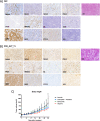
Fig. 6. Histological and immunohistochemical profiles for the ambiguous endometrial carcinoma.
Histological (HE) and immunophenotype of the ambiguous endometrial carcinoma based on a set of proteins described previously to help discriminate between SEC and differentiated EECs: MSH6, MLH1, MSH2, PMS2, PTEN, HMGA2, ER, ki67, IMP2, IMP3, CYC E1 and p53 [24] in AEC patient (A) and PDX derived from tumor 1 (B). Magnification ×20. C Growth of AEC-derived PDX tumors treated with the drugs indicated for 30 days. Tumor volumes were measured three times each week with a digital caliper and the volumes were calculated as: (length × width2)/2 = mm3; *p-value < 0.05, significantly different placebo vs. Carboplatin-Paclitaxel; +p-value < 0.05, significantly different placebo vs. Olaparib analyzed by one-way ANOVA. The data show the mean of 6–7 independent mice for each treatment.
Fig. 7
Fig. 7. Summary of the procedure followed for samples included in the study.
WES whole exome sequencing, P primary tumor, M metastasis, CNA copy number aberrations, PDX patient-derived xenografts.

Similar articles

Cited by

References

    1. Siegel RL, Miller KD, Jemal A. Cancer statistics, 2020. CA Cancer J Clin. 2020;70:7–30. - PubMed
    1. Bokhman JV. Two pathogenetic types of endometrial carcinoma. Gynecol Oncol. 1983;15:10–7. - PubMed
    1. Lax SF, Kurman RJ. A dualistic model for endometrial carcinogenesis based on immunohistochemical and molecular genetic analyses. Verh Dtsch Ges Pathol. 1997;81:228–32. - PubMed
    1. Kandoth C, Schultz N, Cherniack AD, Akbani R, Liu Y, Shen H, et al. Integrated genomic characterization of endometrial carcinoma. Nature. 2013;497:67–73. - PMC - PubMed
    1. Soslow RA. Endometrial carcinomas with ambiguous features. Semin Diagn Pathol. 2010;27:261–73.. - PubMed

Publication types