The role of iliocapsularis in hip pathology: a scoping review
- PMID: 35145711
- PMCID: PMC8826026
- DOI: 10.1093/jhps/hnab057
The role of iliocapsularis in hip pathology: a scoping review
Abstract
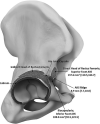
The iliocapsularis is a relatively unheard-of muscle, located deep in the hip covering the anteromedial capsule of the hip joint. Little is known about this constant muscle despite its clinical relevance. The aims of this scoping review are to collate the various research studies reporting on the detailed anatomy and function of iliocapsularis and to demonstrate how inter-individual differences in iliocapsularis can be used as a clinical adjunct in guiding diagnosis and treatment of certain hip joint pathologies. A computer-assisted literature search was conducted based on Preferred Reporting Items for Systematic Reviews and Meta-Analyses guidelines. Our review found 13 studies including 384 cases meeting our inclusion criteria. About 53.8% of the studies involved human cadavers. The current scoping review indicates the relevant anatomy of the iliocapsularis, being a small muscle which arises from the inferior border of the anterior inferior iliac spine and anteromedial capsule of the hip joint, inserting distal to the lesser trochanter. Therefore, based upon these anatomical attachments, iliocapsularis acts as a dynamic stabilizer by tightening the anterior capsule of the hip joint. Implications of this association may be that the muscle is hypertrophied in dysplastic or unstable hips. Determining the size of the iliocapsularis could be of conceivable use in patients with hip symptoms featuring signs of both borderline hip dysplasia and subtle cam-type deformities. Although future research is warranted, this study will aid physicians to understand the clinical importance of the iliocapsularis.
© The Author(s) 2021. Published by Oxford University Press.
Figures


Similar articles
-
An increased iliocapsularis-to-rectus-femoris ratio is suggestive for instability in borderline hips.Clin Orthop Relat Res. 2015 Dec;473(12):3725-34. doi: 10.1007/s11999-015-4382-y. Clin Orthop Relat Res. 2015. PMID: 26088766 Free PMC article.
-
Anatomical features of the iliocapsularis muscle: a dissection study.Surg Radiol Anat. 2022 Apr;44(4):599-608. doi: 10.1007/s00276-022-02905-y. Epub 2022 Feb 26. Surg Radiol Anat. 2022. PMID: 35218407 Free PMC article.
-
Anatomy of the iliocapsularis muscle. Relevance to surgery of the hip.Clin Orthop Relat Res. 2000 May;(374):278-85. doi: 10.1097/00003086-200005000-00025. Clin Orthop Relat Res. 2000. PMID: 10818987
-
[Preoperative MR imaging for hip dysplasia : Assessment of associated deformities and intraarticular pathologies].Orthopadie (Heidelb). 2023 Apr;52(4):300-312. doi: 10.1007/s00132-023-04356-8. Epub 2023 Mar 28. Orthopadie (Heidelb). 2023. PMID: 36976331 Free PMC article. Review. German.
-
Anatomical variation of the Psoas Valley: a scoping review.BMC Musculoskelet Disord. 2020 Apr 10;21(1):219. doi: 10.1186/s12891-020-03241-1. BMC Musculoskelet Disord. 2020. PMID: 32276620 Free PMC article.
Cited by
-
Capsular Management During Hip Arthroscopy.Curr Rev Musculoskelet Med. 2023 Dec;16(12):607-615. doi: 10.1007/s12178-023-09855-x. Epub 2023 Jul 12. Curr Rev Musculoskelet Med. 2023. PMID: 37436651 Free PMC article. Review.
References
-
- Ward WT, Fleisch ID, Ganz R. Anatomy of the iliocapsularis muscle. Relevance to surgery of the hip. Clin Orthop Relat Res 2000; 374: 278–85. - PubMed
-
- Cruveilhier J. Traité D’anatomie Descriptive, 2nd edn. Paris: Ancienne Maison Béchet Jeune, 1843, 130–1.
-
- Ganz R, Klaue K, Vinh TS. et al. A new periacetabular osteotomy for the treatment of hip dysplasias. Technique and preliminary results. Clin Orthop Relat Res 1988; 232: 26–36. - PubMed
-
- Dora C, Houweling M, Koch P. et al. Iliopsoas impingement after total hip replacement: the results of non-operative management, tenotomy or acetabular revision. J Bone Joint Surg Br 2007; 89: 1031–5. - PubMed
-
- Bedi A, Galano G, Walsh C. et al. Capsular management during hip arthroscopy: from femoroacetabular impingement to instability. Arthroscopy 2011; 27: 1720–31. - PubMed
Publication types
LinkOut - more resources
Full Text Sources

