

Netarsudil-associated reticular corneal epithelial edema
- PMID: 35146185
- PMCID: PMC8801350
- DOI: 10.1016/j.ajoc.2022.101287
Netarsudil-associated reticular corneal epithelial edema
Abstract
Purpose: To describe 8 cases of reversible reticular corneal epithelial edema of the cornea that developed after use of the topical Rho-kinase inhibitor netarsudil.
Methods: This is a retrospective chart review case series of 8 patients treated with netarsudil at an academic medical center.
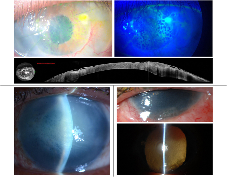
Observations: Patients had predisposing corneal conditions including penetrating keratoplasty, corneal decompensation after trabeculectomy-associated endophthalmitis, congenital glaucoma with Haab striae, aphakic bullous keratopathy, history of Ahmed valve and silicone oil, and Fuchs endothelial corneal dystrophy undergoing Descemet stripping only. One patient did not have clear predisposing corneal disease other than low endothelial cell density and a history of trabeculectomy. All patients developed reticular corneal epithelial edema, which appeared as collections of moderate sized superficial epithelial bullae arranged in a reticular pattern resembling a honeycomb. Most developed these changes within weeks of initiating netarsudil, but unique to this series are 2 cases in which netarsudil was tolerated by the cornea for months before developing reticular corneal epithelial edema after diode laser cyclophotocoagulation. In cases which underwent anterior segment optical coherence tomography, the imaging demonstrated that the corneal stroma was not edematous, and the reticular corneal epithelial edema involved both host and donor corneal epithelium in cases of penetrating keratoplasty. This fully resolved in all cases upon cessation of netarsudil, and this series is the first to document resolution via a pattern in which the individual bullae become smaller and more widely spaced apart.
Conclusion: Netarsudil can cause a reversible reticular corneal epithelial edema.
Keywords: Corneal edema; Honeycomb edema; Netarsudil; Reticular edema; Rhopressa.
© 2022 The Authors.
Conflict of interest statement
No conflicting relationship exists for any author.
Figures


References
-
- Serle J.B., Katz L.J., McLaurin E., et al. Two phase 3 clinical trials comparing the safety and efficacy of netarsudil to timolol in patients with elevated intraocular pressure: rho kinase elevated IOP treatment trial 1 and 2 (ROCKET-1 and ROCKET-2) Am J Ophthalmol. 2018;186:116–127. - PubMed
-
- Kahook M.Y., Serle J.B., Mah F.S., et al. Long-term safety and ocular hypotensive efficacy evaluation of netarsudil ophthalmic solution: rho kinase elevated IOP treatment trial (ROCKET-2) Am J Ophthalmol. 2019;200:130–137. - PubMed
-
- Macsai M.S., Shiloach M. Use of topical rho kinase inhibitors in the treatment of Fuchs dystrophy after Descemet stripping only. Cornea. 2019;38(5):529–534. - PubMed
-
- Davies E. Case series: novel utilization of rho-kinase inhibitor for treatment of corneal edema. Cornea. 2021;40:116–120. - PubMed
-
- Fernandez M.M. Reticular epithelial edema in edematous corneas treated with netarsudil. Ophthalmology. 2018;125:1709. - PubMed
Publication types
LinkOut - more resources
Full Text Sources
