Review
. 2021 Dec 10;29(4):513-525.
doi: 10.53854/liim-2904-3. eCollection 2021.

Neglected and (re-)emergent infections of the CNS i n low-/middle-income countries

Affiliations
Review

Neglected and (re-)emergent infections of the CNS i n low-/middle-income countries

Graciela Cárdenas et al. Infez Med. .

Abstract

Low- and middle-income countries (LMIC) have suffered from long-term health system deficiencies, worsened by poor living conditions, lack of sanitation, a restricted access to health facilities and running water, overcrowding, and overpopulation. These factors favor human displacement and deepen marginalization; consequently, their population endures a high burden of infectious diseases. In this context, the current epidemiological landscape and its impact on health and economic development are not promissory, despite the commitment by the international community to eradicate neglected tropical infections - especially tuberculosis and malaria, by 2030. Neglected and (re)-emerging infectious diseases affecting the central nervous system (CNS) are a major public health concern in these countries, as they cause a great morbidity and mortality; furthermore, survivors often suffer from severe neurological disabilities. Herein, we present a retrospective review focused on some neglected and (re)-emerging infectious diseases, including neurocysticercosis, malaria, rabies, West Nile virus encephalitis, tuberculosis, neuroborreliosis, and SARS-CoV-2 in LMIC. A retrospective review of studies on selected neglected and (re)-emerging infectious diseases in LMIC was performed, including reports by the World Health Organization (WHO) published within the last five years. Data on infection by SARS-CoV-2 were provided by the John Hopkins University Coronavirus Resource Center. CNS neglected and (re)-emerging infectious diseases remain as important causes of disease in LMIC. An alarming increase in the prevalence of malaria, tuberculosis, and cysticercosis is observed in the region, compounded by the recent COVID-19 pandemic. The WHO is currently supporting programs/efforts to cope with these diseases. Herein, we highlight the epidemiological burden of some CNS infections in LMIC, and their clinical and neuroimaging features, to facilitate an accurate diagnosis, considering that most of these diseases will not be eradicated in the short term; instead, their incidence will likely increase along with poverty, inequality, and related socioeconomic problems.

Keywords: Neglected; central nervous system; emergent; infections; low/middle income countries.


Conflict of interest statement

Competing interests statement The authors declare that no conflict of interest exists.

Figures

Figure 1
Figure 1
Neurocysticercosis. A) Axial CT shows parenchymal neurocysticercosis with multiple vesicular lesions, some of them with visible scolex. B) Axial MRI, contrast-enhanced T1-weighted sequence shows parenchymal miliary colloidal lesions with gadolinium enhancement. C) Axial CT shows multiple parenchymal calcified lesions. D) Axial MRI shows a solitary vesicular lesion with a prominent scolex lodged at left frontal region. E) Brain coronal pathological specimen shows vesicular miliary neurocysticercosis. F) Sagittal MRI shows a solitary colloidal cysticercus in the fourth ventricle. G) Axial MRI T1-weighted shows multiple vesicular lesions at subarachnoid space of the base. H) Axial MRI on FIESTA sequence shows subarachnoid vesicular lesions. I) Ventral side of a brain pathological specimen shows multiple vesicular lesions at the basal cisterns.
Figure 2
Figure 2
Malaria. A) Axial MRI T2-weighted sequence shows multiple hyperintense lesions in the hemispheric white matter. B) and C) Axial MRI on FLAIR sequences show hyperintense lesions in the in the hemispheric white matter and basal ganglia nuclei.
Figure 3
Figure 3
Rabies. A 24-year-old woman with a history of being bitten by a grey fox (Urocyon cinereoargenteus) three weeks before hospital admission. Upon emergency admission, A) MRI T1-weighted sequence shows no apparent lesions. B) and C). FLAIR sequences show faint lesions at basal ganglia (globus pallidus). D) Sagittal MRI of the cervical spinal cord shows intramedullary longitudinal hyperintense lesion. E) Axial MRI at a cervical level shows hyperintense lesion in the gray matter. F) Post-mortem hematoxylin-eosin-stained section at the cerebellar cortex shows eosinophilic neuronal cytoplasmic inclusions (Negri bodies).
Figure 4
Figure 4
West Nile virus encephalitis. A) Axial MRI T1-weighted shows bilateral hyperintensities at the basal ganglia, more prominently at the left side. B) Axial contrast-enhanced MRI bilateral enhancement, nodular, at the left side of basal ganglia. C) Axial MRI on diffusion sequence shows bilateral hyperintensities at the basal ganglia. D) Sagittal MRI at thoracic level shows an intramedullary longitudinal hyperintense lesion. E) Axial MRI contrast enhanced T1 sequence at a lumbar level shows enhancement and thickening of the spinal roots the cauda equina. F) Axial pathological specimen of the spinal cord shows right side hemorrhagic lesion.ì
Figure 5
Figure 5
Tuberculosis. A) Coronal MRI at a thoracic level shows an abscess with intervertebral disc involvement (spondylodiscitis/Pott disease). B) Sagittal MRI imaging at a thoracic level shows an abscess involving two vertebral bodies (spondylodiscitis/Pott disease). C) Axial MRI T1-weighted shows a heterogeneous, hypointense left-side basal ganglia lesion. D) Axial MRI T2-weighted shows an isointense capsule and important vasogenic edema involving left frontal and insular regions. E) Axial contrast-enhanced MRI T1-weighted shows basal meningitis (arachnoiditis) and a mycotic aneurism at the right middle cerebral artery. F) Alcohol-acid resistance bacilli are observed in the purulent aspiration of an abscess.
Figure 6
Figure 6
Lyme disease. A) Axial contrast-enhanced MRI at the level of intracanalicular, genicular ganglion and tympanic portions of the facial nerve. Clinically, patients presented an atypical Bell’s palsy. B) Coronal contrast-enhanced MRI shows limbs of the facial nerve. C) and D) Sagittal and coronal contrast-enhanced MRI shows the descending portion of the facial nerve.
Figure 7
Figure 7
(A–L) - Neuro-COVID-19. Cerebrovascular manifestations Case 1. (A to D) A 37-year-old female which developed atypical pneumonia and SARS-CoV-2 infection was confirmed by a positive RT-PCR. After two-weeks of respiratory symptoms, suddenly complained of headache and left hemiparesis. In the MRI, it was observed on FLAIR sequence a hyperintense right temporal lesion (A), hypointense on SWAN sequence (B) which involved superior, media and inferior gyrus, highly suggested of a cerebral infarction with hemorrhage transformation. After the administration of gadolinium, a cortical gyral enhancement (C) and thickening of the infundibulum was observed (D, long yellow arrow). Case 2. (E) A 58-year-old woman, with developed atypical pneumonia by COVID-19, she suddenly developed right anisocoria. CT scan showed ischemic stroke involving the temporal posterior region by obstruction of the right middle cerebral artery without mass effect. Case 3. (F to H) A 48-year male with atypical pneumonia by COVID-19, he developed severe hypoxia with neurological worsening characterized by fixed symmetrical pupils. CT scan showed multiple hyperdense lesions (infra and supratentorial hemorrhages), associated with cytotoxic edema as well as hyperdensity in both M1 segments of middle cerebral artery. Case 4. (I to L) A 70-year-old woman with COVID-10 pneumonia which developed hyperintense (anoxic) brain lesions observed in T1, FLAIR, diffusion, and SWAN.
Figure 7
Figure 7
(M–V) - Neuro-COVID-19. Nerve and pachymeningeal enhancement manifestations Case 5. (M to R) A 29-year-old female which was hospitalized by a psychotic episode and after 14-days, she developed dry cough and atypical pneumonia by SARS-CoV-2. In the MRI, after gadolinium administration it was observed post-contrast enhancement in the olfactory grooves (M; yellow arrow), in the optic nerves and pathway (N, O), in the acoustic pores (P) and geniculate ganglia, as well as global (Q, R) and pre-pontine crura pachymeningeal enhancement. Case 6. (S to V) A 45-year-old male which developed atypical pneumonia by SARS-CoV-2 and showed severe respiratory distress. After one month of recovery, he developed headaches, blurred vision, dizziness, and vertigo. High IgM and low IgG anti-SARS-CoV-2 antibody titers were detected. At MRI, after gadolinium administration it was observed a slight enhancement of the cochlear nerves and vestibular complexes (S and T, yellow circles), as well as the right geniculate ganglia. There was a marked enhancement in the cavernous sinuses and rostral to the cortices of the temporal lobes (U and V).

Similar articles

Cited by

References

    1. Furuse Y. Analysis of research intensity on infectious disease by disease burden reveals which infectious diseases are neglected by researchers. Proc Natl Acad Sci USA. 2019;116(2):478–83. - PMC - PubMed
    1. GBD 2016 DALY and HALE collaborators global, regional, and national disability-adjusted life-years (DALYs) for 333 diseases and injuries and healthy life expectancy (HALE) for 195 countries and territories, 1990–2012. A systematic analysis for the Global Burden of Disease Study 2016. Lancet. 2017;390(10100):1260–344. - PMC - PubMed
    1. World Population Ageing 2020 Highlights. [[accessed November 1, 2021].]. https://www.un.org/development/desa/pd/sites/www.un.org/development.desa... .
    1. World Population 2019. [[accessed November 1, 2021].]. At https://population.un.org/wpp/Publications/Files/WPP2019-wallchart.pdf.
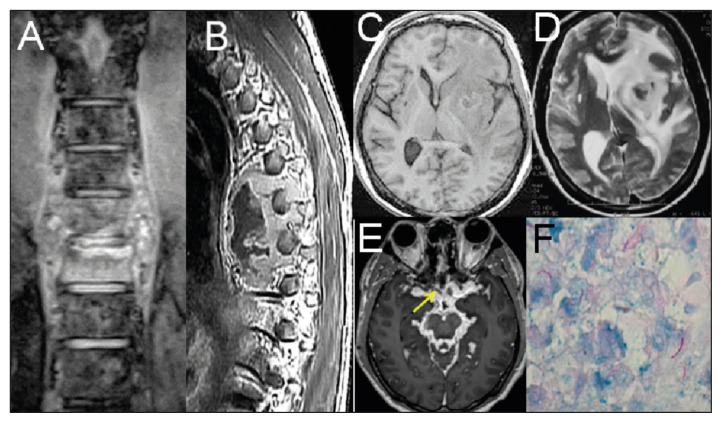
    1. Kimura-Hayama ET, Higuera JA, Corona-Cedillo R, et al. Neurocysticercosis:radiologic-pathologic correlation. Radiographics. 2010;30(6):1705–19. - PubMed

LinkOut - more resources