

Does dentine mineral change with anatomical location, microscopic site and patient age?
- PMID: 35146411
- PMCID: PMC8818708
- DOI: 10.1016/j.yjsbx.2022.100060
Does dentine mineral change with anatomical location, microscopic site and patient age?
Abstract
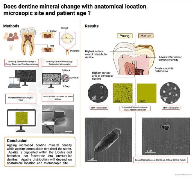
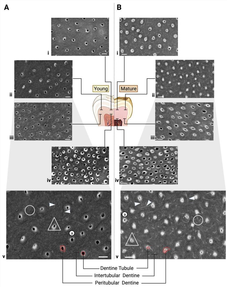
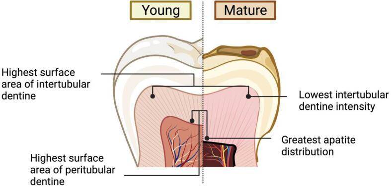
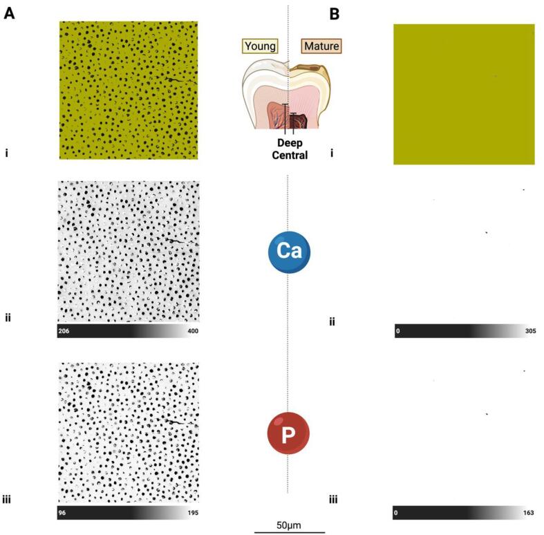
Objective: To determine the effect of patient age (young or mature), anatomical location (shallow/deep and central/peripheral) and microscopic site (intertubular/peritubular) on dentine mineral density, distribution and composition.
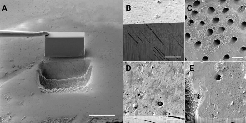
Methods: Extracted posterior teeth from young (aged 19-20 years, N = 4) and mature (aged 54-77 years, N = 4) subjects were prepared to shallow and deep slices. The dentine surface elemental composition was investigated in a SEM using Backscattered Electron (BSE) micrographs, Energy Dispersive X-ray Spectroscopy, and Integrated Mineral Analysis. Qualitative comparisons and quantitative measures using machine learning were used to analyse the BSE images. Quantitative outcomes were compared using quantile or linear regression models with bootstrapping to account for the multiple measures per sample. Subsequently, a Xenon Plasma Focussed Ion Beam Scanning Electron Microscopy (Xe PFIB-SEM) was used to mill large area (100 µm) cross-sections to investigate morphology through the dentine tubules using high resolution secondary electron micrographs.
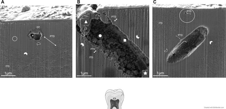
Results: With age, dentine mineral composition remains stable, but density changes with anatomical location and microscopic site. Microscopically, accessory tubules spread into intertubular dentine (ITD) from the main tubule lumens. Within the lumens, mineral deposits form calcospherites in the young that eventually coalesce in mature tubules and branches. The mineral occlusion in mature dentine increases overall ITD density to reflect peritubular dentine (PTD) infiltrate. The ITD observed in micrographs remained consistent for age and observation plane to suggest tubule deposition affects overall dentine density. Mineral density depends on the relative distribution of PTD to ITD that varies with anatomical location.
Significance: Adhesive materials may interact differently within a tooth as well as in different age groups.
Keywords: Age; Apatite; BSE; BSE, Backscatter Electron; Ca, Calcium; Cl, Chloride; DEJ, Dentine-enamel junction; DT, Dentine Tubule; Dentine; EPMA, Electron Probe Microanalyser; Ga, Gallium; H, Hydrogen; Human; ITD, Intertubular Dentine; Intertubular dentine; LA-ICP-MS, Laser Ablation Induction Coupled Plasma Mass Spectroscopy; Mg, Magnesium; Mineral; Na, Sodium; O, Oxygen; Odontoblasts; P, Phosporus; PTD, Peritubular Dentine; Peritubular dentine; SEM, Scanning Electron Microscope; SEM-EDS; SEM-EDS, Scanning Electron Microscope Energy Dispersive X-ray Spectroscopy; TEM, Transmission Electron Microscope; TIMA, Integrated Mineral Analysis; XE PFIB-SEM, Xenon Plasma Focussed Ion Beam Scanning Electron Microscope; Xe PFIB-SEM; β-TCMP, Magnesium-whitlockite.
© 2022 The Author(s).
Conflict of interest statement
The authors declare that they have no known competing financial interests or personal relationships that could have appeared to influence the work reported in this paper.
Figures

Similar articles
-
Effect of dentine site on resin and cement adaptation tested using X-ray and electron microscopy to evaluate bond durability and adhesive interfaces.Eur J Oral Sci. 2022 Oct;130(5):e12890. doi: 10.1111/eos.12890. Epub 2022 Aug 12. Eur J Oral Sci. 2022. PMID: 35959863 Free PMC article.
-
Structure-Function Correlative Microscopy of Peritubular and Intertubular Dentine.Materials (Basel). 2018 Aug 21;11(9):1493. doi: 10.3390/ma11091493. Materials (Basel). 2018. PMID: 30134596 Free PMC article.
-
The composition of bovine peritubular dentin: matching TOF-SIMS, scanning electron microscopy and biochemical component distributions. New light on peritubular dentin function.Cells Tissues Organs. 2009;189(1-4):12-9. doi: 10.1159/000151726. Epub 2008 Aug 26. Cells Tissues Organs. 2009. PMID: 18728348 Free PMC article.
-
Ultrastructure of human dentine 40 years ago--progress and perspectives.Arch Oral Biol. 1999 Dec;44(12):979-84. doi: 10.1016/s0003-9969(99)00109-0. Arch Oral Biol. 1999. PMID: 10669074 Review.
-
Ultrastructure of dentine carious lesions.Arch Oral Biol. 2008 Feb;53(2):124-32. doi: 10.1016/j.archoralbio.2007.08.007. Epub 2007 Oct 29. Arch Oral Biol. 2008. PMID: 17915189 Review.
Cited by
-
Effect of dentine site on resin and cement adaptation tested using X-ray and electron microscopy to evaluate bond durability and adhesive interfaces.Eur J Oral Sci. 2022 Oct;130(5):e12890. doi: 10.1111/eos.12890. Epub 2022 Aug 12. Eur J Oral Sci. 2022. PMID: 35959863 Free PMC article.
References
-
- Arganda-Carreras I., Kaynig V., Rueden C., Eliceiri K.W., Schindelin J., Cardona A., Sebastian Seung H., Murphy R. Trainable Weka Segmentation: a machine learning tool for microscopy pixel classification. Bioinformatics. 2017;33(15):2424–2426. - PubMed
-
- Arnold W.H., Konopka S., Gaengler P. Qualitative and Quantitative Assessment of Intratubular Dentine Formation in Human Natural Carious Lesions. Calcif. Tissue Int. 2001;69:268–273. - PubMed
-
- Bajaj D., Sundaram N., Nazari A., Arola D. Age, dehydration and fatigue crack growth in dentine. Biomaterials. 2006;27(11):2507–2517. - PubMed
-
- Blatz M.B., Chiche G., Bahat O., Roblee R., Coachman C., Heymann H.O. Evolution of aesthetic dentistry. J. Dent. Res. 2019;98(12):1294–1304. - PubMed
LinkOut - more resources
Full Text Sources
