. 2022 Jan 24:3:100087.
doi: 10.1016/j.crphar.2022.100087. eCollection 2022.

GSK2606414 attenuates PERK/p-eIF2α/ATF4/CHOP axis and augments mitochondrial function to mitigate high glucose induced neurotoxicity in N2A cells

Affiliations

GSK2606414 attenuates PERK/p-eIF2α/ATF4/CHOP axis and augments mitochondrial function to mitigate high glucose induced neurotoxicity in N2A cells

Chayanika Gundu et al. Curr Res Pharmacol Drug Discov. .

Abstract

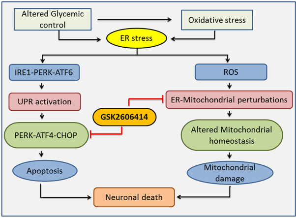
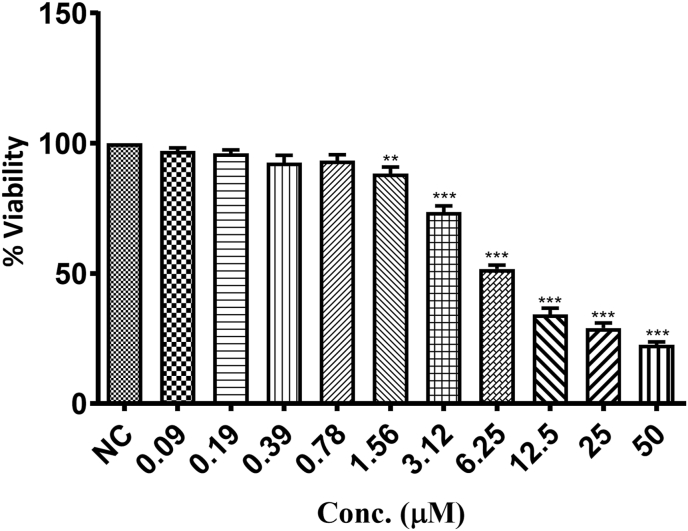
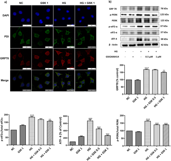
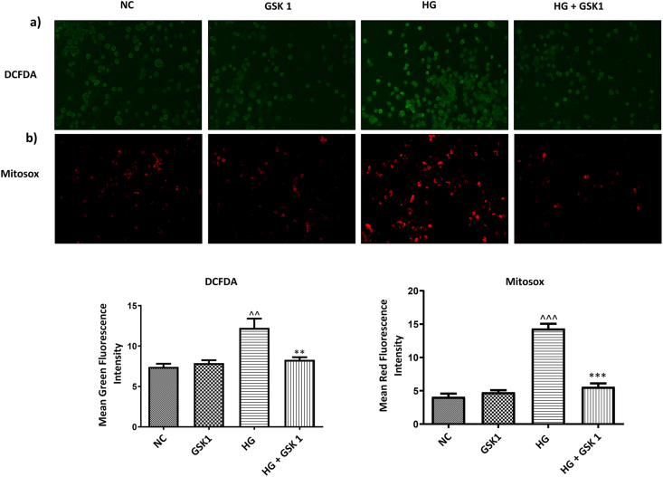
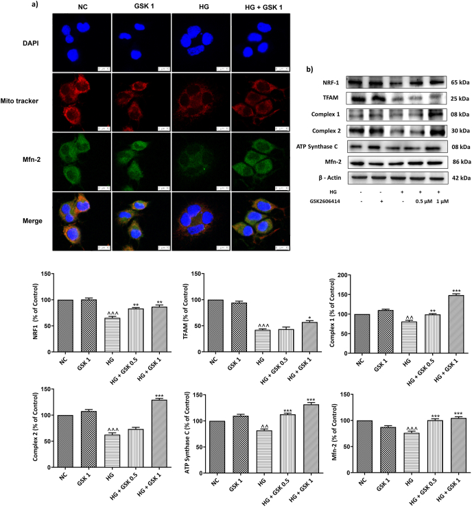
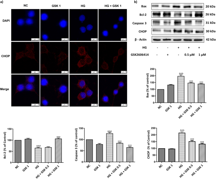
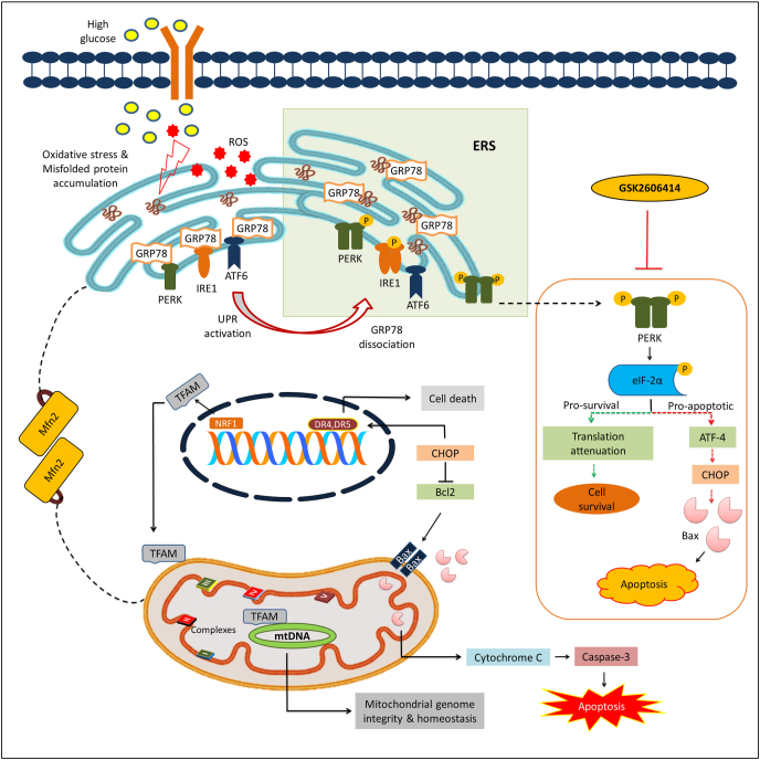
Neuronal dysfunction and subsequent apoptosis under high glucose conditions during diabetes contribute majorly to the manifestation of diabetic peripheral neuropathy (DPN). PERK (protein kinase RNA (PKR)-like ER kinase) one among the three canonical arms of unfolded protein response (UPR), is believed to play a crucial role in determining the cell fate during endoplasmic reticulum stress (ERS/ER stress) conditions. We evaluated the role of PERK inhibitor GSK2606414 in high glucose (30 ​mM) treated neuroblastoma (N2A) cells. High glucose resulted in disruption of ER proteostasis by activation of UPR which is evident through increased (p ​< ​0.001) expression of GRP78, p-PERK, p-eIF2α, ATF-4 and CHOP when compared to normal cells. It is accompanied with enhanced GRP78 localization in Endoplasmic Reticulum (ER) lumen evident from ER labeling Immunofluorescence (IF) staining. PERK activation resulted in altered mitochondrial function evident by increased mitochondrial superoxide production and compromised mitochondrial homeostasis with decrease in Mfn-2 levels. Additionally, ER stress induced neuronal apoptosis was attenuated by GSK2606414 treatment via inhibiting the PERK-eIF2α-ATF4-CHOP axis that not only curtailed the levels of apoptotic proteins like Bax and caspase 3 but also elevated the levels of anti-apoptotic Bcl-2. Collectively, our findings revealed the neuroprotective potential of GSK2606414 against high glucose induced neurotoxicity in N2A cells.

Keywords: ER stress; GSK2606414; High glucose; Mitochondrial function; PERK.


Conflict of interest statement

The authors declare that they have no known competing financial interests or personal relationships that could have appeared to influence the work reported in this paper.

Figures

Image 1
Graphical abstract
Fig. 1
Fig. 1
Effect of GSK2606414 on cell viability. Graphical representation of cell viability of N2A cells treated with different concentrations (0.1–100 ​μM) of GSK2606414 with high glucose (30 ​mM). Values are expressed as mean ​± ​SEM (n ​= ​3). ∗∗P ​< ​0.01 and ∗∗∗P ​< ​0.001 Vs Normal N2A cells (NC).
Fig. 2
Fig. 2
Effect of GSK2606414 on ER stress markers. (a) Immunofluorescence staining of N2A cells, representing co-expression of PDI & GRP78. Photographs were taken at 400× magnification (scale: 25 ​μM) (b) Representative immunoblots of ER stress markers. Values are expressed as mean ​± ​SEM (n ​= ​3). NC: normal N2A cells, GSK 1: normal N2A cells treated with GSK260414 (1 ​μM), HG: hyperglycemic N2A cells (30 ​mM β-D glucose), HG+ ​GSK 0.5: HG cells treated with GSK260414 (0.5 ​μM), HG+ ​GSK1: HG cells treated with GSK260414 (1 ​μM). ^^^P ​< ​0.001 v/s NC and ∗∗P ​< ​0.01, ∗∗∗P ​< ​0.001 v/s HG.
Fig. 3
Fig. 3
Effect of GSK2606414 on ROS and mitochondrial function. Representative images and corresponding bar graphs of (a) DCFDA staining (b) Mitosox staining. Photographs were taken at 200× magnification (scale: 100 ​μM). Values are expressed as mean ​± ​SEM (n ​= ​3). NC: normal N2A cells, GSK 1: normal N2A cells treated with GSK260414 (1 ​μM), HG: hyperglycemic N2A cells (30 ​mM β-D glucose), HG+ ​GSK 0.5: HG cells treated with GSK260414 (0.5 ​μM), HG+ ​GSK1: HG cells treated with GSK260414 (1 ​μM), ^^P ​< ​0.01, ^^^P ​< ​0.001 v/s NC and ∗∗P ​< ​0.01, ∗∗∗P ​< ​0.001 v/s HG.
Fig. 4
Fig. 4
(a) Immunofluorescence staining of Mfn-2 (green) along with mitotracker (red) and (b) Representative immunoblots of proteins depicting mitochondrial function. Photographs were taken at 400× magnification (scale: 10 ​μM). Values are expressed as mean ​± ​SEM (n ​= ​3). NC: normal N2A cells, GSK 1: normal N2A cells treated with GSK260414 (1 ​μM), HG: hyperglycemic N2A cells (30 ​mM β-D glucose), HG+ ​GSK 0.5: HG cells treated with GSK260414 (0.5 ​μM), HG+ ​GSK1: HG cells treated with GSK260414 (1 ​μM), ^^P ​< ​0.01, ^^^P ​< ​0.001 v/s NC and ∗ P ​< ​0.05, ∗∗P ​< ​0.01, ∗∗∗P ​< ​0.001 v/s HG. (For interpretation of the references to color in this figure legend, the reader is referred to the Web version of this article.)
Fig. 5
Fig. 5
Effect of GSK2606414 on apoptosis. (a) Immunofluorescence staining of CHOP (red) (b) Representative immunoblots of proteins involved in apoptosis. Photographs were taken at 400× magnification (scale: 10 ​μM). Values are expressed as mean ​± ​SEM (n ​= ​3). NC: normal N2A cells, GSK 1: normal N2A cells treated with GSK260414 (1 ​μM), HG: hyperglycemic N2A cells (30 ​mM β-D glucose), HG+ ​GSK 0.5: HG cells treated with GSK260414 (0.5 ​μM), HG+ ​GSK1: HG cells treated with GSK260414 (1 ​μM). ^^^P ​< ​0.001 v/s NC and ∗∗∗P ​< ​0.001 v/s HG. (For interpretation of the references to color in this figure legend, the reader is referred to the Web version of this article.)
Fig. 6
Fig. 6
Probable mechanistic basis of GSK2606414 against high glucose induced neurotoxicity. High glucose exposure to N2A cells causes oxidative stress and alters protein folding capacity of ER by accumulation of misfolded/unfolded proteins that triggers UPR. UPR is executed by activation of three transmembrane proteins and their respective downstream signalling events by which ERS response exhibits a dichotomic module. PERK plays a critical role in UPR which decides the cell fate where initial mild or acute stress favors the activation of pro-survival module (represented in green dotted line) that results in either amelioration of initial stress or adaptation to it. However, chronic stress induces cell death by activating pro-apoptotic events (represented in red dotted line). Further ERS potentiates mitochondrial dysfunction by multiple mechanisms including excess generation of ROS, CHOP/Bax/Cytochrome C release and altering ER-mitochondria tethering via modulation of Mfn2. GSK2606414, a synthetic PERK inhibitor, modulates the PERK/p-eIF2α/ATF4/CHOP signalling axis and augments mitochondrial function to mitigate high glucose induced neurotoxicity in N2A cells. GRP78: glucose-regulated protein of 78 ​kDa, ROS: Reactive Oxygen Species, PERK: protein kinase activated by double stranded RNA-like ER kinase, ATF-6: Activating transcription factor 6, ATF-4: Activating transcription factor 4, eIF2α: Eukaryotic Initiation Factor 2 α, IRE1α: Inositol-requiring enzyme 1 alpha, UPR: Unfolded Protein Response, CHOP: C/EBP homologous protein, Mfn2: Mitofusin 2, Bax: BCL2-associated X protein, Bcl2: B-cell lymphoma 2, DR4: Death receptor 4, DR5: Death receptor 5, TFAM: Mitochondrial transcription factor A, NRF1: Nuclear respiratory factor 1, mtDNA: Mitochondrial DNA. (For interpretation of the references to color in this figure legend, the reader is referred to the Web version of this article.)

References

    1. Adams C.J., Kopp M.C., Larburu N., Nowak P.R., Ali M.M. Structure and molecular mechanism of ER stress signaling by the unfolded protein response signal activator IRE1. Front. Mol. Biosci. 2019;6:11. - PMC - PubMed
    1. Arruri V., Komirishetty P., Areti A., Dungavath S.K.N., Kumar A. Nrf2 and NF-κB modulation by Plumbagin attenuates functional, behavioural and biochemical deficits in rat model of neuropathic pain. Pharmacol. Rep. 2017;69:625–632. - PubMed
    1. Arruri V.K., Gundu C., Kalvala A.K., Sherkhane B., Khatri D.K., Singh S.B. Carvacrol abates NLRP3 inflammasome activation by augmenting Keap1/Nrf-2/p62 directed autophagy and mitochondrial quality control in neuropathic pain. Nutr. Neurosci. 2021:1–16. doi: 10.1080/1028415X.2021.1892985. - DOI - PubMed
    1. Augusto L., Martynowicz J., Staschke K.A., Wek R.C., Sullivan W.J., Jr. Effects of PERK eIF2α kinase inhibitor against Toxoplasma gondii. Antimicrob. Agents Chemother. 2018;62 e01442-01418. - PMC - PubMed
    1. Axten J.M., Medina J.s.R., Feng Y., Shu A., Romeril S.P., Grant S.W., Li W.H.H., Heerding D.A., Minthorn E., Mencken T. Discovery of 7-methyl-5-(1-{[3-(trifluoromethyl) phenyl] acetyl}-2, 3-dihydro-1 H-indol-5-yl)-7 H-pyrrolo [2, 3-d] pyrimidin-4-amine (GSK2606414), a potent and selective first-in-class inhibitor of protein kinase R (PKR)-like endoplasmic reticulum kinase (PERK) J. Med. Chem. 2012;55:7193–7207. - PubMed