. 2022 Apr;388(1):19-32.
doi: 10.1007/s00441-022-03594-0. Epub 2022 Feb 11.

Morphologies, dimensions and targets of gastric nitric oxide synthase neurons

Affiliations

Morphologies, dimensions and targets of gastric nitric oxide synthase neurons

Madeleine R Di Natale et al. Cell Tissue Res. 2022 Apr.

Abstract

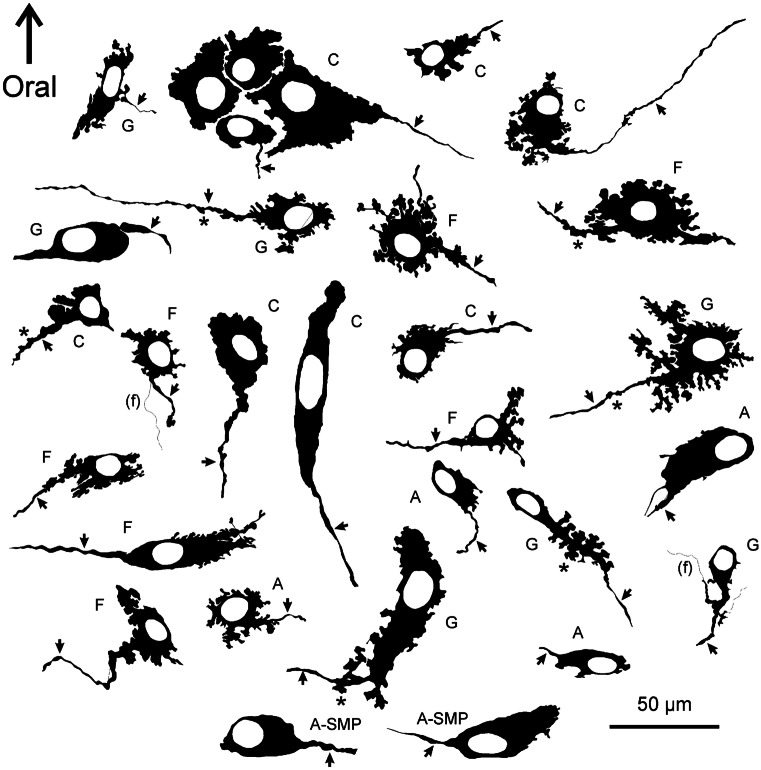
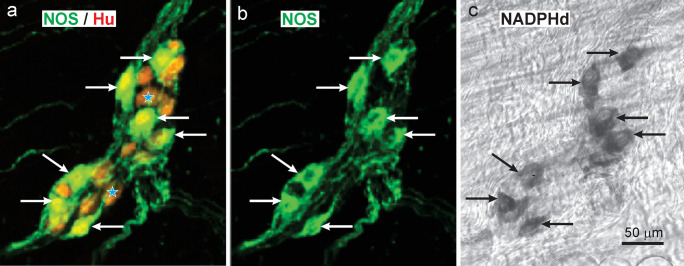
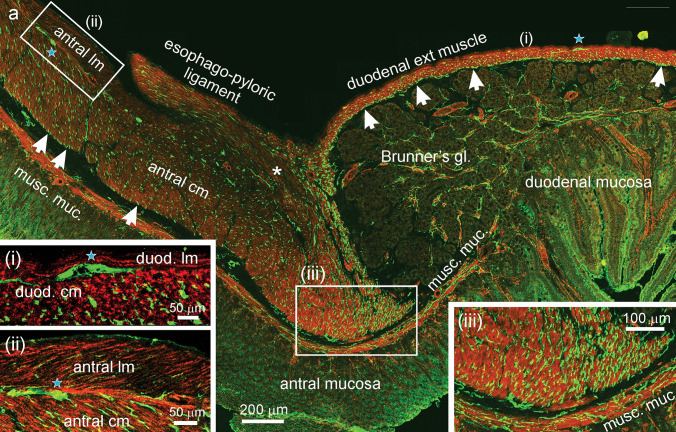
We investigated the distributions and targets of nitrergic neurons in the rat stomach, using neuronal nitric oxide synthase (NOS) immunohistochemistry and nicotinamide adenine dinucleotide phosphate (NADPH) diaphorase histochemistry. Nitrergic neurons comprised similar proportions of myenteric neurons, about 30%, in all gastric regions. Small numbers of nitrergic neurons occurred in submucosal ganglia. In total, there were ~ 125,000 neuronal nitric oxide synthase (nNOS) neurons in the stomach. The myenteric cell bodies had single axons, type I morphology and a wide range of sizes. Five targets were identified, the longitudinal, circular and oblique layers of the external muscle, the muscularis mucosae and arteries within the gastric wall. The circular and oblique muscle layers had nitrergic fibres throughout their thickness, while the longitudinal muscle was innervated at its inner surface by fibres of the tertiary plexus, a component of the myenteric plexus. There was a very dense innervation of the pyloric sphincter, adjacent to the duodenum. The muscle strands that run between mucosal glands rarely had closely associated nNOS nerve fibres. Both nNOS immunohistochemistry and NADPH histochemistry showed that nitrergic terminals did not provide baskets of terminals around myenteric neurons. Thus, the nitrergic neuron populations in the stomach supply the muscle layers and intramural arteries, but, unlike in the intestine, gastric interneurons do not express nNOS. The large numbers of nNOS neurons and the density of innervation of the circular muscle and pyloric sphincter suggest that there is a finely graded control of motor function in the stomach by the recruitment of different numbers of inhibitory motor neurons.

Keywords: Enteric nervous system; Gastric motor neurons; Inhibitory neurons; Nitrergic neurons; Nitric oxide; Stomach.


Conflict of interest statement

The authors declare no competing interests.

Figures

Fig. 1
Fig. 1
Neuronal NOS neurons revealed by NADPHd histochemistry in wholemounts of the rat stomach. (a): Fundus. Nerve cells were found in myenteric ganglia, in which they appeared to be randomly distributed, here and throughout the stomach. Some single nerve cells were also observed (asterisks). NADPHd was seen in fibres of internodal strands (i.s.) that connect the ganglia and in residual circular muscle. (b): Corpus. Myenteric ganglia viewed at higher power. It is apparent that there are no pericellular baskets of NADPHd positive nerve fibres around either the NADPHd positive or NADPHd negative nerve cells. Central regions of ganglia which contain unreactive nerve cells, but no perineuronal endings, are indicated. On the other hand, positively stained nerve fibres can be seen innervating the circular muscle (c.m. axons), in internodal strands (i.s.) and in the tertiary plexus (tert plex). (c): A submucosal neuron in the corpus revealed by NADPHd histochemistry. Typically, submucosal ganglia were very small and single NADPHd neurons were observed in the ganglia. Refractile connective tissue can be seen in the background
Fig. 2
Fig. 2
The appearance of the myenteric plexus of the lesser curvature stained for NADPHd histochemistry. (a): Along the lesser curvature and for 1.5–2 mm lateral to the lesser curvature on the adjacent dorsal and ventral gastric surfaces, there was an absence, or very few myenteric ganglia. (b): An area of panel a, indicated by the rectangle, shown at greater magnification. A region of ganglionated plexus is adjacent to regions largely free of ganglia. Small adherent fat deposits that are found along the attachment of the lesser omentum are seen. (c): Two small myenteric ganglia found close to the lesser curvature (enlargement of region in panel a)
Fig. 3
Fig. 3
Camera lucida tracings of NADPHd positive nerve cells from different gastric regions. These images make it possible to see the fine details of nerve cell morphologies. From the myenteric plexus are neurons labelled A, antrum; C, corpus; F, fundus; and G, the gradient region in which the mucosa of the corpus and antrum merge. Neurons labelled A-SMP are from submucosal ganglia of the antrum. The neurons have single axons (arrows) and commonly have elaborate lamellar dendrites. Examples of axonal spines (asterisks) are indicated. Some fine fibres that cross cell bodies are included (f). There was a wide range of sizes (see also Fig. 5e)
Fig. 4
Fig. 4
Localisation of nNOS immunoreactivity (b) and NADPH diaphorase activity (c) in the same neurons of a myenteric ganglion in the rat antrum. Wholemounts were first incubated with a mixture of anti-nNOS (green) and anti-Hu (red) antibodies and processed for immunohistochemistry. Tissue was mounted on microscope slides and selected ganglia were photographed (a, b). The wholemounts were then removed from the slides, washed, and reacted for NADPH diaphorase histochemistry (c). Neurons with nNOS immunoreactivity were also NADPHd positive and vice-versa (arrows). There were slight changes of relative neuron positions caused by unmounting and remounting the tissue
Fig. 5
Fig. 5
Colocalisation of immunoreactivity for nNOS and Hu in myenteric ganglia of the fundus (a), corpus (b) and antrum (c). Hu immunoreactivity (red) can be seen in the nuclei of nNOS neurons, whose cytoplasms appear yellow/green. Hu is red in the nuclei and cytoplasm of nNOS negative neurons. The nNOS cell bodies are scattered in what appears to be a random fashion in the ganglia. Neuronal NOS is in nerve fibres in the internodal strands (i.s.) that connect ganglia, in fibres in the circular muscle (cm) and in the tertiary plexus (stars). Nerve cell bodies, examples of which are indicated by arrows, do not have pericellular nNOS fibre baskets. There is a wide range of sizes of nNOS neuron cell bodies; examples of small, medium (med) and large neurons are indicated. The micrographs are maximum intensity projections from z-series images. (d): Percentages of Hu immunoreactive nerve cell bodies that had nNOS immunoreactivity in the myenteric plexus of different gastric regions (see Supplementary methods) and in male and female rats (each n = 4). GC, greater curvature; LC, lesser curvature. The ventral antrum includes a transition (gradient) between corpus and antrum. (e): Profile areas of nNOS immunoreactive neurons measured from myenteric ganglia visualised in wholemounts. Each bar is a 50-µm interval, mid values indicated for every second bar
Fig. 6
Fig. 6
Distributions of nNOS nerve terminals in the rat stomach; nNOS in green, smooth muscle in red (e, d, g), Hu in red (b). (a): Section from the corpus external muscle, lateral to the esophago-gastric junction, shows terminals throughout the thickness of the circular (cm) and oblique muscle (om). Very rare fibres were present in the longitudinal muscle (lm). The dashed lines mark the boundaries of the layers. Some fibre bundles are in the submucosa (sm). (b): In wholemounts, nNOS nerve terminals were found in the tertiary plexus. This plexus is believed to innervate the longitudinal muscle. An internodal strand (int strand) and a ganglion of the myenteric plexus (myent gang) are in the same plane as the tertiary plexus (Hu positive neurons are red). (c). External muscle of the fundus. nNOS fibres are seen throughout the thickness of the circular (cm), but not the longitudinal muscle (lm). Some fibres are adjacent to the inner surface of the circular muscle (arrows). (d): nNOS fibres are related to both the circular (cm) and longitudinal muscle (lm) layers of the muscularis mucosae (image from antrum). (e): Smooth muscle strands (actin, red) in the mucosa of the corpus (asterisks). These seldom had close approaches of nNOS fibres (arrows). (f): nNOS innervation of the esophago-pyloric ligament. Nerve fibres within the ligament run parallel to its length (double headed arrow). A network of small fibre bundles is at the ligament surface (arrows). (g): nNOS immunoreactive fibres around a small artery within the gastric wall. Fibresclose to the arterial muscle are indicated by the stars
Fig. 7
Fig. 7
Distributions of nNOS terminals at the gastro-duodenal junction. In all the images, nNOS immunoreactivity is in green, smooth muscle actin immunoreactivity is in red. (a) (Main panel): low power view of the junction. The pyloric end of the esophago-pyloric ligament and its insertion at the pyloric sphincter region (asterisk) can be seen. The antral muscle thickens slightly to form the pyloric sphincter following which is a rapid transition from the thick antral muscle to the thin external muscle of the duodenum. The duodenum immediately adjacent to the pylorus is dominated by the submucosal glands of Brunner (Brunner’s gl.). There is a prominent network of nNOS fibres at the internal surface of the circular muscle of the duodenum (arrows in (a), at right). This is the deep muscular plexus. nNOS positive myenteric neurons can be seen in the layers between the longitudinal and circular muscle (blue stars). The longitudinal muscle was only about 10-µm thick in the duodenum. Neuronal NOS fibres (arrowed) are also seen at the inner surface of the antral circular muscle (arrows in a, at left). There were very few nNOS immunoreactive axons in the antral and duodenal longitudinal muscle (insets, i, ii). The antral (pyloric sphincter) muscle adjacent to the duodenum is densely innervated (inset, iii). Fibres immunoreactive for nNOS were rare in the mucosa
Fig. 8
Fig. 8
Sizes and distributions of rat gastric ganglia. (a), (b) and (c) show images of the myenteric plexus in the corpus, antrum and fundus, from wholemounts stained for nNOS. (d): Average areas of myenteric ganglia of the three regions. Ganglia were largest in the antrum. (e, f): Total numbers of myenteric (e) and submucosal (f) neurons in the regions. (g): Representation of the rat stomach to illustrate the distributions and relative sizes of myenteric ganglia

Similar articles

Cited by

References

    1. Anetsberger D, Kürten S, Jabari S, Brehmer A. Morphological and immunohistochemical characterization of human intrinsic gastric neurons. Cells Tissues Organs. 2018;206:183–195. doi: 10.1159/000500566. - DOI - PubMed
    1. Boeckxstaens GE, Pelckmans PA, Bogers JJ, Bult H, De Man JG, Oosterbosch L, Herman AG, Van Maercke YM. Release of nitric oxide upon stimulation of nonadrenergic noncholinergic nerves in the rat gastric fundus. J Pharmacol Exp Ther. 1991;256:441–447. - PubMed
    1. Boeckxstaens GE, Pelckmans PA, De Man JG, Bult H, Herman AG, Van Maercke YM. Evidence for a differential release of nitric oxide and vasoactive intestinal polypeptide by noradrenic noncholinergic nerves in the rat gastric fundus. Arch Int Pharmacodyn Ther. 1992;318:107–115. - PubMed
    1. Bossolani GDP, Pintelon I, Detrez JD, Buckinx R, Thys S, Zanoni JN, De Vos WH, Timmermans J-P (2018) Comparative analysis reveals Ce3D as optimal clearing method for in toto imaging of the mouse intestine. Neurogastroenterol Motil 31:e13560 - PubMed
    1. Brehmer A, Schrödl F, Neuhuber W. Morphological classifications of enteric neurons- 100 years after Dogiel. Anat Embryol. 1999;200:125–135. doi: 10.1007/s004290050267. - DOI - PubMed

LinkOut - more resources