

Recurrence of signs consistent with cervical intervertebral disc extrusion in dogs
- PMID: 35146745
- PMCID: PMC9303190
- DOI: 10.1111/jsap.13480
Recurrence of signs consistent with cervical intervertebral disc extrusion in dogs
Abstract
Objectives: Report the rate of recurrent clinical signs following successful treatment of cervical intervertebral disc extrusion, and explore the association between treatment method and recurrence.
Materials and methods: Medical records of dogs with MRI- or CT-confirmed cervical intervertebral disc extrusion were reviewed to verify that they recovered. Type of treatment, site of initial extrusion and whether dogs re-presented with recurrent clinical signs were recorded. Recurrence was considered presumed if based on clinical signs or confirmed if based on repeat cross-sectional imaging.
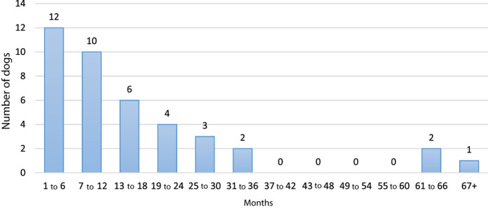
Results: Complete recovery was documented following medical (36/119, 30.3%) or surgical (83/119, 69.7%) management of initial cervical disc extrusion. There was a recurrence of consistent clinical signs in 40 of 119 (34%) cases, of which 27 of 83 (33%) were initially managed surgically and 13 of 36 (36%) medically. In 24 of 40 (60%) cases, there was imaging confirmation of recurrent extrusion; in medically managed dogs, recurrence mostly occurred at the same site, whereas after surgery, recurrence most commonly involved an adjacent disc. Of the 40 recurrences, 32 (80%) occurred within 2 years of diagnosis. Rate of recurrence was similar between treatment methods in both univariable and multivariable time-to-event analyses (hazard ratio 1.03; 95% confidence interval: 0.67 to 1.53; P=0.87).
Clinical significance: Following successful initial medical or surgical treatment, clinical signs consistent with recurrent cervical disc extrusion occurred with similar frequency. Medically treated cases tended to have recurrence at the same site as initial presentation, whereas surgical treatment prevented this. Recurrence usually occurred within 2 years. The retrospective study design, small number of recurrences and lack of imaging confirmation of every recurrence should be considered when interpreting the results.
© 2022 The Authors. Journal of Small Animal Practice published by John Wiley & Sons Ltd on behalf of British Small Animal Veterinary Association.
Conflict of interest statement
None of the authors of this article has a financial or personal relationship with other people or organisations that could inappropriately influence or bias the content of the paper.
Figures


References
-
- Aikawa, T. , Fujita, H. , Shibata, M. , et al. (2012) Recurrent thoracolumbar intervertebral disc extrusion after hemilaminectomy and concomitant prophylactic fenestration in 662 chondrodystrophic dogs. Veterinary Surgery 41, 381‐390 - PubMed
-
- Brisson, B. A. (2010) Intervertebral disc disease in dogs. Veterinary Clinics of North America: Small Animal Practice 40, 829‐858 - PubMed
-
- Brisson, B. A. (2017) Intervertebral disc fenestration. In: Current Techniques in Canine and Feline Neurosurgery. Eds Shores A. and Brisson B. A.. New York, United States, John Wiley & Sons Inc.
-
- Brisson, B. A. , Holmberg, D. L. , Parent, J. , et al. (2011) Comparison of the effect of single‐site and multiple‐site disk fenestration on the rate of recurrence of thoracolumbar intervertebral disk herniation in dogs. Journal of the American Veterinary Medical Association 238, 1593‐1600 - PubMed
MeSH terms
LinkOut - more resources
Full Text Sources
Medical
