Review
. 2022 Mar;306(1):25-42.
doi: 10.1111/imr.13055.

Chemo- and mechanosensing by dendritic cells facilitate antigen surveillance in the spleen

Affiliations
Review

Chemo- and mechanosensing by dendritic cells facilitate antigen surveillance in the spleen

Dan Liu et al. Immunol Rev. 2022 Mar.

Abstract

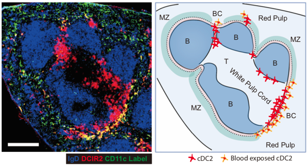
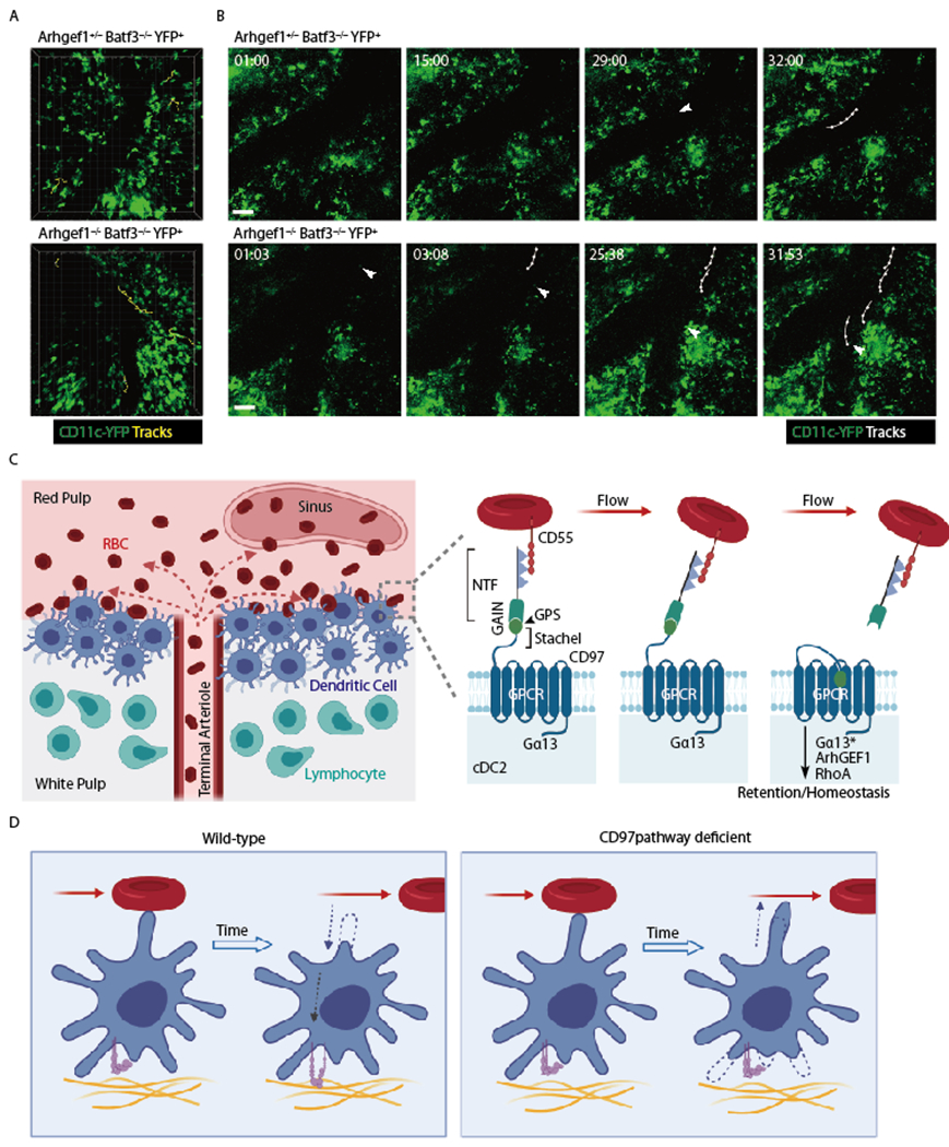
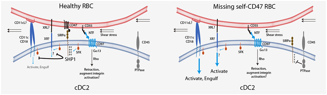
Spleen dendritic cells (DC) are critical for initiation of adaptive immune responses against blood-borne invaders. Key to DC function is their positioning at sites of pathogen entry, and their abilities to selectively capture foreign antigens and promptly engage T cells. Focusing on conventional DC2 (cDC2), we discuss the contribution of chemoattractant receptors (EBI2 or GPR183, S1PR1, and CCR7) and integrins to cDC2 positioning and function. We give particular attention to a newly identified role in cDC2 for adhesion G-protein coupled receptor E5 (Adgre5 or CD97) and its ligand CD55, detailing how this mechanosensing system contributes to splenic cDC2 positioning and homeostasis. Additional roles of CD97 in the immune system are reviewed. The ability of cDC2 to be activated by circulating missing self-CD47 cells and to integrate multiple red blood cell (RBC)-derived inputs is discussed. Finally, we describe the process of activated cDC2 migration to engage and prime helper T cells. Throughout the review, we consider the insights into cDC function in the spleen that have emerged from imaging studies.

Keywords: cell surface molecules; cell trafficking; chemokines; dendritic cells; integrins; spleen.


Conflict of interest statement

Conflict of Interest

J.G.C. is a Scientific Advisory Board member of MiroBio and BeBio and owns stock in ALX Oncology. The other authors have no financial or personal relationships that could be viewed as a conflict of interest.

Figures

Figure 1.
Figure 1.. Section of mouse spleen showing DCIR2 distribution and labeling of blood exposed cells.
Left panel: Spleen section from a mouse that had been i.v. injected with anti-CD11c-PE (green) 3 minutes before organ isolation, stained to detect IgD (blue) and DCIR2 (red). Right panel: Sketch of the main white pulp cord in the spleen image, with principal zones labeled, distribution of cDC2 shown and cDC2 that are blood exposed highlighted.
Figure 2.
Figure 2.. Expression of Flt3l, Dll1, and oxysterol synthesizing enzymes in spleen stromal cells.
Analysis of mouse splenic fibroblastic stromal cell scRNA-seq data from (38). Umap plots generated using Scanpy (50). Upper left plot shows cluster assignments according to the marker genes expressed by each cluster. The remaining Umap plots show the expression of the indicated individual genes (expression level shown as log normalized count) or co-expression of the two indicated genes. Assignment of co-expression required that each cell has at least 1 UMI count of each gene. FDC, follicular dendritic cells; MRC, marginal reticular cells (likely includes marginal sinus lining cells); SMC, smooth muscle cells (or TNC1, triple negative cells-1 (38)); Mesothelial cells are from the spleen capsule and CD34+Lumican+ cells are thought to be predominantly subcapsular fibroblasts but this cluster may also include perivascular reticular cells (PRC) (38); remaining clusters are labeled by marker genes.
Figure 3:
Figure 3:. CD97 pathway restrains cDC2 motility and promotes retention in the spleen.
(A, B) Images from intravital two-photon microscopy of CD11c-YFP+ control (Arhgef1+/−) or Arhgef1−/− cDC in the spleen of mice that lack cDC1 (Batf3−/−). The view in A shows tracks of all YFP+ cells (cDC2) in the imaging volume that moved more than 30μm during the ~30min imaging period. The time series in B show YFP+ cells (cDC2) that enter a large sinus (arrowhead), with each such cell then being tracked over time (min:sec). Imaging was performed as in (17). (C) Left diagram shows cDC2 enriched in a bridging channel (BC) at the interface of the white pulp and red pulp, with cells facing the red pulp being exposed to RBC that are released from an open-ended terminal arteriole. Right panel shows a model of CD97 activation by engagement with CD55 on an RBC (not to scale) that is in fluid flow. The pulling force exerted by CD55 on the CD97 NTF causes its extraction and exposure of the putative tethered ligand domain (stachel sequence) and activation of the GPCR leading to Rho activation and cDC2 retention within the spleen. NTF, N-terminal fragment; Gα13*, activated form of Gα13 that has separated from Gβγ and engaged ArhGEF1. (D) Model of WT versus CD97 pathway deficient cDC2 behavior following encounter with an RBC in flow. Left: Contact with RBC triggers cDC2 to retract a membrane extension and increase integrin mediated adhesion to its tissue microenvironment. Right: In the absence of the CD55-CD97 signal, the cDC2 is shown extending into a region of blood flow.
Figure 4.
Figure 4.. Model for missing self-CD47 sensing by splenic cDC2.
Encounter between cDC2 and healthy RBC leads to close membrane juxtaposition, CD47 binding to SIRPα and exclusion of the bulky CD45 transmembrane PTPase from the interface. SFK-mediated phosphorylation of SIRPα ITIMs allows retention of the SH2-domain containing SHP1 PTPase at the interface and antagonism of SFK-mediated signaling via other receptors, possibly including CD11c/CD18 and an unknown receptor (XR). The existence of XR is postulated because the integrin may be too bulky to locate near SIRPα (and away from CD45) at the cell-cell contact site. The ligands for CD11c/CD18 (CD11cL) and XR (XRL) are unclear, but shear stress may contribute to activation of the CD11c/CD18 integrin. Contemporaneously with CD47 engagement of SIRPα we suggest that CD55 engages CD97 leading to extraction of the CD97 NTF and activation of signaling via Rho to promote cDC2 membrane retraction. This signal may also contribute to inside-out priming of CD11c/CD18 function. In the case of cells with defective or missing self-CD47, SIRPα is not recruited to the interface and remains unphosphorylated due to CD45 PTPase activity. The lack of SHP1 activity at the interface may allow SFK to support signaling via CD11c/CD18 and XR to trigger cDC2 activation and RBC engulfment.

References

    1. Mebius RE, Kraal G. Structure and function of the spleen. Nat Rev Immunol.2005;5:606–616. - PubMed
    1. Lewis SM, Williams A, Eisenbarth SC. Structure and function of the immune system in the spleen. Sci Immunol.2019;4:eaau6085. - PMC - PubMed
    1. Yamamoto K, Kobayashi T, Murakami T. Arterial terminals in the rat spleen as demonstrated by scanning electron microscopy of vascular casts. Scan Electron Microsc.1982;Pt 1:455–458. - PubMed
    1. Schmidt EE, MacDonald IC, Groom AC. Comparative aspects of splenic microcirculatory pathways in mammals: the region bordering the white pulp. Scanning Microsc.1993;7:613–628. - PubMed
    1. Bajenoff M, Glaichenhaus N, Germain RN. Fibroblastic reticular cells guide T lymphocyte entry into and migration within the splenic T cell zone. J Immunol.2008;181:3947–3954. - PMC - PubMed

Publication types