. 2022 Aug;24(4):511-518.
doi: 10.1007/s11307-022-01708-2. Epub 2022 Feb 11.

EGFR-Targeted ImmunoPET of UMUC3 Orthotopic Bladder Tumors

Affiliations

EGFR-Targeted ImmunoPET of UMUC3 Orthotopic Bladder Tumors

Tran T Hoang et al. Mol Imaging Biol. 2022 Aug.

Abstract

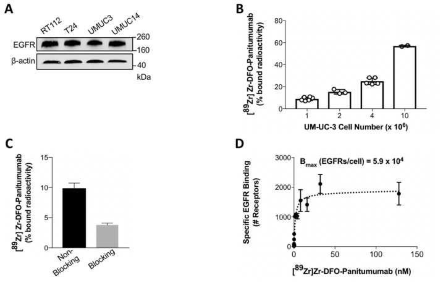
Purpose: Immuno-positron emission tomography (immunoPET) combines the specificity of an antibody with the sensitivity of PET to image dysregulated pathways in cancer. This study examines the performance of immunoPET using the radioimmunoconjugate [89Zr]Zr-DFO-Panitumumab to detect epidermal growth factor receptor (EGFR) expression in an orthotopic model of bladder cancer (BCa).

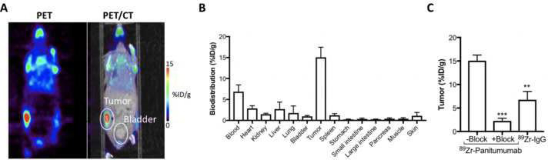
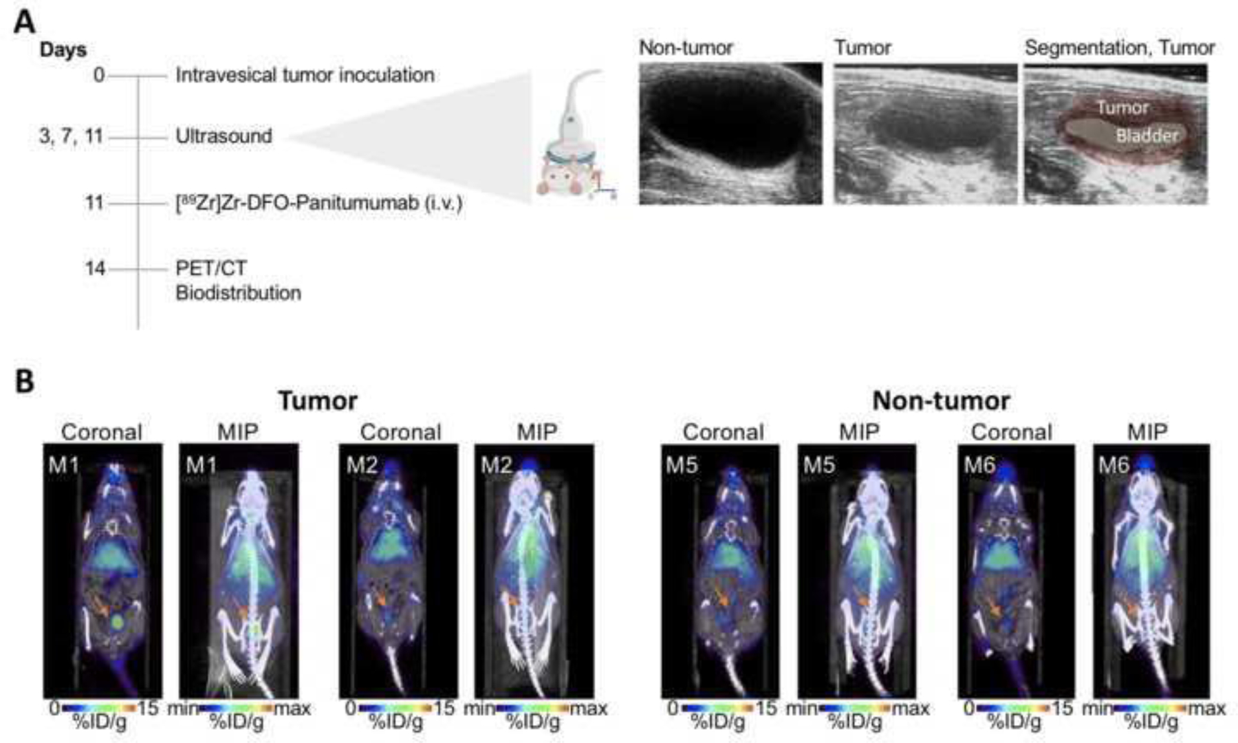
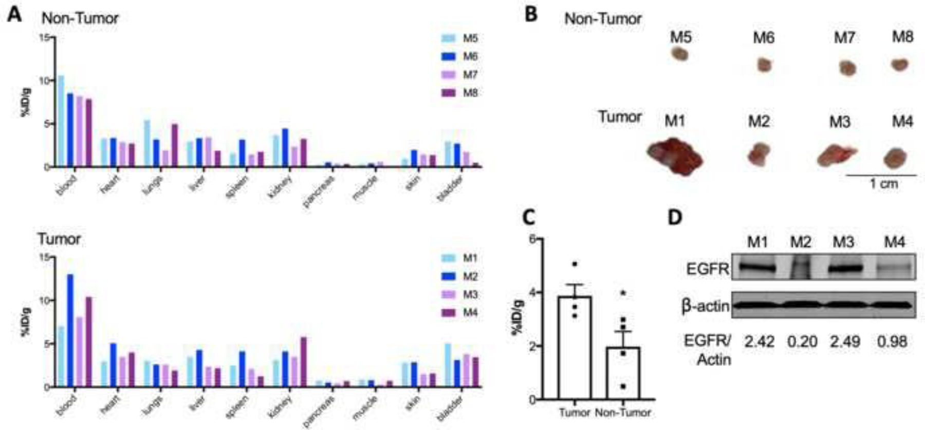
Procedures: Expression and quantification of EGFR receptors were confirmed in four different BCa cell lines. Binding assays validated [89Zr]Zr-DFO-Panitumumab specificity for EGFR-expressing UMUC3 BCa cells. Subcutaneous and orthotopic UMUC3 xenografts were then used for PET imaging and ex vivo biodistribution of the radioimmunoconjugate. Control cohorts included non-tumor mice, 89Zr-labeled non-specific IgG, and blocking experiments.

Results: [89Zr]Zr-DFO-Panitumumab binds specifically to EGFR-expressing UMUC3 cells with a Bmax value of 5.9 × 10⁴ EGFRs/cell in vitro. ImmunoPET/CT images show localization of the antibody in subcutaneous UMUC3 xenografts and murine bladder tumors. In the orthotopic model, the immunoPET signal correlates with the respective tumor volume. Ex vivo biodistribution analysis further confirmed imaging results.

Conclusion: The preclinical data presents a proof of concept for utilizing EGFR-targeted immunoPET to image BCa with altered EGFR protein levels.

Keywords: Bladder Cancer; EGFR; ImmunoPET.


Conflict of interest statement

CONFLICT OF INTEREST STATEMENT

J. S. Lewis is an Editor-in-Chief for the Molecular Imaging and Biology journal. He recused himself from all aspects of the review of this manuscript and all decisions made in regard to revisions/acceptance.

Figures

Figure 1.
Figure 1.
(A) Western blot analysis of EGFR protein levels in RT112, T24, UMUC3, and UMUC14 bladder cancer cell lines. (B, C) 89Zr-labeled Panitumumab binding to EGFR-expressing UMUC3 bladder cancer cells in the absence (B) and presence (C) of excess Panitumumab. UMUC3 bladder cancer cells (1, 2, 4, or 10 million) were incubated with 37 KBq of [89Zr]Zr-DFO-Panitumumab (0.33–0.36 μg) for 1 h at 4°C. For blocking of 89Zr-labeled Panitumumab binding to cancer cells, cells were incubated with 89Zr-labeled Panitumumab in the presence of 33–36 μg DFO-Panitumumab. Data are presented as mean ± S.E.M, n = 6 (2 independent experiments). (D) UMUC3 cells were incubated with [89Zr]Zr-DFO-Panitumumab (0–125 nM) for 3 h at 4°C. Specific binding of [89Zr]Zr-DFO-Panitumumab and non-linear regression curve fit are represented in black spheres and dotted lines. Data are presented as mean ± S.E.M, n = 3.
Figure 2.
Figure 2.
(A) Representative coronal PET images and (B) biodistribution data of [89Zr]Zr-DFO-Panitumumab in athymic nude mice bearing subcutaneous UMUC3 tumors. PET images and biodistribution were performed at 72 h after tail vein injection of [89Zr]Zr-DFO-Panitumumab (8.25–9.18 MBq, 58–65 μg protein). Bars, n = 4 mice per group, mean ± S.E.M. (C) 89Zr-labeled IgG or 89Zr-labeled Panitumumab accumulation in UMUC3 subcutaneous tumors. Tumors were collected at 72 h after tail vein injection of [89Zr]Zr-DFO-Panitumumab or [89Zr]Zr-DFO-IgG (8.25–9.18 MBq, 58–65 μg protein). Blocking experiments were performed by administering [89Zr]Zr-DFO-Panitumumab in the presence of 2.32 mg DFO-Panitumumab. Data are presented as mean ± S.E.M, n = 4 mice per group. **P < 0.01, based on a Student’s t-test. %ID/g, percentage of injected dose per gram.
Figure 3.
Figure 3.
(A) Schematic of PET imaging and biodistribution studies of [89Zr]Zr-DFO-Panitumumab in orthotopic UMUC3 tumors. Right panel shows representative ultrasound images of murine bladders at 11 days after UMUC3 cells’ implantation in the bladder. (B) Representative coronal and MIP PET images of [89Zr]Zr-DFO-Panitumumab in athymic nude mice bearing orthotopic UMUC3 tumors or mice without tumors (M1, M2, M5, and M6 are mouse identifications of 2 mice included in the respective cohorts). PET images were collected at 72 h after tail vein injection of [89Zr]Zr-DFO-Panitumumab (8.25–9.18 Mq, 58–65 μg protein). %ID/g, percentage of injected dose per gram.
Figure 4.
Figure 4.
(A) Biodistribution data of [89Zr]Zr-DFO-Panitumumab in athymic nude mice without tumors (top panel, labeled as M5-M8 where each bar represents an individual mouse) or with UMUC3 orthotopic tumors (bottom panel, labeled as M1-M4 where each bar represents an individual mouse). Biodistribution was performed at 72 h after tail vein injection of [89Zr]Zr-DFO-Panitumumab (8.25–9.18 MBq, 58–65 μg protein). Each bar represents data collected for one mouse of each cohort. (B) Bladder tumors at 14 days after UMUC3 cells’ implantation in the bladder. (C) 89Zr-labeled Panitumumab accumulation in UMUC3 orthotopic tumors or non-tumor bladders at 14 days after UMUC3 cells’ implantation in the bladder. Bladders were collected at 72 h after tail vein injection of [89Zr]Zr-DFO-Panitumumab (8.25–9.18 MBq, 58–65 μg protein). Data are presented as mean ± S.E.M, n = 4 mice per group. *P < 0.05, based on a Student’s t-test. (D) EGFR protein levels in UMUC3 orthotopic bladder tumors. Tumors were collected and EGFR expression analyzed by western blot for mice labeled as M1, M2, M3, and M4. β-actin is a loading control.

References

    1. Siegel RL, Miller KD, Jemal A. Cancer statistics, 2019. CA Cancer J Clin 2019;69(1):7–34. - PubMed
    1. Lenis AT, Lec PM, Chamie K, Mshs MD. Bladder Cancer: A Review. JAMA 2020;324(19):1980–91. - PubMed
    1. Grasso M Bladder Cancer: A major public health issue. Eur Urol Suppl 2008;7(7):510–5.
    1. Wong VCK, Ganeshan D, Jensen CT, Devine CE. Imaging and management of bladder cancer. Cancers 2021;13(6). - PMC - PubMed
    1. Escorcia FE, Steckler JM, Abdel-Atti D, Price EW, Carlin SD, Scholz WW, et al. Tumor-specific 89Zr immuno-PET imaging in a human bladder cancer model. Mol Imaging Biol 2018;20(5):808–15. - PMC - PubMed

Publication types