. 2022:2438:217-230.
doi: 10.1007/978-1-0716-2035-9_14.

Two-Photon Cell and Tissue Level Laser Ablation Methods to Study Morphogenetic Biomechanics

Affiliations

Two-Photon Cell and Tissue Level Laser Ablation Methods to Study Morphogenetic Biomechanics

Abigail R Marshall et al. Methods Mol Biol. 2022.

Abstract

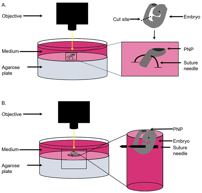
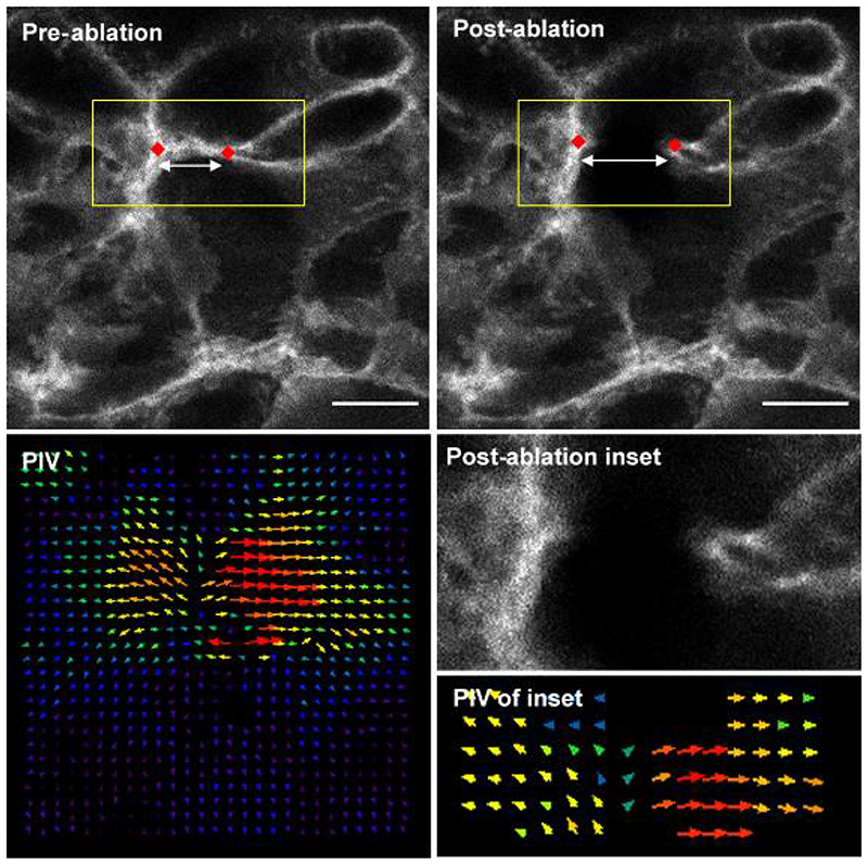
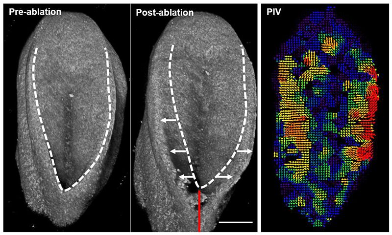
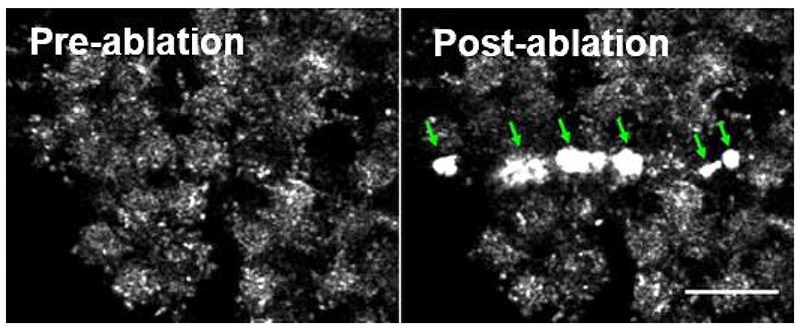
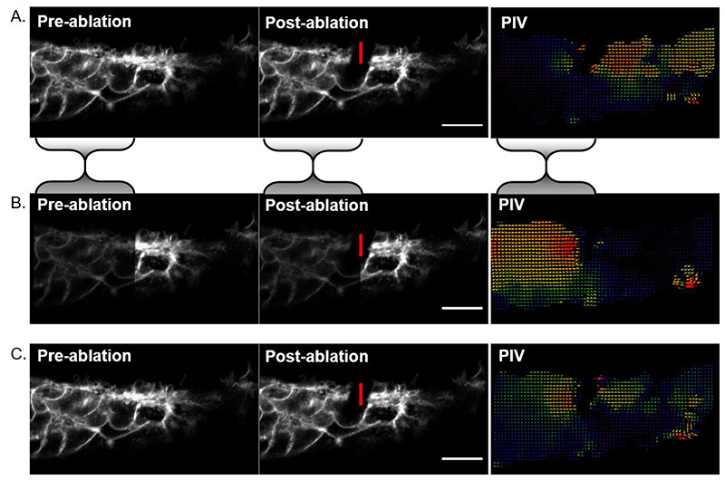
Laser ablation is routinely performed to infer mechanical tension in cells and tissues. Here we describe our method of two-photon laser ablation at the cellular and tissue level in mouse embryos. The primary outcome of these experiments is initial retraction following ablation, which correlates with, and so can be taken as a measure of, the tensile stress that structure was under before ablation. Several experimental variables can affect interpretation of ablation tests. Pre-test factors include differences in physical properties such as viscoelasticity between experimental conditions. Factors relevant during the test include viability of the cells at the point of ablation, image acquisition rate and the potential for overzealous ablations to cause air bubbles through heat dissipation. Post-test factors include intensity-biased image registration that can artificially produce apparent directionality. Applied to the closing portion of the mouse spinal neural tube, these methods have demonstrated long-range biomechanical coupling of the embryonic structure and have identified highly contractile cell populations involved in its closure process.

Keywords: Biomechanics; Laser ablation; Mouse; Neural tube; Two photon.


Figures

Figure 1
Figure 1. Schematic to demonstrate positioning of embryos for single cell and tissue laser ablation.
A) For single cell ablations, the caudal region of the embryo is cut using forceps and positioned by piercing the ventral half of the tissue using a curved suture needle. The tissue is then held in place with both ends of the suture needle fixed in the agarose. B) For tissue level ablation, whole embryos are positioned by creating a hole in the agarose and piercing the embryo through the body to the walls of the hole using a suture needle.
Figure 2
Figure 2. Representative single cell border laser ablation of surface ectoderm rostral to the mouse PNP.
The distance between two reference points (red diamonds) can be compared before and after ablation as a measure of initial recoil. Particle Image Velocimetry (PIV) reveals the extent of movement in the regions around the border. Direction of arrows represent direction of pixel movement. Length and size of arrows represents magnitude of movement. Scale bars = 10 μm.
Figure 3
Figure 3. 3D projections of representative tissue-level ablation rostral to the mouse PNP fusion point.
The caudal end of the embryo is at the top of the image. The dashed line indicates the outline of the PNP before ablation, white arrows indicate PNP widening due to lateral tissue recoil and the red line indicates the ablated region. Recoil magnitude varies between genotypes and developmental stages (20). PIV analysis enables visualisation of PNP deformation several hundred microns caudal to the ablation site as previously reported (4). Scale bar = 100 μm.
Figure 4
Figure 4. Air bubbles (arrows) produced following laser ablation.
These bubbles are visible as brightly reflective surfaces on reflection imaging as shown here. They can also be seen under a stereomicroscope and can be dislodged, causing them to float off the tissue. Bubble formation precludes analysis of cell border recoil adjacent to them. Scale bar = 20 μm.
Figure 5
Figure 5. Asymmetrical image brightness alters intensity-based registration output.
A) Cell-level laser ablation registered using rigid body registration. The bulk of the signal is on the left of the image so registration reduces apparent displacement in that portion relative to the right side of the image. This makes it look like the post-ablation recoil was directionally biased to the right in the PIV analysis. B) As an illustrative example, image intensity was halved in the region bounded by the shaded brackets and rigid body registration was repeated. Now the right side of the image is brighter, so its apparent deformation is minimised at the expense of greater discrepancy on the left. PIV shows leftward bias. C) Intensity-independent landmark-based registration suggests the recoil is symmetrical and largely restricted to the region surrounding the ablation. Vertical red lines indicate the ablated border, scale bars = 20 μm.

References

    1. Nikolopoulou E, Galea GL, Rolo A, Greene ND, Copp AJ. Neural tube closure: cellular, molecular and biomechanical mechanisms. Development. 2017;144:552–566. - PMC - PubMed
    1. Vijayraghavan DS, Davidson LA. Mechanics of neurulation: From classical to current perspectives on the physical mechanics that shape, fold, and form the neural tube. Birth Defects Res. 2017;109:153–168. - PMC - PubMed
    1. Butler MB, et al. Rho kinase-dependent apical constriction counteracts M-phase apical expansion to enable mouse neural tube closure. J Cell Sci. 2019;132 - PMC - PubMed
    1. Galea GL, et al. Biomechanical coupling facilitates spinal neural tube closure in mouse embryos. Proc Natl Acad Sci U S A. 2017;114:E5177–E5186. - PMC - PubMed
    1. Zulueta-Coarasa T, Fernandez-Gonzalez R. Tension (re)builds: Biophysical mechanisms of embryonic wound repair. Mech Dev. 2017;144:43–52. - PubMed

Publication types

LinkOut - more resources